Abstract
Ameloblastoma is a benign locally invasive tumor with a high tendency to recur. It is considered rare in the pediatric population, with most cases diagnosed in the third to fifth decades of life. Approximately 80% of ameloblastomas occur in the molar and ramus region of the mandible, while 20% of cases occur in the maxillary posterior region. This report presents a case of plexiform ameloblastoma in an uncommon location in an 8-year-old child. The lesion was initially thought to be a dentigerous cyst, based on its location and radiographic appearance. The clinical and radiographic features, histopathology, and treatment of solid, plexiform, maxillary ameloblastoma are reviewed, with an added emphasis on a literature review of ameloblastoma in children. This report emphasize the importance of long-term follow-up, since recurrence may occur many years after initial tumor removal.
Keywords: Ameloblastoma, Child, Cone-Beam Computed Tomography, Maxilla
Ameloblastoma is the most common tumor of the odontogenic epithelium, representing roughly 1% of all oral odontogenic epithelial tumors and 11% of all odontogenic tumors.1 Ameloblastomas are persistent, grow slowly, are locally invasive, and demonstrate benign growth characteristics.1
Ameloblastoma is considered a rarity in children, who account for only approximately 10–15% of all reported cases of ameloblastoma.2 Most cases are diagnosed in the third to fifth decades of life, but the lesion can be found in any age group.3 Ameloblastoma affects males and females with equal frequency, but some authors found the rate of occurrence to be higher in males.3,4,5,6 Approximately 80%–85% of ameloblastomas occur in the molar and ramus region of the mandible, followed by the mandibular symphyseal area. The remaining 15%–20% of cases occur in the maxilla, usually in the posterior region. Ameloblastomas in the maxilla may extend into the maxillary sinus and nasal floor.3,7
Based on the 2005 World Health Organization histological classification, ameloblastomas are divided into 4 types: conventional solid or multicystic, unicystic, peripheral (extraosseous), and desmoplastic. The conventional solid or multicystic type is the most common variant, accounting for 75%–86% of all cases.1,7 The follicular and plexiform patterns are the most common histopathological variants of the solid or multicystic type.8
Radiographically, these lesions appear as unilocular or multilocular radiolucencies with a soap-bubble or honeycombed appearance. In some cases, ameloblastomas appear as a circumscribed radiolucency surrounding the crown of an unerupted tooth, resembling a dentigerous cyst. Resorption of the adjacent tooth is not uncommon.9 Diagnosis is confirmed through the radiographic appearance of the lesion, its clinical behavior, and most definitively, biopsy of the lesion.10
This case report presents a case of plexiform ameloblastoma in an uncommon location in an 8-year old child. The lesion was initially diagnosed as a dentigerous cyst, based on its location and radiographic appearance. The importance of long-term follow-up is demonstrated. The clinical and radiographic features, histopathology, and treatment of solid, plexiform, maxillary ameloblastoma are reviewed, with an added emphasis on a literature review of ameloblastoma in children.
Case Report
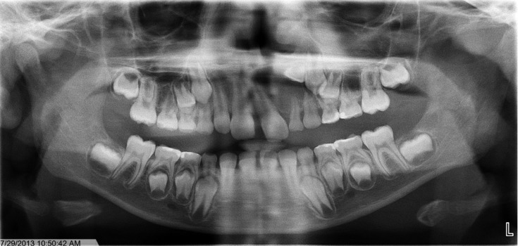
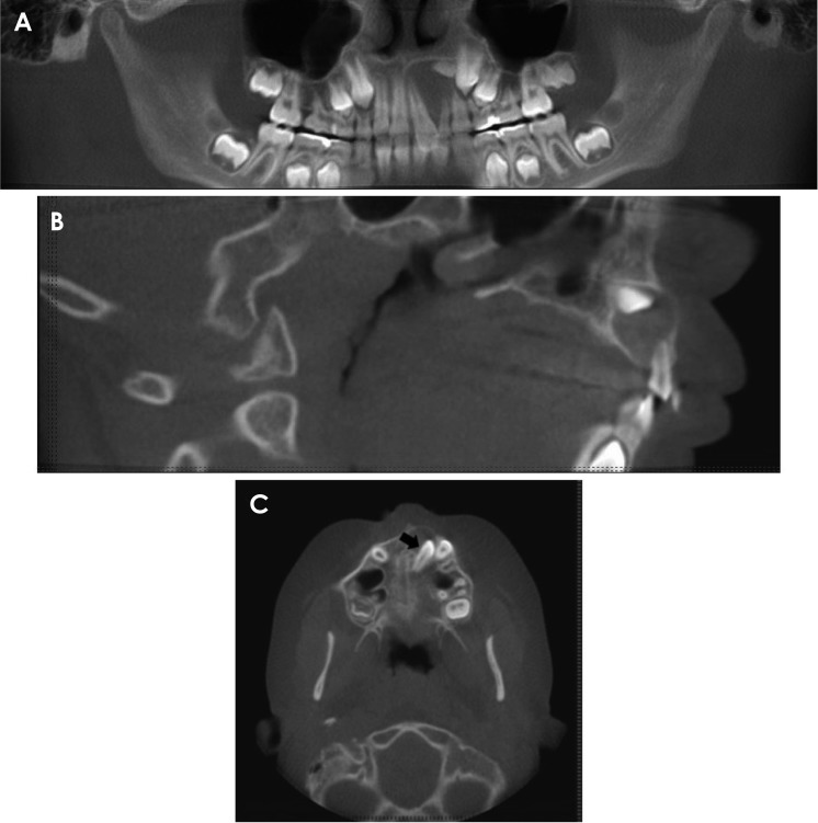
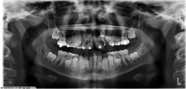
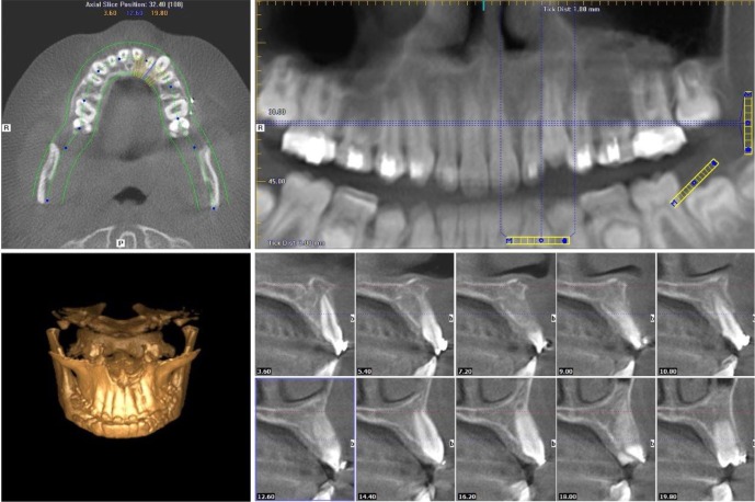
An 8-year-old boy presented to the Rutgers School of Dental Medicine with the chief complaint of pain and swelling of the upper anterior region for the past month. The patient's mother reported that 1 year prior, he had swelling in the same area and underwent a decompression procedure under intravenous sedation. No biopsy was taken at that time. According to the patient and his mother, the present enlargement was larger than the previous one. Intraoral examination revealed expansion of the left maxillary vestibule, with tenderness to palpation, from the permanent maxillary left central incisor to the primary maxillary left first molar. Diastema was present between the maxillary central incisors. An initial panoramic radiograph demonstrated a small, oval radiolucency around the impacted maxillary left lateral incisor (Fig. 1). A conebeam computed tomography (CBCT) examination was performed (iCAT Next Gen; Imaging Sciences, Hatfield, PA, USA), with a dose area product of 312.9 mGy·cm2 and settings of 120 kVp, 5 mA, and a 0.3-mm voxel size. The panoramic and multiplanar CBCT reconstructions revealed that the maxillary left lateral incisor was impacted and horizontally positioned. The crown of this tooth was surrounded by a corticated radiolucency measuring 1.5 cm×1.5 cm×1.8 cm. The buccal and palatal cortices were expanded in this area and the maxillary left central incisor was displaced buccally, with its root directed towards the midline. The unerupted maxillary left canine was displaced distally. A hypertrophic left inferior nasal concha was noted, as well as deviation of the nasal septum to the right (Fig. 2).
Fig. 1. Panoramic radiograph taken on the first visit. A small oval radiolucency can be observed around the impacted maxillary left lateral incisor.
Fig. 2. Cone-beam computed tomography (CBCT) images taken on the first visit. A. CBCT panoramic reconstruction shows a low-density area surrounding the permanent maxillary left central and lateral incisor and causing displacement of the teeth. B. Sagittal CBCT view shows expansion of the buccal and palatal cortices in the anterior maxilla. C. Axial CBCT view at the level of the maxilla. The arrow indicates the impacted maxillary left lateral incisor.
Based on the clinical and radiographic findings, a provisional diagnosis of dentigerous cyst associated with an impacted tooth (the maxillary left lateral incisor) was made. The patient was referred to the Department of Orthodontics for evaluation for treatment in anticipation of surgical removal of the lesion and placement of a space maintainer and arch wire. The goal of this intervention was to bring the impacted tooth into the arch. Informed consent from the parent was obtained before all procedures.
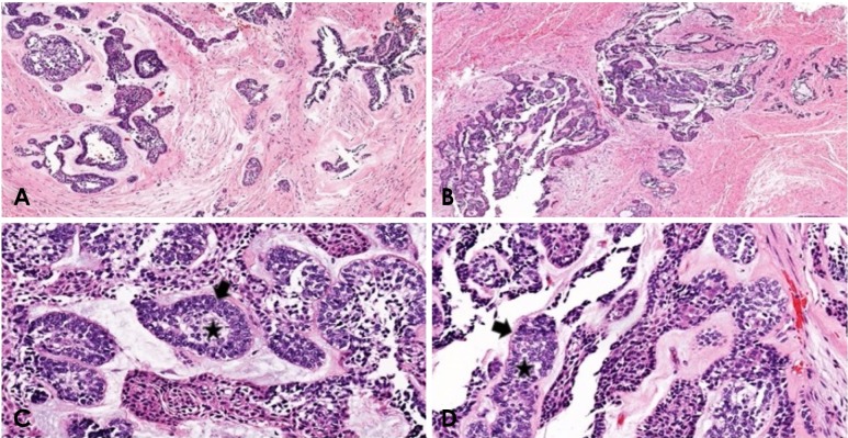
Prior to the surgical removal of the lesion, the primary maxillary left lateral incisor and canine were extracted without any complications. Excision of the low-density lesion was performed under local anesthesia and the impacted maxillary left lateral incisor was exposed. At the time of biopsy, it was noted that the lesion was a solid tumor with no appreciable amount of fluid, as would be expected with a dentigerous cyst. The lesion was removed in fragments in order to avoid compromising the maxillary left lateral incisor (Fig. 3). The entire lesion was placed in formalin and sent to an oral pathologist for a histopathological examination. The maxillary left lateral incisor was exposed, bonded, and ligated at the time of the procedure. The patient was referred back to the Department of Orthodontics for repositioning of the impacted maxillary left lateral incisor. The histopathological examination revealed cords of epithelial elements within the stroma without cystic degeneration identified within the specimen. The epithelial elements were composed of well-differentiated palisaded cells with nuclei that were polarized away from the basement membrane and showed budding proliferative cords. All these features are characteristic of the plexiform variant of ameloblastoma (Fig. 4).
Fig. 3. Clinical photographs taken at the time of surgical enucleation. A and B. Impacted maxillary left lateral incisor exposed, bonded, and ligated at the time of the procedure. C. Fragments of the lesion and extracted primary maxillary left lateral incisor and canine.
Fig. 4. Hematoxylin and eosin stained sections of the lesion demonstrating cords and sheets of anastomosing odontogenic epithelial cells consistent in appearance with the plexiform variant of ameloblastoma. Note the epithelial cells, which show reverse polarization away from the basement membrane (arrowheads), and the stellate reticulum-like cells and suprabasal cells, which compose loosely arranged angular cells (star). A and B are ×4 magnification fields, C and D are ×10 magnification fields.
The oral and maxillofacial surgery team devised the following 3 treatment options and discussed them with the patient's mother: 1) observation, serial extraction, continued treatment, and long-term follow-up with a high risk of recurrence; 2) extraction of the maxillary left central incisor, lateral incisor, and canine and limited ostectomy, with a moderate risk of recurrence; and 3) resection with extraction of multiple teeth in the anterior maxilla.
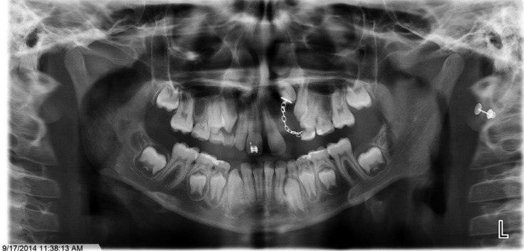
As the patient's parents were not willing to consent to further surgical treatment at this time, they agreed to the first option. The postoperative healing was uneventful. The patient is being followed for 5 years, with annual CBCT scans and panoramic radiography to check for any signs of recurrence (Fig. 5). A postoperative panoramic radiograph was taken after 1 year, and it showed no evidence of recurrence. However, the panoramic radiograph obtained after 2 years of follow-up showed a radiolucency distal to the maxillary left lateral incisor (Fig. 6).
Fig. 5. Postoperative panoramic radiograph (2014) taken after 1 year shows the maxillary left lateral incisor protruding through the mucosa.
Fig. 6. Panoramic radiograph (2015) shows a radiolucency distal to the maxillary left lateral incisor.
A CBCT examination was prescribed and conducted in 2015, subsequent to the panoramic radiograph, to rule out recurrence. A slightly concave appearance of the buccal cortex was seen in the region of interest. An enlarged periodontal ligament space with partially missing buccal and palatal cortices, as well as altered trabecular architecture, was found around the maxillary left lateral incisor. The periodontal enlargement around the maxillary left lateral incisor may have been related to orthodontic tooth movement (Fig. 7).
Fig. 7. Cone-beam computed tomography cross-sectional images show an enlarged periodontal ligament space, missing buccal plate, and bone loss around the maxillary left lateral incisor.
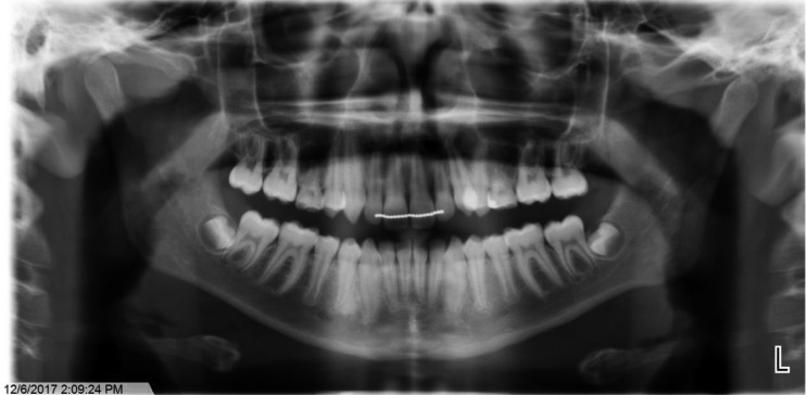
The CBCT and panoramic examinations were repeated after 1 year and the reorganized trabecular pattern was almost identical to the previous scan. The defect appeared smaller, with corticated margins giving a pseudo-canal appearance. The periodontal ligament around the maxillary left lateral incisor was no longer widened, and there was no distinct radiographic evidence of recurrence (Fig. 8).
Fig. 8. Panoramic radiograph (2017) shows no distinct radiographic evidence of recurrence.
An oval radiolucency in the interdental bone between the maxillary left canine and first premolar was noted. The roots of these teeth were displaced. Due to the patient's history, recurrence of ameloblastoma was strongly suspected. A CBCT examination and incisional biopsy were recommended.
The most recent CBCT scan showed no changes from the previous CBCT scan, suggesting the absence of any signs of recurrence. The patient was advised to present for regular annual clinical and radiographic follow-ups.
Discussion
Ameloblastoma is a benign, but locally invasive tumor with a high tendency to recur. These lesions may derive from the remnants of dental lamina, from a developing enamel organ, from the epithelial lining of a preexisting odontogenic cyst, or from the basal cells of the oral mucosa.11
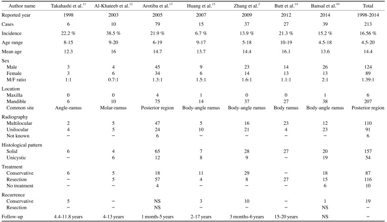
Ameloblastoma is considered rare in the pediatric population, so a literature review was conducted to determine whether any correlations of this tumor with various factors in children and adolescents are known to exist. The search used the PubMed database for published articles on ameloblastoma, with an emphasis on its presentation in children. The MeSH terms used in the search were “ameloblastoma” AND “children.” Only case series of ameloblastoma in the pediatric population that were reported in the last 20 years (1997–2017) were included (Table 1).
Table 1. Literature review of case series of ameloblastoma in children and adolescents.
NS: not stated
According to the literature review, the overall proportion of ameloblastoma incidence in patients less than 20 years old was 16.6% (213 cases out of 1286 total cases), which is similar to the proportions of 15.2% reported by Bansal et al. and 13.9% found by Zhang et al.2,4 However, Takahashi et al. (22.2%),12 Al Khateeb et al. (38.5%),13 Arotiba et al. (21.9%),5 and Butt et al. (21.3%)6 reported higher proportions. The majority of lesions (90.1%; 192 of 213) in this age group occurred in patients between the ages of 11 and 20 years; only 9.9% (21 of 213) of the cases were found in patients 10 years of age or younger. The included articles documented 124 affected males and 89 affected females, yielding a male-to-female ratio of 1.39:1. Takahashi et al. and Butt et al. reported an equal sex distribution.6,12 A gradually growing painless swelling of the jaw was the chief complaint of the majority of the patients. The site distribution in the published literature showed that ameloblastomas have a marked predilection for the mandible. Roughly 97% of cases were found in the mandible, meaning that the mandible (207 of 213) was affected 35 times more often than the maxilla (6 of 213). The molar-ramus region was the most common mandibular site, followed by the symphyseal region. Arotiba et al. reported that this tumor has a site predilection for the symphyseal region of the mandible in the African population.5 A similar observation was made by Chukwuneke et al. recently in a study conducted in the Nigerian population, as 58% of the lesions were reported to be in the anterior mandible or symphyseal region.14
In terms of radiological findings, it has been reported that unilocular ameloblastomas tend to occur more commonly in younger age groups.2,4,12,15 This is in agreement with the findings of Takahashi et al. (66.67%), Huang et al. (66.67%), Zhang et al. (56.76%), and Bansal et al. (59%). However, the results of the literature review revealed that multilocular radiolucent lesions (54.6%) predominated over unilocular lesions (44%). Butt et al. reported that the majority (85.2%) of their cases exhibited the typical soap-bubble or multilocular radiological pattern.6 Arotiba et al. noted a higher rate of root resorption with multilocular lesions (21.3%) than unilocular lesions (16.7%).5 In the present case, the lesion appeared as a unilocular radiolucency with an impacted maxillary left lateral incisor mimicking a dentigerous cyst. Even though the clinical examination and radiographic evaluation provide important clues, the diagnosis and treatment plan are ultimately dependent on the histopathological evaluation.
The follicular and plexiform patterns are the most common histopathological variants of ameloblastoma. Less common histopathological patterns include acanthomatous, granular cells, desmoplastic, and basal cell types.8 Solid or multicystic lesions are more aggressive and demonstrate a higher rate of recurrence than the unicystic variant. Histologically, the solid/multicystic type (157 out of 213) predominated over the unicystic type. Some authors have reported a higher proportion of unicystic ameloblastoma in pediatric population.13,15,16 By correlating radiographic findings with histological type, 32 of 157 cases of solid multicystic tumors presented as unilocular radiolucencies. This was reported by Takahashi et al., Huang et al., Zhang et al., and Bansal et al.2,4,12,15 The present case was also visualized radiographically as a solid tumor with a unilocular radiolucency. Therefore, when ameloblastomas appear as unilocular lesions radiographically, the solid type should be included in the differential diagnosis.
Management of ameloblastoma in children is controversial, because surgical resection and reconstruction can affect maxillofacial development. There are 2 approaches to treatment: conservative and radical. The conservative approach involves enucleation in conjunction with other adjuncts, such as the use of liquid nitrogen, cryotherapy, chemical cautery, or curettage with peripheral ostectomy.11,16,17,18 Radical approach includes surgical resection with wide margins of uninvolved bone and soft tissue. The literature review suggested that the recommended bone margins are 1.5–2.0 cm for the solid/multicystic histological type.11,18 Takahashi et al.12 believed that plexiform ameloblastomas behave less aggressively and recommended conservative treatment in children. Huang et al.15 suggested initially performing a decompression procedure to reduce the tumor volume and to obtain optimal specimens for histopathological examinations in cases of the cystic type of ameloblastoma. Most authors have recommended conservative treatment such as enucleation, followed by curettage and liquid nitrogen cryospray or Carnoy's solution for the unicystic variant. However, recent studies have revealed that type 3 unicystic ameloblastomas are aggressive and should be treated as radically as solid ameloblastomas.2,4,11,16 Pogrel and Montes16 found that simple enucleation alone plays no role in the management of solid/multicystic ameloblastoma, because of its high recurrence rate (60%–80%). In this case, surgical resection with 1.5–2 cm of bone margin and extraction of multiple upper anterior teeth was the most appropriate treatment option. However, upon the request of the patient's parents, no surgical resection was performed. Since reappearance of the initial lesion can even occur 20 years after initial treatment, long-term follow-up is essential. Several clinicians recommend annual radiographic evaluations for a minimum of 10 years.
This report of a rare case of ameloblastoma focused on the occurrence of a solid, plexiform, unilocular maxillary ameloblastoma in an 8-year-old child. This case is unusual in light of the literature on ameloblastoma in children because of the patient's age, tumor location, and radiographic and histopathological findings. None of the cases found in the literature review had all the characteristics of the present case. Clinically, ameloblastoma demonstrates a range of appearances, ranging from a small cyst-like lesion to an extensive multilocular lesion affecting the whole jaw. Pediatric patients' age, tumor size, location, histological type, and craniofacial development should be considered prior to treatment. Long-term follow-up is necessary, because recurrence may occur many years after tumor removal.
References
- 1.Neville BW, Damm DD, Allen CM, Bouquot JE. Oral and maxillofacial pathology. 3rd ed. St. Louis: Saunders/Elsevier; 2009. [Google Scholar]
- 2.Zhang J, Gu Z, Jiang L, Zhao J, Tian M, Zhou J, et al. Ameloblastoma in children and adolescents. Br J Oral Maxillofac Surg. 2010;48:549–554. doi: 10.1016/j.bjoms.2009.08.020. [DOI] [PubMed] [Google Scholar]
- 3.Kreppel M, Zöller J. Ameloblastoma - clinical, radiological, and therapeutic findings. Oral Dis. 2018;24:63–66. doi: 10.1111/odi.12702. [DOI] [PubMed] [Google Scholar]
- 4.Bansal S, Desai RS, Shirsat P, Prasad P, Karjodkar F, Andrade N. The occurrence and pattern of ameloblastoma in children and adolescents: an Indian institutional study of 41 years and review of the literature. Int J Oral Maxillofac Surg. 2015;44:725–731. doi: 10.1016/j.ijom.2015.01.002. [DOI] [PubMed] [Google Scholar]
- 5.Arotiba GT, Ladeinde AL, Arotiba JT, Ajike SO, Ugboko VI, Ajayi O. Ameloblastoma in Nigerian children and adolescents: a review of 79 cases. J Oral Maxillofac Surg. 2005;63:747–751. doi: 10.1016/j.joms.2004.04.037. [DOI] [PubMed] [Google Scholar]
- 6.Butt FM, Guthua SW, Awange DA, Dimba EA, Macigo FG. The pattern and occurrence of ameloblastoma in adolescents treated at a university teaching hospital, in Kenya: a 13-year study. J Craniomaxillofac Surg. 2012;40:e39–e45. doi: 10.1016/j.jcms.2011.03.011. [DOI] [PubMed] [Google Scholar]
- 7.Giraddi GB, Arora K, Saifi AM. Ameloblastoma: a retrospective analysis of 31 cases. J Oral Biol Craniofac Res. 2017;7:206–211. doi: 10.1016/j.jobcr.2017.08.007. [DOI] [PMC free article] [PubMed] [Google Scholar]
- 8.Kashyap B, Reddy PS, Desai RS. Plexiform ameloblastoma mimicking a periapical lesion: a diagnostic dilemma. J Conserv Dent. 2012;15:84–86. doi: 10.4103/0972-0707.92614. [DOI] [PMC free article] [PubMed] [Google Scholar]
- 9.Singer SR, Mupparapu M, Philipone E. Cone beam computed tomography findings in a case of plexiform ameloblastoma. Quintessence Int. 2009;40:627–630. [PubMed] [Google Scholar]
- 10.Castro-Silva II, Israel MS, Lima GS, de Queiroz Chaves Lourenço S. Difficulties in the diagnosis of plexiform ameloblastoma. Oral Maxillofac Surg. 2012;16:115–118. doi: 10.1007/s10006-011-0265-x. [DOI] [PubMed] [Google Scholar]
- 11.Payne SJ, Albert T, Lighthall JG. Management of ameloblastoma in the pediatric population. Oper Tech Otolaryngol Head Neck Surg. 2015;26:168–174. [Google Scholar]
- 12.Takahashi K, Miyauchi K, Sato K. Treatment of ameloblastoma in children. Br J Oral Maxillofac Surg. 1998;36:453–456. doi: 10.1016/s0266-4356(98)90462-4. [DOI] [PubMed] [Google Scholar]
- 13.Al-Khateeb T, Ababneh KT. Ameloblastoma in young Jordanians: a review of the clinicopathologic features and treatment of 10 cases. J Oral Maxillofac Surg. 2003;61:13–18. doi: 10.1053/joms.2003.50002. [DOI] [PubMed] [Google Scholar]
- 14.Chukwuneke FN, Anyanechi CE, Akpeh JO, Chukwuka A, Ekwueme OC. Clinical characteristics and presentation of ameloblastomas: an 8-year retrospective study of 240 cases in Eastern Nigeria. Br J Oral Maxillofac Surg. 2016;54:384–387. doi: 10.1016/j.bjoms.2015.08.264. [DOI] [PubMed] [Google Scholar]
- 15.Huang IY, Lai ST, Chen CH, Chen CM, Wu CW, Shen YH. Surgical management of ameloblastoma in children. Oral Surg Oral Med Oral Pathol Oral Radiol Endod. 2007;104:478–485. doi: 10.1016/j.tripleo.2007.01.033. [DOI] [PubMed] [Google Scholar]
- 16.Pogrel MA, Montes DM. Is there a role for enucleation in the management of ameloblastoma? Int J Oral Maxillofac Surg. 2009;38:807–812. doi: 10.1016/j.ijom.2009.02.018. [DOI] [PubMed] [Google Scholar]
- 17.Iordanidis S, Makos C, Dimitrakopoulos J, Kariki H. Ameloblastoma of the maxilla. Case report. Aust Dent J. 1999;44:51–55. doi: 10.1111/j.1834-7819.1999.tb00536.x. [DOI] [PubMed] [Google Scholar]
- 18.McClary AC, West RB, McClary AC, Pollack JR, Fischbein NJ, Holsinger CF, et al. Ameloblastoma: a clinical review and trends in management. Eur Arch Otorhinolaryngol. 2016;273:1649–1661. doi: 10.1007/s00405-015-3631-8. [DOI] [PubMed] [Google Scholar]


