Abstract
Use of 3,4-methylenedioxymethamphetamine (MDMA; ecstasy) has increased dramatically in recent years, yet little is known about its effects on the developing brain. Neonatal rats were administered MDMA on days 1–10 or 11–20 (analogous to early and late human third trimester brain development). MDMA exposure had no effect on survival but did affect body weight gain during treatment. After treatment, body weight largely recovered to 90–95% of controls. MDMA exposure on days 11–20 resulted in dose-related impairments of sequential learning and spatial learning and memory, whereas neonatal rats exposed on days 1–10 showed almost no effects. At neither stage of exposure did MDMA-treated offspring show effects on swimming ability or cued learning. Brain region-specific dopamine, serotonin, and norepinephrine changes were small and were not correlated to learning changes. These findings suggest that MDMA may pose a previously unrecognized risk to the developing brain by inducing long-term deleterious effects on learning and memory.
Keywords: methylenedioxymethamphetamine, development, MDMA, ecstasy, amphetamines, serotonin, dopamine, learning and memory, spatial learning, sequential learning
3,4-Methylenedioxymethamphetamine (MDMA) is a ring-substituted derivative of methamphetamine whose use is increasing. It is often used by young adults at social gatherings known as “raves” (Peroutka et al., 1988; Henry et al., 1992; Randall, 1992). Emergency room presentations and fatalities resulting from MDMA abuse have been reported (Dowling et al., 1987; Henry et al., 1992;Randall, 1992; Screaton et al., 1992). MDMA has also been associated with residual effects, including reports of anxiety, depression, panic, perceptual changes, and sleep disturbances (Peroutka et al., 1988;Kosten and Price, 1992; Allen et al., 1993; Schifano and Magni, 1994;McCann et al., 1994). Nonetheless, the perception of individuals taking MDMA is that it is safe (Randall, 1992).
MDMA administration to adult animals causes reductions in brain serotonin (5-HT) and its metabolite, 5-hydroxyindoleacetic acid (5-HIAA) (Commins et al., 1987; Schmidt, 1987; Ricaurte et al., 1988; Slikker et al., 1988, 1989). The activity of tryptophan hydroxylase, the rate-limiting 5-HT synthetic enzyme, is also reduced (Schmidt and Taylor, 1987). Receptor binding and enzyme kinetic studies of the 5-HT transporter show a loss of reuptake sites after MDMA treatment (Battaglia et al., 1987; Schmidt, 1987). MDMA administration also results in a loss of 5-HT-labeled immunoreactive fibers and related changes (Commins et al., 1987; O'Hearn et al., 1988; Scallet et al., 1988; Slikker et al., 1988).
MDMA may affect the developing brain differently, because 5-HT has neurotrophic effects before the maturation of its neurotransmitter function (Lauder and Krebs, 1978; Lauder et al., 1983; Lauder, 1988;Whitaker-Azmitia, 1991). For example, neonatal treatment [on postnatal day 10 (P10)-P20] with the tryptophan hydroxylase inhibitorp-chlorophenylalanine induces changes in olfactory and radial-arm learning and reduces MAP2 immunoreactivity in the offspring as adults (Mazer et al., 1997). However, previous developmental studies of MDMA have shown only transient effects on monoamines and behavior (Winslow and Insel, 1990; St. Omer et al., 1991; Broening, 1994). There are no studies on the long-term effects of developmental exposure to MDMA on learning and memory. Understanding such effects may be important because as the use of MDMA increases, there will inevitably be increases in the number of users who are pregnant. Here we report the first evidence that exposure to MDMA in rats during stages analogous to early and late third trimester human fetal brain development (Dobbing and Sands, 1979; Rodier, 1980; Morgane et al., 1992; Bayer et al., 1993; Rodier, 1994) induces specific types of long-term learning and memory impairments.
MATERIALS AND METHODS
Subjects
Male and female Sprague Dawley rats were mated; the day sperm plugs were detected was designated embryonic day 0. At parturition, litters were culled to eight pups (four males and four females) using a random number table. Progeny were individually identified with foot tattoos. Parturition was termed P0. Dams were allowed to wean their offspring naturally (Redman and Sweney, 1976;Blass and Teicher, 1980); pups were separated on P28 and identified by ear punch. Rats were housed in a vivarium accredited by the Association for the Assessment and Accreditation of Laboratory Animal Care and in compliance with all Federal animal care and use guidelines. The protocols described here were approved by the Institutional Animal Care and Use Committee at our institution.
Experimental procedures
Solutions of d,l-MDMA·HCl (National Institute on Drug Abuse, Bethesda, MD) were prepared weekly in sterile isotonic saline and calculated to represent the free base for doses of 0, 5, 10, or 20 mg/kg MDMA in a volume of 5 ml/kg. MDMA was administered by subcutaneous injection twice daily, 8 hr apart. Each dose was administered to one male and one female within each litter. Two dosing periods were investigated: MDMA administration on P1–P10 and MDMA administration on P11–P20. Thirty litters were used: 15 were treated on P1–P10, and 15 were treated on P11–P20.
Behavioral methods
Straight channel trials were performed at an average age of P60 (range, P56–P65). Testing in the multiple-T maze began at an average age of P63 (range, P59–P68). Testing in the visible platform version of the Morris water maze began at an average age of P70 (range, P66–P75). Testing in the hidden platform version of the Morris water maze began at an average age of P77 (range, P73–P82).
Straight channel. Straight channel swimming was performed to (1) acclimate rats to swimming, (2) determine whether there were any motoric deficits before maze testing, and (3) determine whether the test subjects are motivationally comparable. The trials were performed in a 15 × 150 cm water-filled channel with a stainless steel ladder at one end. Each rat received four timed trials to escape the channel after being placed in the opposite end facing away from the ladder (water temperature, ∼22°C).
Multiple-T maze. The water maze for assessing sequential learning was a nine unit multiple-T maze (“Cincinnati” maze) that has been described previously (Vorhees, 1987; Vorhees et al., 1991). The apparatus was constructed of black acrylic and was placed in a large tank of water maintained at ∼22°C. Rats were administered two trials per day for 4 d [path B; intertrial interval (ITI) was 30 min]. Rats failing to escape from the maze in 5 min were removed. The dependent measures were errors of commission (whole-body entries into cul-de-sacs of the Ts) and latency to escape. The maze and the straight channel were located in the same room.
Morris water maze. The Morris hidden platform maze was used as described previously (Brandeis et al., 1989; Vorhees and Minck, 1989; Davis et al., 1992). The water tank was 183 cm in diameter, and the platform was 10 × 10 cm and submerged 2 cm beneath the water surface. The inside of the tank was flat black, and the platform was camouflaged by being made of clear acrylic. Rats received four acquisition trials per day (limit 2 min per trial to find the platform) for 6 d with a 30 sec ITI on the platform. The start position was randomized with the restriction that all four cardinal start positions were used within each set of four trials. On days 2, 4, and 6, rats received one probe trial of memory with the platform removed (60 sec). Water temperature was maintained at ∼22°C. The performance of the rat was tracked automatically using a video tracking system (Polytrack System; San Diego Instruments, San Diego, CA). For acquisition trials, the dependent measures were latency, path length to the platform, and cumulative distance from the platform. For memory (probe) trials, the dependent measures were percentage of time in the target quadrant, average distance from the platform site, and number of platform site crossings. The Morris maze was in a different room than the straight channel and Cincinnati maze.
Hidden platform Morris maze testing was divided into three phases: acquisition, reversal, and reduced (double reversal). For reversal, the platform was placed in the opposite quadrant from acquisition. For reduced, the platform was placed in the opposite quadrant from reversal (i.e., back in the acquisition quadrant). The reduced platform was 5 × 5 cm.
Cued learning. The visible platform procedure was similar to that of the hidden platform version described above. The difference was that the platform was 2 cm above water level to make its location visible. Also, the platform location and the start position were changed on every trial and no probe trials were given. The time limits and number of trials given per day were the same as for the hidden platform condition. The dependent measure was latency to reach the platform.
Growth
Offspring were weighed at the time of each injection and weekly thereafter for the remainder of the experiment.
Neurotransmitter assays
At P105, rats were decapitated, and brains were rapidly removed and dissected over ice into frontal cortex and hippocampus using a brain block (Zivic-Miller, Pittsburgh, PA) that first divided the brain into 1 mm slices. Each slice had the region of interest carved out, and sections of the same region were pooled bilaterally and stored at −70°C until assay. Tissues were thawed, weighed, and diluted with 20 vol of 0.2N perchloric acid containing 400 nm of 3,4-dihydroxybenzylamine as an internal standard. Tissues were ultrasonified and centrifuged at 1000 × g for 10 min. A 200 μl aliquot of supernatant was then removed and filtered through a 0.45 μm pore nylon-66 microfilter, and 25 μl of filtrate was injected into an HPLC apparatus with electrochemical detector. The HPLC system was composed of a Bioanalytical Systems (West Lafayette, IN) PM80 HPLC pump, a Rheodyne (Cotati, CA) 7125 injector, a Supelcosil LC18 3 μm 4.6 × 250 mm reversed-phase analytical column (Supelco, Bellefonte, PA), a Bioanalytical Systems LC-4B amperometric detector, and a reference electrode maintained at an oxidation potential of +0.70 V. The mobile phase consisted of 1 vol of methanol mixed with 9 vol of buffer (0.10m monobasic potassium phosphate, pH 3.0, 1.0 mm 1-heptane sulfonic acid sodium salt, and 40 mg/L EDTA disodium salt). The flow rate was 1.0 ml/min. Chromatograms were recorded and integrated, and neurotransmitter concentrations were calculated from standard curves generated for each analyte. Tissue concentrations were determined for dopamine (DA), DOPAC, homovanillic acid, serotonin, 5-HIAA, and norepinephrine (NE). Patterns for neurotransmitters and metabolites were similar; therefore, only neurotransmitters are reported.
Statistical methods
Behavioral and body weight data were analyzed by mixed-model split-plot ANOVA using the general linear modeling procedure. Main effects were treatment group (dose of MDMA), exposure period, gender, day, and trial. Treatment age was a between factor in all analyses, whereas treatment group, gender, day, and trial were within factors. The experimental unit was the litter. Significant (p < 0.05) treatment-related interactions were analyzed further using simple-effect ANOVAs. Significant treatment main effects or simple effects were analyzed further by the step-down ANOVA method (Kirk, 1995) to control for multiple comparisons. Step-down ANOVAs were continued until no significant differences were encountered or until the level of two-group comparisons. For repeated-measure factors, a test for sphericity was performed to ensure the symmetry of the variance–covariance matrix. The Greenhouse–Geisser correction was used in instances in which these matrices were significantly nonspherical. Neurotransmitters were also analyzed by factorial ANOVA followed by step-down ANOVAs in which treatment effects were obtained.
RESULTS
Growth and survival
There were no effects of MDMA treatment on offspring survival and no body weight differences before or during the first 2–3 d of treatment. After the third treatment day, MDMA produced a dose-dependent reduction in the rate of body weight gain regardless of whether treatment began on P1 or P11. These effects peaked on P14 in the P1–P10 treated groups and diminished thereafter. Similarly, the effects in the P11–P20 treated groups peaked shortly after the end of treatment and then progressively diminished. Both early and late treated MDMA groups showed a small residual body weight reduction (∼10% below controls in the MDMA-20 group and ∼5% below controls in the MDMA-10 and MDMA-5 groups) throughout the experiment. These body weight differences did not correlate with behavior as evidenced by the fact that, first, both P1–P10 and P11–P20 treated groups showed comparable body weight changes, but the learning and memory effects were concentrated in the P11–P20 treated MDMA groups. Second, even among the P11–20 treated MDMA groups, no effects of altered weight were seen on straight channel performance or cued learning, reinforcing the fact that swimming tasks are not affected by body weight differences (Cravens, 1974). Third, the MDMA effects were specific to the phase of Morris maze testing (acquisition and reduced platform trials), whereas weight differences were constant. Fourth, larger developmental weight reductions induced by protein–calorie malnutrition do not impair Morris maze performance (Goodlett et al., 1986; Campbell and Bedi, 1989; Bedi, 1992; Levitsky and Strupp, 1995;Strupp and Levitsky, 1995) (but see Tonkiss et al., 1994, 1997).
Straight channel
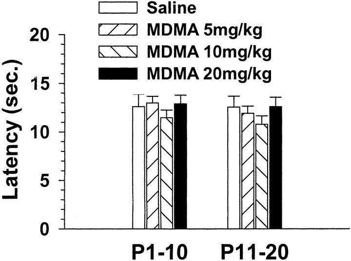
Animals were tested as adults first in a straight channel to determine whether there were effects on swimming performance. MDMA treatment produced no alterations in latency to escape from a straight swimming channel (Fig. 1). This finding indicates that developmental MDMA treatment did not impair swimming performance or alter motivation to escape from water.
Fig. 1.
Latency (seconds) to escape from a straight swimming channel averaged across four trials (mean ± SEM). A one-between, three-within factor ANOVA (age by treatment group by gender by trial) showed no effect of treatment, age, or gender and no interaction between treatment and other factors.
Multiple-T water maze
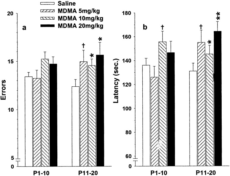
Next, animals were evaluated for performance in a test of sequential learning (Fig. 2). Age at treatment (P1–P10 vs P11–P20) significantly interacted with the group effect of MDMA treatment in all analyses; therefore, the treatment ages were analyzed separately. Significant increases in errors occurred among the MDMA group treated at P11–P20, but not among the MDMA groups treated at P1–P10 (Fig. 2a). For latency, there was a significant MDMA by day effect for the P1–P10 age group. Additional analyses showed a nonsignificant trend toward longer latency in the MDMA-10 group on several days. Rats treated with MDMA on P11–P20 showed a consistent increase in latency to find the escape at all dose levels (Fig. 2b).
Fig. 2.
Errors (a) and latency (b) (seconds) to escape from the multiple-T (Cincinnati) water maze (mean ± SEM averaged across days, trials, and gender). Overall ANOVAs with age as a factor showed that age was part of a significant interaction with treatment; therefore, follow-up ANOVAs within each age group were performed. For errors, the P1–P10 ANOVA showed no treatment effect and one interaction (treatment by day by trial; F(9,126) = 1.99;p < 0.05). The P11–P20 ANOVA showed a treatment effect (F(3,42) = 3.04;p < 0.05) and interactions between treatment by day (F(9,126) = 2.68;p < 0.01), treatment by day by gender (F(9,126) = 1.98; p< 0.05), and treatment by trial by gender (F(3,42) = 3.04; p< 0.05). These interactions all reflected MDMA-induced increases in errors, but the effects were larger on certain days and trials and in females. For simplicity of presentation, the main effect of treatment is shown, because it captures the essence of the principal effects seen in the MDMA groups. For latency, the P1–P10 ANOVA showed a treatment main effect (F(3,42) = 3.17;p < 0.05) and a treatment by day interaction (F(9,126) = 2.76; p< 0.01). These effects represented differences among the MDMA groups (data not shown). The P11–P20 ANOVA for latency showed a treatment effect (F(3,42) = 3.89;p < 0.02) and no interactions. *p < 0.05, **p < 0.01, †p < 0.10 compared with saline controls.
Morris water maze
Cued learning
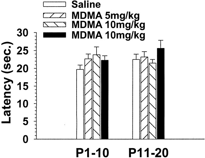
Animals were tested in the Morris water maze for cued learning with the platform above the water to provide proximal cues. Developmental MDMA exposure did not induce alterations in latency to find the visible platform among groups treated at either P1–P10 or P11–P20, indicating that performance in the Morris maze was not impaired when local (visual) cues were available (Fig.3).
Fig. 3.
Latency (seconds) to escape from the Morris water maze during cued learning trials (4 trials per day averaged across 5 d; mean ± SEM). A one-between, four-within ANOVA (age by treatment group by gender by day by trial) showed no main effect of treatment, age, or gender and no treatment interactions with day or trial. Data are shown for males and females combined.
Spatial learning
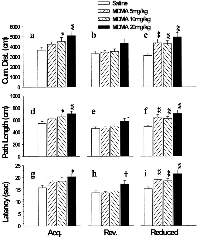
In the hidden platform (spatial) version of the Morris maze, each phase (acquisition, reversal, reduced) contained both learning and memory (probe) trials. Learning phases are presented first. There was a significant interaction of MDMA with treatment age on all three phases of testing; therefore, separate analyses were conducted on the P1–P10 and P11–P20 groups. No MDMA effects were found after treatment on P1–P10. MDMA effects were reliably found after treatment on P11–P20 on the acquisition phase on learning trial measures of cumulative distance from the platform, path length, and latency (Fig.4a,d,g). During reduced platform trials, a similar pattern was observed, (i.e., no effects after P1–P10 treatment but consistent MDMA effects after P11–P20 exposure) (Fig. 4c,f,i). A similar trend was seen during the reversal phase, but with the exception of latency, the other measures were not significant (Fig. 4b,e,h). Group comparisons among the P11–P20 treated groups showed increased cumulative distance from the platform in the MDMA-10 and MDMA-20 groups during acquisition (Fig.4a) and in all three MDMA groups during reduced platform trials (Fig. 4c). A similar pattern was observed for path length; i.e., the MDMA-10 and MDMA-20 P11–P20 groups had longer path lengths on acquisition (Fig. 4d), and all three MDMA groups had longer path lengths on reduced platform trials (Fig.4f). For latency, only the MDMA-20 P11–P20 group had significantly longer latencies on acquisition (Fig. 4g). On reversal trials, the MDMA-20 P11–P20 group showed a trend toward longer latencies (Fig. 4h). On reduced platform-size trials, all three MDMA P11–P20 groups had longer latencies than controls (Fig.4i).
Fig. 4.
Morris water maze spatial learning results in the P11–P20 treatment groups (mean ± SEM) averaged across four trials per day and 5 d. a–c, Cumulative distance from the hidden platform during acquisition (a), reversal (b), and reduced platform (c) trials. d–f, Path length to find the platform during the same three phases of testing.g–i, Latency (seconds) to find the platform during the three phases of testing. No treatment effects were found in the Morris water maze in the P1–P10 treatment groups (data not shown). Treatment effects were obtained on measures of cumulative distance during acquisition trials (a) (F(3,42) = 4.88; p< 0.01) and reduced platform trials (c) (F(3,42) = 8.74, p< 0.0001), but not on reversal. A similar pattern was obtained for path-length analyses (acquisition:F(3,42) = 4.70; p< 0.01; reduced: F(3,42) = 7.55;p < 0.001; reversal was not significant). For latency, treatment effects were obtained on all three phases (acquisition: F(3,42) = 3.31;p < 0.05; reversal:F(3,42) = 3.05; p< 0.05; reduced: F(3,42) = 7.90;p < 0.001). Data for males and females are combined for presentation. Several interactions between treatment and day and gender were also obtained. These indicated that the effects of MDMA were largest in females and on early and middle days and smallest on the last day of testing. *p < 0.05, **p < 0.01, †p < 0.10 compared with saline control.
Memory trials
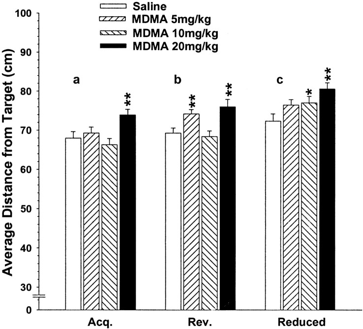
On probe trials [trials with the platform removed to test spatial preference (memory) for the previous location of the platform], there were treatment age by MDMA interactions obtained on measures of platform site crossings and average distance from the platform but not for time in the target quadrant. These effects were found during acquisition, reversal, and reduced platform probe trials. There were no MDMA effects on probe trials among those animals treated on P1–P10. However, there were MDMA effects among those animals treated on P11–P20 for platform site crossings and average distance from the platform site on acquisition, reversal, and reduced platform trials. Group comparisons showed increased average distance from the platform site on probe trials conducted during acquisition in the MDMA-20 group (Fig. 5a). Both the MDMA-5 and the MDMA-20 groups had longer average distances from the platform site on reversal probe trials (Fig. 5b), whereas on reduced platform probe trials the increases were significant in the MDMA-10 and MDMA-20 groups (Fig. 5c). Platform site crossings showed a similar pattern, with reduced site crossings in the MDMA-5 and MDMA-20 groups on acquisition and reversal (data not shown). On reduced platform trials, there was an MDMA by trial interaction at the P11–P20 treatment age; the MDMA-20 group had significantly fewer platform site crossings on probe trial 1, whereas the MDMA-10 group had fewer crossings on probe trial 3.
Fig. 5.
Morris water maze results of memory (probe) trials in the P11–P20 treatment groups (mean ± SEM) averaged across trials; one probe trial was administered on days 2, 4, and 6. There were no treatment main effects among the P1–P10 treatment groups (data not shown). For the P11–P20 treatment groups, treatment main effects were found on average distance from the target during the probe trials for acquisition (F(3,42) = 6.17;p < 0.002), reversal (F(3,42) = 5.88; p< 0.002), and reduced (F(3,42) = 5.58;p < 0.01). Data for males and females were combined for presentation. No interactions with treatment were obtained. *p < 0.05, **p < 0.01 compared with saline controls. Similar patterns were found for the P11–P20 treatment groups on platform site crossings and for the percentage of time spent in the target quadrant (data not shown).
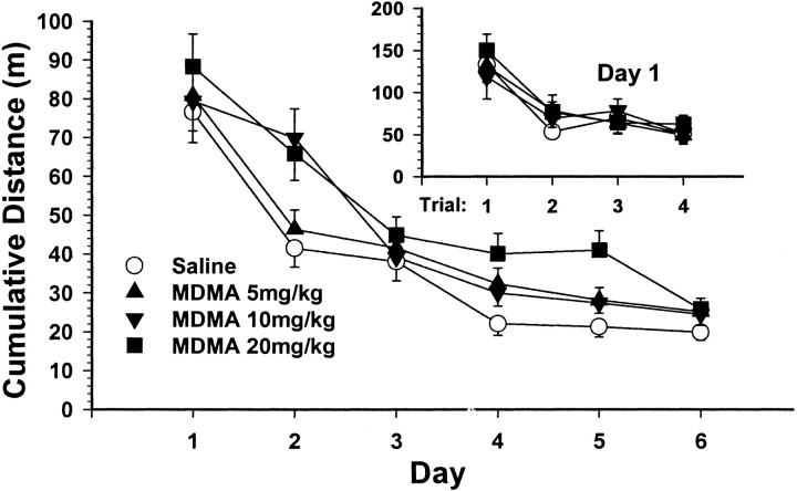
Learning curves for the P11–P20 groups are shown in Figure6. The data are shown for the acquisition phase for cumulative distance from the platform, a measure of proximity to the goal, measured every 55 msec. The differences on day 1 were not significant. A more detailed analysis of day 1 may be seen in Figure 6(inset). There were no group differences on day 1, trial 1, demonstrating that there were no pre-existing differences among the groups before finding the platform for the first time. Clear group differences did not emerge until day 2. Subsequently, controls reached asymptotic performance by day 4, whereas MDMA-20 animals did not approach this level of performance until day 6. The MDMA-10 and MDMA-5 groups showed an intermediate rate of improvement across days and approached control levels of performance on day 5.
Fig. 6.
Cumulative distance from the platform learning curves for the P11–P20 groups for each day of testing during the acquisition phase of Morris water maze testing (mean ± SEM) averaged across genders. Inset, Additional details for each trial for day 1 of testing. There were no significant group differences on day 1 and no differences on trial 1 of day 1, demonstrating that MDMA animals showed no pre-existing performance differences before finding the hidden platform for the first time.
Neurotransmitter findings
After the completion of cognitive testing, animals were killed on P105; brains were removed and frozen for later analysis of monoamine content. There were no main effects or interactions with gender; therefore, only the male data are presented. However, all significant effects were found in both males and females. In the frontal cortex, no changes in dopamine or norepinephrine were obtained. Serotonin content was not changed in the P1–P10 MDMA groups but was affected in the P11–P20 MDMA groups (p < 0.01). Group comparisons showed significant reductions in all three MDMA groups. Among males, the differences ranged from 5 to 11% and were not dose-dependent (Table 1).
Table 1.
Effects of developmental MDMA on adult brain monoamine concentrations in males expressed as nanomoles per gram tissue wet weight (mean ± SEM)
| Dose | P1–10 | P11–20 | ||
|---|---|---|---|---|
| Hippocampus | 5-HT | 0 | 2.07 ± 0.05 | 2.21 ± 0.06 |
| 5 | 1.82 ± 0.041-160 | 2.03 ± 0.061-160 | ||
| 10 | 1.72 ± 0.051-160 | 1.94 ± 0.051-160 | ||
| 20 | 1.82 ± 0.081-160 | 2.08 ± 0.07* | ||
| NE | 0 | 2.76 ± 0.12 | 2.88 ± 0.11 | |
| 5 | 2.88 ± 0.10 | 3.23 ± 0.111-160 | ||
| 10 | 3.00 ± 0.091-160 | 3.20 ± 0.121-160 | ||
| 20 | 2.94 ± 0.121-160 | 3.35 ± 0.081-160 | ||
| Frontal cortex | 5-HT | 0 | 3.16 ± 0.10 | 3.15 ± 0.09 |
| 5 | 3.23 ± 0.10 | 3.00 ± 0.101-160 | ||
| 10 | 3.01 ± 0.10 | 2.80 ± 0.151-160 | ||
| 20 | 2.99 ± 0.09 | 2.93 ± 0.091-160 | ||
| NE | 0 | 2.49 ± 0.08 | 2.44 ± 0.10 | |
| 5 | 2.53 ± 0.06 | 2.59 ± 0.07 | ||
| 10 | 2.56 ± 0.09 | 2.43 ± 0.13 | ||
| 20 | 2.52 ± 0.07 | 2.64 ± 0.07 | ||
| DA | 0 | 1.28 ± 0.23 | 1.70 ± 0.22 | |
| 5 | 1.08 ± 0.10 | 1.24 ± 0.14 | ||
| 10 | 1.41 ± 0.11 | 1.32 ± 0.24 | ||
| 20 | 1.42 ± 0.14 | 1.56 ± 0.44 |
Rats were 105 d old at the time of assay. There were no significant effects of gender; therefore, only data for males are presented.
p < 0.05,
F1-160: p < 0.01 compared with control within each region and neurotransmitter. Group sizes = 15 per cell. Group sizes were the same for females (n = 15 per cell). Dose is milligrams per kilogram per treatment with two treatments per day.
In the hippocampus, serotonin was also reduced (p < 0.01). This effect occurred in both the P1–P10 and P11–P20 age groups (both p < 0.01). In both age groups, all three MDMA groups showed significant reductions. Among males, these ranged from 12 to 17% reductions in the P1–P10 groups and from 6 to 12% in the P11–P20 groups, but the reductions were not dose-dependent in either age group. Norepinephrine in the hippocampus was significantly increased (p < 0.05). This increase was significant for both age groups (bothp < 0.01). Individual group comparisons showed significant increases only in the MDMA-10 and MDMA-20 groups among the P1–P10 age groups but in all MDMA dose groups among the P11–P20 age groups. Among males, the P1–P10 age groups showed NE increases ranging from 6 to 9%, whereas the P11–P20 age groups showed NE increases ranging from 11 to 16%. As with 5-HT, the NE changes were not dose-dependent. In sum, developmental MDMA treatment produced small long-term reductions in brain 5-HT in the hippocampus in both males and females in both P1–P10 and P11–P20 treated offspring, but the effects were not dose-dependent and did not match the cognitive changes, which only occurred after P11–P20 treatment. Similarly, developmental MDMA treatment produced small long-term increases in brain NE in the hippocampus in both males and females in both P1–P10 and P11–P20 treated offspring, but the effects were neither dose-dependent nor correlated with the cognitive changes. There were, however, developmental MDMA-associated reductions in frontal cortex 5-HT that were only seen among the P11–P20 treated MDMA groups. This pattern matched the cognitive changes, but the reductions were small and not dose-dependent.
To determine whether the age-specific frontal cortex 5-HT reductions were quantitatively related to the spatial learning deficits, Pearson product–moment correlation coefficients were calculated between two key indices of spatial learning (average cumulative distance scores for acquisition and reduced platform trials) and 5-HT concentrations. Cumulative distance acquisition and reduced platform scores were chosen because they showed the clearest pattern of MDMA-related changes. Neither the correlation between acquisition (r = 0.15) nor reduced platform scores (r = 0.04) and frontal cortex 5-HT approached significance.
DISCUSSION
The findings demonstrate that developmental MDMA exposure disrupts both sequential and spatial reference memory-based learning. These learning deficits reflect a developmentally specific vulnerability in that they were selective, affecting only those animals treated on P11–P20 and not those treated on P1–P10. The effects were also long-term in that they were seen in the offspring as adults. Third, the effects were not related to any long-term changes in 5-HT, DA, or NE.
The manifestations of developmental MDMA treatment are different from those seen in animals treated with MDMA as adults. In adult animals, MDMA primarily affects the serotonergic system. Administration results in a reproducible profile that consists of lasting reductions in 5-HT content in the forebrain (Commins et al., 1987; Schmidt, 1987; Johnson et al., 1988; Ricaurte et al., 1992). This is accompanied by the loss of tryptophan hydroxylase activity and a reduction in the number of serotonin reuptake sites (Battaglia et al., 1987; Johnson et al., 1988). Some markers of neuronal damage are also seen after adult MDMA exposure. Astrogliosis as reflected by increased GFAP straining and argyrophilia as reflected by silver degeneration staining are reported in the striatum and cortex after high-dose MDMA treatment (Commins et al., 1987; Slikker et al., 1988; O'Callaghan and Miller, 1994, 2000). In addition, an attenuation of the serotonin syndrome is reported after acute adult MDMA administration after a pretreatment regimen with MDMA, suggesting that long-term reductions in 5-HT have functional consequences under certain circumstances (Shankaran and Gudelsky, 1999). In developing animals, by contrast, only modest reductions in frontal cortex and hippocampal serotonin were seen, and norepinephrine in the hippocampus was increased. The magnitude of the MDMA-induced 5-HT reductions is only a fraction of that seen in adult animals treated with MDMA. In adults, serotonin reductions in forebrain regions of ≥50% are reported (Shankaran and Gudelsky, 1999), whereas neonatal treatment produced serotonin reductions of <15%. Others have reported similarly small reductions after developmental MDMA treatment, and no reductions in serotonin reuptake sites are seen after developmental MDMA (Broening et al., 1994, 1995; Aguirre et al., 1998). This raises the possibility that developmental MDMA exposure may induce cognitive deficits through mechanisms other than through injury to serotonin nerve terminals.
The learning deficits found here in the Morris maze are not attributable to malnutrition during development because undernutrition does not induce changes in spatial learning in the Morris maze (Goodlett et al., 1986; Campbell and Bedi, 1989; Bedi, 1992; Levitsky and Strupp, 1995) (but see Tonkiss et al., 1997). Furthermore, the MDMA-induced Morris maze deficits most likely do not occur because of stress (Holscher, 1999) or because of the absence of relevant maze experience, as has been suggested in experiments using NMDA receptor inhibitors (Bannerman et al., 1995; Saucier and Cain, 1995), because of the present design. In the present design, animals received neonatal handling, a manipulation known to attenuate stress effects and improve later Morris maze performance (Meaney et al., 1988). In addition, the animals received straight channel swimming, multiple-T maze swimming, and cued Morris maze swimming before entering the hidden platform (spatial) version of the maze. Similar such experiences are known to reduce or eliminate group differences in spatial learning if either stress or transfer of training were contributing factors to these deficits (Bannerman et al., 1995; Saucier and Cain, 1995; Saucier et al., 1996; Cain, 1997). Given that this was not the case, the findings suggest that MDMA has selective effects on cognitive development that are not confounded by these other factors.
The present study sought to control the rearing environment by using a split-litter design in which all treatment groups were represented within each litter. This controls for litter effects (Holson and Pearce, 1992). However, this design cannot control for differential maternal responsiveness to treated compared with control offspring (Ruppert et al., 1983; Booze and Mactutus, 1985). It is unknown whether MDMA induces differential maternal responses; however, mitigating this concern is the fact that MDMA-induced spatial learning deficits have been replicated using a between-litter design (our unpublished observations). In the latter design, entire litters receive the same treatment, thereby eliminating within-litter differential maternal care as a factor. The finding that the learning impairments are the same using both designs suggests that maternal factors do not significantly alter the effects of MDMA on brain development. Similarly, spatial learning deficits are seen after P11–P20 treatment with methamphetamine regardless of whether a between-litter (Vorhees et al., 1994, 2000) or split-litter design is used (our unpublished observations).
The differences between the responses of adult versus developing rats after MDMA administration most likely occur because of the maturational stage of the CNS at the time of treatment, although other factors such as differences in metabolism or drug disposition have not been ruled out. It has been shown that MDMA and other amphetamines require presynaptic uptake to cause neurotransmitter effects (Schmidt and Gibb, 1985; Schmidt, 1987; Schmidt and Taylor, 1987; Hekmatpanah and Peroutka, 1990; Marek et al., 1990a,b; Battaglia et al., 1991; Berger et al., 1992; Pu et al., 1994). Serotonin and dopamine transporters have distinct developmental profiles (Broening and Slikker, 1998) and are found at lower concentrations during early development. Depending on how 5-HT transporter activity is measured, it is still developing during the period under investigation in the present study (Broening and Slikker, 1998). If this mechanism were critical, the P11–P20 group should have been the most affected, because the 5-HT transporter would be more developed at this age, thereby allowing more MDMA to enter 5-HT terminals. This is consistent with the present findings, but fails to explain why 5-HT content was not more affected if more MDMA was gaining entry into 5-HT terminals during the P11–P20 treatment period compared with the P1–P10 treatment period.
The age-dependent vulnerability seen here may not be unique to MDMA. We have observed spatial learning and memory deficits in rats exposed tod-methamphetamine on P11–P20 but not after exposure on P1–P10 (Vorhees et al., 1994). This effect also occurs in the absence of effects on cued learning (Vorhees et al., 2000). However, in contrast to what is seen after MDMA, neither P1–P10 nor P11–P20 treatment with d-methamphetamine impairs sequential learning (Vorhees et al., 1994). Yet the P11–P20 MDMA-treated offspring showed consistent deficits in sequential learning measured in terms of errors and latency to escape. The fact that the multiple-T maze test of sequential learning does not require spatial cues (animals can learn this task in the dark; M. T. Williams and C. V. Vorhees, unpublished observations) suggests that developmental MDMA treatment has effects on nonhippocampally dependent behaviors as well as on hippocampally dependent ones such as the Morris maze.
Together, the developmental data on substituted amphetamines reveal that MDMA and d-methamphetamine have different but overlapping effects on learning and memory. Both drugs share the feature that P11–P20 is a period of greater vulnerability compared with P1–P10 exposure. This developmental period is analogous to the third trimester in humans in terms of neuroanatomical development (Bayer et al., 1993; Rice and Barone, 2000). Hence, the present data raise concerns about the safety of MDMA when exposure occurs during stages of brain development analogous to the human late fetal period.
Footnotes
This work was supported by National Institutes of Health Research Grant DA11902 (C.V.V.), by individual National Research Service Award DA05740 (H.W.B.), and by Institutional Training Grant ES07051 (S.L.I.-W. and L.L.M.).
Correspondence should be addressed to Dr. Charles V. Vorhees, Division of Developmental Biology, Children's Hospital Research Foundation, 3333 Burnet Avenue, Cincinnati, OH 45229-3039. E-mail:charles.vorhees@chmcc.org.
REFERENCES
- 1.Aguirre N, Barrionuevo M, Lasheras B, Del Rio J. The role of dopaminergic systems in the perinatal sensitivity to 3,4-methylenedioxymethamphetamine-induced neurotoxicity in rats. J Pharmacol Exp Ther. 1998;286:1159–1165. [PubMed] [Google Scholar]
- 2.Allen RP, McCann UD, Ricaurte GA. Persistent effects of (+/−)3,4-methylenedioxymethamphetamine (MDMA, “Ecstasy”) on human sleep. Sleep. 1993;16:560–564. doi: 10.1093/sleep/16.6.560. [DOI] [PubMed] [Google Scholar]
- 3.Bannerman DM, Good MA, Butcher SP, Ramsay M, Morris RGM. Distinct components of spatial learning revealed by prior training and NMDA receptor blockade. Nature. 1995;378:182–186. doi: 10.1038/378182a0. [DOI] [PubMed] [Google Scholar]
- 4.Battaglia G, Yeh SY, O'Hearn E, Molliver ME, Kuhar MJ, DeSouza EB. 3,4-Methylenedioxymethamphetamine and 3,4-methlenedioxyamphetamine destroy serotonin terminals in rat brain: quantification of neurodegeneration by measurement of [3H]paroxetine-labeled serotonin uptake sites. J Pharmacol Exp Ther. 1987;242:911–916. [PubMed] [Google Scholar]
- 5.Battaglia G, Sharkey J, Kuhar MJ, De Souza EB. Neuroanatomic specificity and time course of alterations in rat brain serotonergic pathways induced by MDMA (3,4-methylenedoxymethamphetamine): assessment using quantitative autoradiograph. Synapse. 1991;8:249–260. doi: 10.1002/syn.890080403. [DOI] [PubMed] [Google Scholar]
- 6.Bayer SA, Altman J, Russo RJ, Zhang X. Timetables of neurogenesis in the human brain based on experimentally determined patterns in the rat. Neurotoxicology. 1993;14:83–144. [PubMed] [Google Scholar]
- 7.Bedi KS. Spatial learning ability of rats undernourished during early postnatal life. Physiol Behav. 1992;51:1001–1007. doi: 10.1016/0031-9384(92)90084-f. [DOI] [PubMed] [Google Scholar]
- 8.Berger UV, Gu XF, Azmitia EC. The substituted amphetamines 3,4-methylenedoxymethamphetamine, methamphetamine, p-chloroamphetamine, and fenfluramine induce 5-hydroxytroptamine release via a common mechanism blocked by fluoxetine and cocaine. Eur J Pharmacol. 1992;215:153–160. doi: 10.1016/0014-2999(92)90023-w. [DOI] [PubMed] [Google Scholar]
- 9.Blass EM, Teicher MH. Suckling. Science. 1980;210:15–22. doi: 10.1126/science.6997992. [DOI] [PubMed] [Google Scholar]
- 10.Booze RM, Mactutus CF. Experimental design considerations: a determinant of acute neonatal toxicity. Teratology. 1985;31:187–191. doi: 10.1002/tera.1420310203. [DOI] [PubMed] [Google Scholar]
- 11.Brandeis R, Brandys Y, Yehuda S. The use of the Morris water maze in the study of memory and learning. Int J Neurosci. 1989;48:29–69. doi: 10.3109/00207458909002151. [DOI] [PubMed] [Google Scholar]
- 12.Broening HW. Developmental age modulates sensitivity to the serotonergic neurotoxicant 3,4-methylenedioxymethamphetamine. Dissertation. University of Arkansas for Medical Sciences; 1994. [Google Scholar]
- 13.Broening HW, Slikker W., Jr . Ontogeny of neurotransmitters: monoamines. In: Slikker W Jr, editor. Handbook of developmental neurotoxicity. Academic; San Diego: 1998. pp. 245–256. [Google Scholar]
- 14.Broening HW, Bacon L, Slikker W., Jr Age modulates the long-term but not the acute effects of the serotonergic neurotoxicant 3,4-methenedioxymethamphetamine. J Pharmacol Exp Ther. 1994;271:285–293. [PubMed] [Google Scholar]
- 15.Broening HW, Bowyer JF, Slikker W., Jr Age dependent sensitivity of rats to the long-term effects of the serotonin neurotoxicant (+/−)-3,4-methylenedioxymethamphetamine (MDMA) correlates with the magnitude of the MDMA-induced thermal response. J Pharmacol Exp Ther. 1995;275:325–333. [PubMed] [Google Scholar]
- 16.Cain DP. LTP, NMDA, genes, and learning. Curr Opin Neurobiol. 1997;7:235–242. doi: 10.1016/s0959-4388(97)80012-8. [DOI] [PubMed] [Google Scholar]
- 17.Campbell LF, Bedi KS. The effects of undernutrition during early life on spatial learning. Physiol Behav. 1989;45:883–890. doi: 10.1016/0031-9384(89)90210-2. [DOI] [PubMed] [Google Scholar]
- 18.Commins DL, Vosmer G, Virus RM, Wooverton WL, Schuster CR, Seiden LS. Biochemical and histological evidence that methylenedioxymethamphetamine (MDMA) is toxic to neurons in the rat brain. J Pharmacol Exp Ther. 1987;241:338–345. [PubMed] [Google Scholar]
- 19.Cravens RW. Effects of maternal undernutrition on offspring behavior: incentive value of a food reward and ability to escape from water. Dev Psychobiol. 1974;7:61–69. doi: 10.1002/dev.420070110. [DOI] [PubMed] [Google Scholar]
- 20.Davis S, Butcher SP, Morris RGM. The NMDA receptor antagonist d-2-amino-5-phophonopentanoate (d-AP5) impairs spatial learning and LTP in vivo at intracerebral concentrations comparable with those that block LTP in vitro. J Neurosci. 1992;12:21–34. doi: 10.1523/JNEUROSCI.12-01-00021.1992. [DOI] [PMC free article] [PubMed] [Google Scholar]
- 21.Dobbing J, Sands J. Comparative aspects of the brain growth spurt. Early Hum Dev. 1979;3:79–83. doi: 10.1016/0378-3782(79)90022-7. [DOI] [PubMed] [Google Scholar]
- 22.Dowling GP, McDonough ET, Bost RO. “Eve” and “Ecstasy”: a report of five deaths associated with the use of MDEA and MDMA. JAMA. 1987;257:1615–1617. doi: 10.1001/jama.257.12.1615. [DOI] [PubMed] [Google Scholar]
- 23.Goodlett CR, Valentineo ML, Morgane PJ, Resnick O. Spatial cue utilization in chronically malnourished rats: task-specific learning deficits. Dev Psychobiol. 1986;19:1–15. doi: 10.1002/dev.420190102. [DOI] [PubMed] [Google Scholar]
- 24.Hekmatpanah CR, Peroutka SJ. 5-Hydroxytryptamine uptake blockers attenuate the 5-hydroxytryptamine-releasing effect of 3,4-methylenedioxymethamphetamine and related agents. Eur J Pharmacol. 1990;177:95–98. doi: 10.1016/0014-2999(90)90555-k. [DOI] [PubMed] [Google Scholar]
- 25.Henry JA, Jeffreys KJ, Dawling S. Toxicity and deaths from 3,4-methylenedioxymethamphetamine (“ecstasy”). Lancet. 1992;340:384–387. doi: 10.1016/0140-6736(92)91469-o. [DOI] [PubMed] [Google Scholar]
- 26.Holscher C. Stress impairs performance in spatial watermaze learning tasks. Behav Brain Res. 1999;100:225–235. doi: 10.1016/s0166-4328(98)00134-x. [DOI] [PubMed] [Google Scholar]
- 27.Holson RR, Pearce B. Principles and pitfalls in the analysis of prenatal treatment effects in multiparous species. Neurotoxicol Teratol. 1992;14:221–228. doi: 10.1016/0892-0362(92)90020-b. [DOI] [PubMed] [Google Scholar]
- 28.Johnson M, Letter AA, Merchant K, Hanson GR, Gibb JW. Effects of 3,4-methylenedioxyamphetamine and 3,4-methylenedioxymethamphetamine isomers on central serotonergic, dopaminergic, and nigral neurotensin systems of the rat. J Pharmacol Exp Ther. 1988;244:977–982. [PubMed] [Google Scholar]
- 29.Kirk RE. Experimental design: procedures for the behavioral sciences. Brooks/Cole Publishing Company; Pacific Grove, CA: 1995. [Google Scholar]
- 30.Kosten TR, Price LH. Phenomenology and sequelae of 3,4-methylenedioxymethamphetamine use. J Nerv Ment Dis. 1992;180:353–354. doi: 10.1097/00005053-199206000-00001. [DOI] [PubMed] [Google Scholar]
- 31.Lauder JM. Neurotransmitters as morphogens. Prog Brain Res. 1988;73:365–388. doi: 10.1016/S0079-6123(08)60516-6. [DOI] [PubMed] [Google Scholar]
- 32.Lauder JM, Krebs H. Serotonin as a differentiation signal in early neurogenesis. Dev Neurosci. 1978;1:15–30. doi: 10.1159/000112549. [DOI] [PubMed] [Google Scholar]
- 33.Lauder JM, Wallace JA, Wilkie MB, DiNome A, Krebs H. Roles for serotonin in neurogenesis. Monogr Neural Sci. 1983;9:3–10. doi: 10.1159/000406871. [DOI] [PubMed] [Google Scholar]
- 34.Levitsky DA, Strupp BJ. Malnutrition and the brain: changing concepts and changing concerns. J Nutr. 1995;125:2212S–2220S. doi: 10.1093/jn/125.suppl_8.2212S. [DOI] [PubMed] [Google Scholar]
- 35.Marek GJ, Vosmer G, Seiden LS. Dopamine uptake inhibitors block long-term neurotoxic effects of methamphetamine upon dopaminergic neurons. Brain Res. 1990a;513:274–279. doi: 10.1016/0006-8993(90)90467-p. [DOI] [PubMed] [Google Scholar]
- 36.Marek GJ, Vosmer G, Seiden LS. The effects of monoamine uptake inhibitors and methamphetamine on neostriatal 6-hydroxydopamine (6-OHDA) formation, short-term monoamine depletions, and locomotor activity in the rat. Brain Res. 1990b;516:1–7. doi: 10.1016/0006-8993(90)90889-j. [DOI] [PubMed] [Google Scholar]
- 37.Mazer C, Muneyyirci J, Taheny K, Raio N, Borella A, Whitaker-Azmitia P. Serotonin depletion during synaptogenesis leads to decreased synaptic density and learning deficits in the adult rat: a possible model of neurodevelopmental disorders with cognitive deficits. Brain Res. 1997;760:68–73. doi: 10.1016/s0006-8993(97)00297-7. [DOI] [PubMed] [Google Scholar]
- 38.McCann UD, Ridenour A, Shaham Y, Ricaurte GA. Serotonin neurotoxicity after (+/−)-3,4-methylenedoxymethamphetamine (MDMA; “Ecstasy”): a controlled study in humans. Neuropsychopharmacology. 1994;10:129–138. doi: 10.1038/npp.1994.15. [DOI] [PubMed] [Google Scholar]
- 39.Meaney MJ, Aitken DH, van Berkel C, Bhatnager S, Sapolsky RM. Effect of neonatal handling on age-related impairments associated with the hippocampus. Science. 1988;239:766–768. doi: 10.1126/science.3340858. [DOI] [PubMed] [Google Scholar]
- 40.Morgane PJ, Austin-LaFrance RJ, Bronzino JD, Tonkiss J, Galler JR. Malnutrition and the developing central nervous system. In: Isaacson RL, Jensen KF, editors. The vulnerable brain and environmental risks, Vol 1, Malnutrition and hazard assessment. Plenum; New York: 1992. pp. 3–44. [Google Scholar]
- 41.O'Callaghan JP, Miller DB. Neurotoxicity profiles of substituted amphetamines in the C57BL/6J mouse. J Pharmacol Exp Ther. 1994;270:741–751. [PubMed] [Google Scholar]
- 42. O'Callaghan JP, Miller DB. Neurotoxic effects of substituted amphetamines in rats and mice: challenges to the current dogma. Handbook of neurotoxicity Massaro E, Broderick PA. 2001. Humana; New York, in press. [Google Scholar]
- 43.O'Hearn E, Battaglia G, DeSouza EB. Methylenedioxyamphetamine evaluation (MDA) and methylenedioxymethamphetamine (MDMA) cause selective ablation of serotonergic axon terminals in forebrain: immunocytochemical evidence for neurotoxicity. J Neurosci. 1988;8:2788–2803. doi: 10.1523/JNEUROSCI.08-08-02788.1988. [DOI] [PMC free article] [PubMed] [Google Scholar]
- 44.Peroutka SJ, Newman H, Harris H. Subjective effects of 3,4-methylenedioxymethamphetamine in recreational users. Neuropsychopharmacology. 1988;1:273–277. [PubMed] [Google Scholar]
- 45.Pu C, Fisher JE, Cappon GD, Vorhees CV. The effects of amfonelic acid, a dopamine uptake inhibitor, on methamphetamine-induced dopaminergic terminal degeneration and astrocytic response in rat striatum. Brain Res. 1994;649:217–224. doi: 10.1016/0006-8993(94)91067-7. [DOI] [PubMed] [Google Scholar]
- 46.Randall T. Ecstasy-fueled “Rave” parties become dances of death for English youths. JAMA. 1992;268:1505–1506. doi: 10.1001/jama.268.12.1505. [DOI] [PubMed] [Google Scholar]
- 47.Redman RS, Sweney LR. Changes in diet and patterns of feeding activity of developing rats. J Nutr. 1976;106:615–626. doi: 10.1093/jn/106.5.615. [DOI] [PubMed] [Google Scholar]
- 48.Ricaurte GA, DeLanney LE, Irwin I, Langston JW. Toxic effects of MDMA on central serotonergic neurons in the primate: importance of route and frequency of drug administration. Brain Res. 1988;446:165–168. doi: 10.1016/0006-8993(88)91309-1. [DOI] [PubMed] [Google Scholar]
- 49.Ricaurte GA, Martello AL, Katz JL, Martello MB. Lasting effects of (+/−)-3,4-methylenedioxymethamphetamine (MDMA) on central serotonergic neurons in nonhuman primates: neurochemical observations. J Pharmacol Exp Ther. 1992;261:616–622. [PubMed] [Google Scholar]
- 50.Rice D, Barone S., Jr Critical periods of vulnerability for the developing nervous system: evidence from humans and animal models. Environ Health Perspect. 2000;108:511–533. doi: 10.1289/ehp.00108s3511. [DOI] [PMC free article] [PubMed] [Google Scholar]
- 51.Rodier PM. Chronology of neuron development: animal studies and their clinical implications. Dev Med Child Neurol. 1980;22:525–545. doi: 10.1111/j.1469-8749.1980.tb04363.x. [DOI] [PubMed] [Google Scholar]
- 52.Rodier PM. Comparative postnatal neurologic development. In: Needleman HL, Bellinger D, editors. Prenatal exposure to toxicants: developmental consequences. Johns Hopkins; Baltimore: 1994. pp. 3–23. [Google Scholar]
- 53.Ruppert PH, Dean KF, Reiter LW. Comparative developmental toxicity of triethyltin using split-litter and whole-litter dosing. J Toxicol Environ Health. 1983;12:73–87. doi: 10.1080/15287398309530408. [DOI] [PubMed] [Google Scholar]
- 54.Saucier D, Cain DP. Spatial learning without NMDA receptor-dependent long-term potentiation. Nature. 1995;378:186–189. doi: 10.1038/378186a0. [DOI] [PubMed] [Google Scholar]
- 55.Saucier D, Hargreaves EL, Boon F, Vanderwolf CH, Cain DP. Detailed behavioral analysis of water maze acquisition under systemic NMDA or muscarinic antagonism: nonspatial pretraining eliminates spatial learning deficits. Behav Neurosci. 1996;110:103–116. [PubMed] [Google Scholar]
- 56.Scallet AC, Lipe GW, Ali SF, Holson RR, Frith CH, Slikker W., Jr Neuropathological evaluation by combined immunohistochemistry and degeneration-specific methods: application to methylenedioxymethamphetamine. Neurotoxicology. 1988;9:529–538. [PubMed] [Google Scholar]
- 57.Schifano F, Magni G. MDMA (“ecstasy”) abuse: psychopathological features and craving for chocolate: a case series. Biol Psychiatry. 1994;36:763–767. doi: 10.1016/0006-3223(94)90088-4. [DOI] [PubMed] [Google Scholar]
- 58.Schmidt CJ. Neurotoxicity of the psychedelic amphetamine, methylenedioxymethamphetamine. J Pharmacol Exp Ther. 1987;240:1–7. [PubMed] [Google Scholar]
- 59.Schmidt CJ, Gibb JW. Role of dopamine uptake carrier in the neurochemical response to MA: effects of amfonelic acid. Eur J Pharmacol. 1985;109:73–80. doi: 10.1016/0014-2999(85)90541-2. [DOI] [PubMed] [Google Scholar]
- 60.Schmidt CJ, Taylor VL. Depression of rat brain tryptophan hydroxylase activity following the acute administration of methylenedioxymethamphetamine. Biochem Pharmacol. 1987;36:4095–4102. doi: 10.1016/0006-2952(87)90566-1. [DOI] [PubMed] [Google Scholar]
- 61.Screaton GR, Cairns HS, Sarner M, Singer M, Thrasher A, Cohen SL. Hyperpyrexia and rhabdomyolysis after MDMA (“ecstasy”) abuse. Lancet. 1992;339:677–678. doi: 10.1016/0140-6736(92)90834-p. [DOI] [PubMed] [Google Scholar]
- 62.Shankaran M, Gudelsky GA. A neurotoxic regimen of MDMA suppresses behavioral, thermal, and neurochemical responses to subsequent MDMA administration. Psychopharmacology. 1999;147:66–72. doi: 10.1007/s002130051143. [DOI] [PubMed] [Google Scholar]
- 63.Slikker W, Jr, Ali SF, Scallet AC, Frith CH, Newport GD, Bailey JR. Neurochemical and neurohistological alterations in the rat and monkey produced by orally administered methylenedioxymethamphetamine (MDMA). Toxicol Appl Pharmacol. 1988;94:448–457. doi: 10.1016/0041-008x(88)90285-2. [DOI] [PubMed] [Google Scholar]
- 64.Slikker W, Jr, Holson RR, Ali SF, Kolta MG, Paule MG, Scallet AC, McMillen DE, Bailey JR, Hong JS, Scalzo FM. Behaval and neurochemical effects of orally administered MDMA in the rodent and nonhuman primate. Neurotoxicology. 1989;10:529–542. [PubMed] [Google Scholar]
- 65.St. Omer VE, Ali SF, Holson RR, Scalzo FM, Slikker W., Jr Behaval and neurochemical effects of prenatal methylenedioxymethamphetamine (MDMA) exposure in rats. Neurotoxicol Teratol. 1991;13:13–20. doi: 10.1016/0892-0362(91)90022-o. [DOI] [PubMed] [Google Scholar]
- 66.Strupp BJ, Levitsky DA. Enduring cognitive effects of early malnutrition: a theoretical reappraisal. J Nutr. 1995;125:2221.S–2232S. doi: 10.1093/jn/125.suppl_8.2221S. [DOI] [PubMed] [Google Scholar]
- 67.Tonkiss J, Shultz P, Galler JR. An analysis of spatial navigation in prenatally protein malnourished rats. Physiol Behav. 1994;55:217–224. doi: 10.1016/0031-9384(94)90126-0. [DOI] [PubMed] [Google Scholar]
- 68.Tonkiss J, Shultz PL, Shumsky JS, Galler JR. Development of spatial navigation following prenatal cocaine and malnutrition in rats: lack of additive effects. Neurotoxicol Teratol. 1997;19:363–372. doi: 10.1016/s0892-0362(97)90027-1. [DOI] [PubMed] [Google Scholar]
- 69.Vorhees CV. Maze learning in rats: a comparison of performance in two water mazes in progeny prenatally exposed to different doses of phenytoin. Neurotoxicol Teratol. 1987;9:235–241. doi: 10.1016/0892-0362(87)90008-0. [DOI] [PubMed] [Google Scholar]
- 70.Vorhees CV, Minck DR. Long-term effects of prenatal phenytoin exposure on offspring behavior in rats. Neurotoxicol Teratol. 1989;11:295–305. doi: 10.1016/0892-0362(89)90072-x. [DOI] [PubMed] [Google Scholar]
- 71.Vorhees CV, Weisenburger WP, Acuff-Smith KD, Minck DR. An analysis of factors influencing complex water maze learning in rats: effects of task complexity, path order, and escape assistance on performance following prenatal exposure to phenytoin. Neurotoxicol Teratol. 1991;13:213–222. doi: 10.1016/0892-0362(91)90013-m. [DOI] [PubMed] [Google Scholar]
- 72.Vorhees CV, Ahrens KG, Acuff-Smith KD, Schilling MA, Fisher JE. Methamphetamine exposure during early postnatal development in rats: I. Acoustic startle augmentation and spatial learning deficits. Psychopharmacology. 1994;114:392–401. doi: 10.1007/BF02249328. [DOI] [PubMed] [Google Scholar]
- 73.Vorhees CV, Inman-Wood SL, Morford LL, Broening HW, Fukumura M, Moran MS. Adult learning deficits after neonatal exposure to d-methamphetamine: selective effects on spatial navigation and memory. J Neurosci. 2000;20:4732–4739. doi: 10.1523/JNEUROSCI.20-12-04732.2000. [DOI] [PMC free article] [PubMed] [Google Scholar]
- 74.Whitaker-Azmitia PM. IV. Role of serotonin and other neurotransmitter receptors in brain development: basis for developmental pharmacology. Pharmacol Rev. 1991;43:553–561. [PubMed] [Google Scholar]
- 75.Winslow JT, Insel TR. Serotonergic modulation of rat pup ultrasonic vocal development: studies with 3,4-methylendioxymethamphetamine. J Pharmacol Exp Ther. 1990;254:212–220. [PubMed] [Google Scholar]


