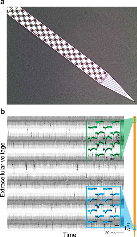
Figure 2. A state-of-the-art rigid probe.
(a) Photograph of the distal 68 sites of the 960 sites on a shank of a Neuropixels probe. The shank is 10 mm long, 70 µm wide, and 24 µm thick, with 12 µm by 12 µm TiN recording sites pitched at 2 per 20 µm of shank length. (b) Two example recordings from Neuropixels probe pads. The probe was chronically implanted in rat prefrontal cortex one day prior to data acquisition. Blue traces and green traces are 30 raw traces in the vicinity of a spike near the top (green) and bottom (blue) of the probe; black lines are average of those traces. Adapted from Jun et al. (2017).

