Abstract
ZNF521 is a transcription co-factor with recognized regulatory functions in haematopoietic, osteo-adipogenic and neural progenitor cells. Among its diverse activities, ZNF521 has been implicated in the regulation of medulloblastoma (MB) cells, where the Hedgehog (HH) pathway, has a key role in the development of normal cerebellum and of a substantial fraction of MBs. Here a functional cross-talk is shown for ZNF521 with the HH pathway, where it interacts with GLI1 and GLI2, the major HH transcriptional effectors and enhances the activity of HH signalling. In particular, ZNF521 cooperates with GLI1 and GLI2 in the transcriptional activation of GLI (glioma-associated transcription factor)-responsive promoters. This synergism is dependent on the presence of the N-terminal, NuRD-binding motif in ZNF521, and is sensitive to HDAC (histone deacetylase) and GLI inhibitors. Taken together, these results highlight the role of ZNF521, and its interaction with the NuRD complex, in determining the HH response at the level of transcription. This may be of particular relevance in HH-driven diseases, especially regarding the MBs belonging to the SHH (sonic HH) subgroup where a high expression of ZNF521 is correlated with that of HH pathway components.
Subject terms: Biochemistry, Transcription factors
Introduction
The Hedgehog (HH) pathway is a master regulator of developmental processes whose dysregulation has been linked to a variety of cancers, including those arising from the cerebellum, skin, pancreas, prostate and lung1,2. This pathway is activated by the binding of HH ligands to the 12-transmembrane domain protein Patched1 (PTCH1), which relieves the repression of smoothened (SMO) and induces its migration to the primary cilium. This results in the dissociation of the suppressor of fused (SUFU)-glioma-associated transcription factor (GLI) complex, allowing the GLI proteins to migrate into the nucleus and act on transcriptional targets, which control cell growth, survival and differentiation3–6.
The GLI factors (GLI1, GLI2 and GLI3) represent the major transcriptional effectors of HH signalling. These proteins share conserved homology of their zinc finger domains and bind to a consensus motif (GACCACCCA) in the promoters of target genes7. It is generally considered that GLI1 acts exclusively as an activator (GLI1A), while GLI2 and GLI3 can act either as activators (GLI2A, GLI3A) or repressors (GLI2R, GLI3R). The combination of activating and repressive forms of the GLI proteins, which act in concert with different signalling pathways in addition to that of HH, has led to the concept of a “GLI code”, where multiple integrated signals contribute to the control of cell fate3,8.
A considerable wealth of information has been accumulated on the activity of the SHH (sonic HH) pathway and of the GLI factors, and on their interactions with other intracellular signalling networks. These data are derived from a variety of cases where the imbalance of one pathway will perturb other signalling mechanisms, thus modulating the control of cellular functions. These include, for example, the RAS-MEK-AKT cascade, the EGF pathway, signalling by TGFB-SMADS and the WNT pathway1–3.
The SHH pathway is a central regulator of cerebellar development and its dysregulation has been implicated in the generation of a substantial fraction of the cerebellar medulloblastomas (MBs), which have been classified in four different molecular subgroups, WNT, SHH, group 3 and group 49,10. The SHH group, characterized by inappropriate expression or aberration of SHH pathway genes, originates from committed granule neuron precursor cells (GNPCs) of the external granular layer of the cerebellum11. In normal physiology, the SHH signal produced by the adjacent Purkinje cells promotes GNPC proliferation and prevents differentiation; once the stimulus is terminated, cells exit the cell cycle and differentiate12–14. SHH inhibitors are considered promising agents for the development of targeted therapeutic strategies in MBs belonging to the SHH subgroup15–20.
The present study has been focussed on ZNF521 (also known as EHZF or Evi3)21,22, a 30 zinc finger transcription co-factor, an important regulator of the homeostasis of the immature hematopoietic cells23–27. ZNF521/Zfp521 has also been implicated in the control of neural development28–32, adipocyte differentiation33–35, maintenance of chondrocyte identity36 and bone formation37,38. Importantly, ZNF521 is highly expressed in the external granule layer of the cerebellum, where the GNPCs considered the cells-of-origin of MBs of the SHH subgroup are located23. Consistently, ZNF521 is particularly abundant in the SHH subtype of MB and has been shown to play a critical regulatory role in MB cells39.
These features prompted us to investigate if a functional cross-talk exists between ZNF521 and the SHH pathway. Our data, illustrated here, delineate a direct interaction of ZNF521 with the GLI1 and GLI2 transcription factors, which enhances the transcriptional activation of GLI target promoters through a mechanism that requires the presence of the N-terminal motif of ZNF521 and the recruitment of the nucleosome remodelling and HDAC (NuRD) complex.
Results
Correlation between the expression of ZNF521 and SHH target genes in MB
A set of 736 MB cases (R2 analysis platform, public database Tumour Medulloblastoma - Cavalli - 763 - rma_sketch - hugene11t40) was analysed for the expression of ZNF521 in the different subgroups, where highest expression was found associated with the SHH subgroup, followed by the WNT subgroup and then group 4, with lowest expression in group 3 (Fig. S1A).
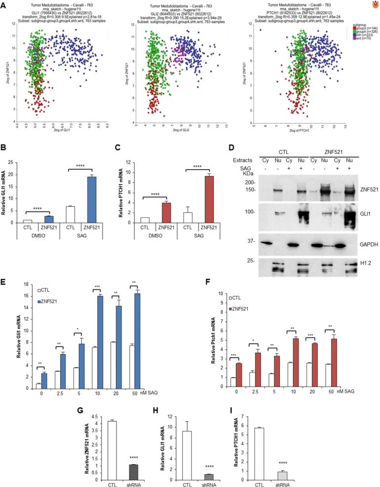
In addition, an analysis was carried out to establish whether a correlation exists between the expression of ZNF521 and that of individual components of the SHH pathway, including GLI1, GLI2 and PTCH1. To this end, the messenger RNA (mRNA) levels of GLI1, GLI2 and PTCH1 were plotted against those of ZNF521. The scatter profile XY plot shows that the expression of GLI1, GLI2 and PTCH1 is particularly associated with the presence of high amounts of ZNF521 transcript (Fig. 1a). There is an overall positive correlation (Fig. S1B) among ZNF521 and the 763 clinical cases expressing GLI1, GLI2 and PTCH1. However, when individual groups are considered, a negative correlation emerges (Fig. S1C). It is interesting to note that high expression of ZNF521, although found predominantly in cases of MB with expression of the SHH pathway, is also high in WNT cases (Fig. 1a, Fig. S1D). The WNT genes were also tested for a direct correlation with ZNF521 in the MB cases and it was found that only WNT5A and LEF1 had a significant correlation, whereas no association was found with other WNT genes (Fig. S3).
Fig. 1. ZNF521 expression is associated with the SHH subgroup of MBs and promotes the activation of SHH pathway.
a Association of ZNF521 with SHH pathway genes in subgroups of MBs. Co-expression (2 log XY plots) of ZNF521 (8022612 reporter probe) associated with either GLI1 (7956430 reporter probe), GLI2 (8044933 reporter probe) or PTCH1 (8162533 reporter probe). The different MB subgroups are indicated as WNT (pink dots), SHH (blue dots), group 3 (red dots) and group 4 (green dots). b–h Enforced overexpression of ZNF521 activates the SHH pathway. DAOY cells transduced with either ZNF521 or FUIGW control vector (CTL) were stimulated with 200 nM SAG, the SHH agonist, for 48 h or without (DMSO). RT-qPCR analysis shows the increase in GLI1 (b) and PTCH1 (c) mRNA with ZNF521 transduction and an additional increase with SAG. d These cells once transduced with ZNF521 showed an increase in GLI1 protein predominantly in the nucleus, which was overexpressed when the cells are treated with 200 nM SAG. The cytosolic (Cy) and nuclear (Nu) extracts were controlled by the enrichment of GAPDH and histone H1.2, respectively. NIH3T3 transduced with ZNF521 and incubated for 48 h with 0–50 nM SAG were analysed by RT-qPCR for Gli1 (e) and Ptch1 (d) mRNA. UW228 MB cells were silenced for ZNF521 using a lentiviral shRNA vector and analysed for ZNF521 (g), GLI1 (h) and PTCH1 (i) mRNA expression by RT-qPCR. *p < 0.05, **p < 0.01, ***p < 0.001, ****p < 0.0001
Enforced overexpression of ZNF521 stimulates the expression of SHH target genes and phenocopies SHH signalling
In the light of the abundance of ZNF521 mRNA in SHH MB and the association between its expression and that of SHH pathway mediators, depicted in Fig. 1 and Fig. S1, we sought to further characterize the possible existence of a cooperative cross-talk between ZNF521 and SHH signalling. As shown in Fig. 1b, c, lentiviral-mediated enforced expression of ZNF521 in DAOY cells enhanced by approximately two-fold the expression of the HH target genes, GLI1 and PTCH1. Moreover, the presence of ZNF521 reinforced the GLI1 and PTCH1 up-regulation upon treatment with the SMO agonist, SAG41. Additional analyses of cytoplasmic and nuclear extracts confirmed that ZNF521 induced a small increase of nuclear GLI1 protein, which is enhanced once stimulated with SAG (Fig. 1d). The same trend was also observed in NIH3T3 cells in which ZNF521-overexpressing cells displayed Gli1 and Ptch1 mRNA up-regulation, which became more prominent upon SAG treatment in a dose-dependent manner (Fig. 1e, f).
The UW228 MB cell line was used to test for the role of endogenous ZNF521 in the SHH pathway. These cells express high amounts of ZNF521 and it was previously shown that once ZNF521 was silenced by lentiviral transduction of short hairpin RNA (shRNA), they displayed reduced growth, colony formation and migration39. Here it is found that once endogenous ZNF521 was silenced there was a marked decrease in both of the SHH pathway targets, GLI1 and PTCH1 mRNA (Fig. 1g–i).
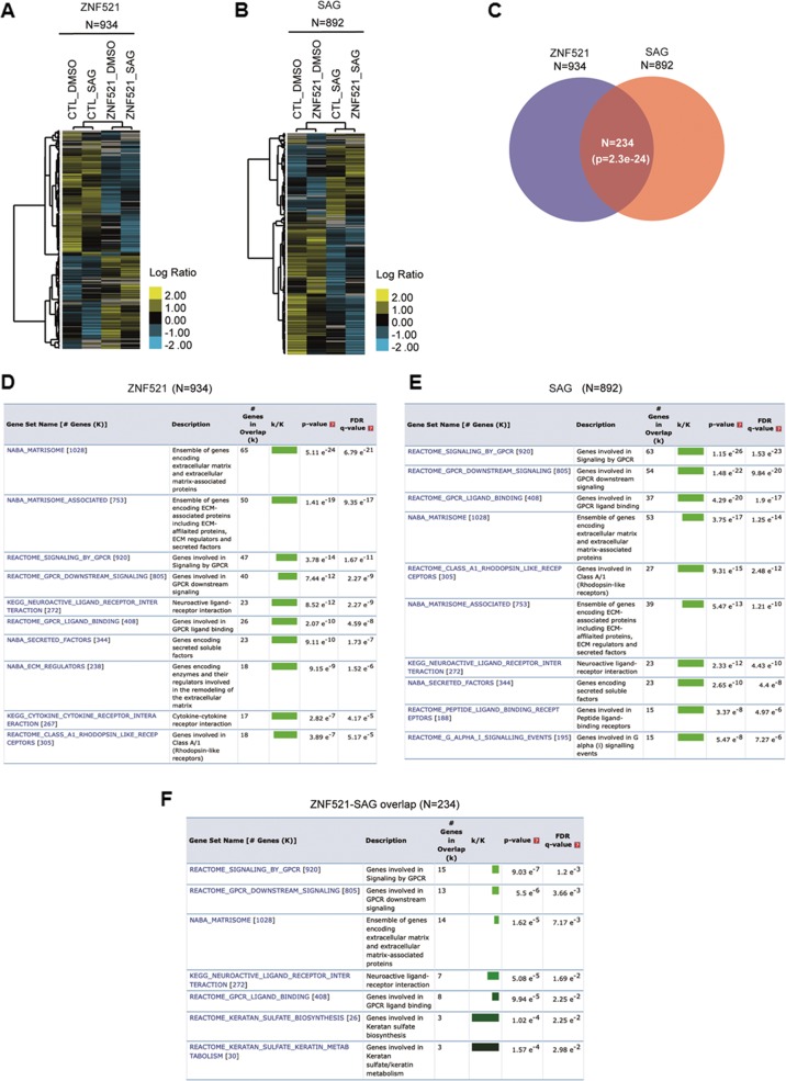
We next analysed how ZNF521 modulate HH signalling and the overall transcriptome of DAOY cells. Gene expression profile analysis by RNA-sequencing (RNA-Seq) of DAOY cells revealed that the ectopic expression of ZNF521 modulates a total of 934 genes (p ≤ 0.05) with respect to control cells (Fig. 2a; Supplementary Table 1). On the other hand, by treating DAOY cells with SAG, a total of 892 genes (p ≤ 0.05) resulted significantly modulated when the SHH signalling is activated, regardless of the presence of ZNF521 with respect to the control treatment group (Fig. 2b; Supp. Table 2). Interestingly, we found 234 genes (p = 2.3e−24) overlapping between the 934 and 892 gene sets (Fig. 2c; Supplementary Table 3). Strikingly, when we analysed the overrepresentation of canonical pathways among these sets of genes (i.e. the 934, the 892, and the 234), we found that the top-scoring pathways in terms of significance (false discovery rate (FDR) q value ≤ 0.05) were related to SHH mechanisms, such as extracellular matrix remodelling, G protein-coupled receptor signalling and downstream signalling (Fig. 2d–f). Taken together, these results showed that ZNF521 expression modulation largely impacts the transcriptional profile of genes involved in SHH signalling.
Fig. 2. Gene expression profile analysis of DAOY cells.
a Hierarchical clustering analysis of 934 genes differentially expressed (p value ≤ 0.05) in ZNF521-overexpressing cells versus control cells (CTL). b Hierarchical clustering analysis of 892 genes differentially expressed (p value ≤ 0.05) in SAG-treated cells versus untreated control cells (DMSO). Scale bars, the log 2 ratio of expression of mean centred genes. c Venn diagram using the 934 and 892 gene sets. A total of 234 genes were found overlapping. P value was calculated using the hypergeometric distribution test. d–f The top 10 overlapping gene sets (K) representing MSigDB canonical pathways (see also Methods) are shown for the 934, 892 or 234 genes (k). Colour bar shading from light green to black, where lighter colours indicate more significant “false discovery rate” (FDR) q values (<0.05) and black indicates less significant FDR q values (≥0.05)
ZNF521 interacts with Gli1 and Gli2 proteins
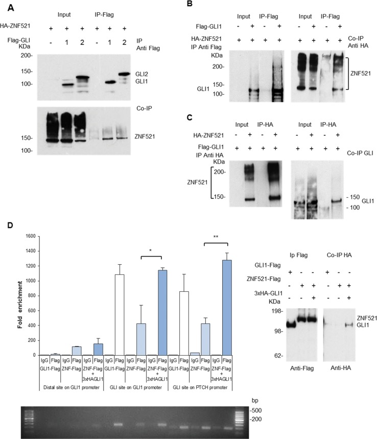
To test whether a physical interaction occurs between ZNF521 and mediators of the SHH pathway, we performed co-immunoprecipitation (Co-IP) assays in HEK293T cells. Following co-transfection of cDNAs encoding tagged ZNF521, GLI1 and GLI2, ZNF521 co-immunoprecipitated with Flag-tagged GLI1 or GLI2 (Fig. 3a). In complementary Co-IP experiments, Flag-tagged GLI1 pulled down HA-ZNF521 (Fig. 3b), and conversely HA-ZNF521 co-precipitated Flag-GLI1 (Fig. 3c).
Fig. 3. Interaction of ZNF521 and GLI1/ GLI2 demonstrated by Co-IP.
a IP of Flag-GLI1 or Flag-GLI2 results in the Co-IP of HA-ZNF521. b IP of Flag-GLI1 results in the Co-IP of HA-ZNF521. c IP of HA-ZNF521 results in the Co-IP of Flag-GLI1. d ChIP demonstrates the pull down of ZNF521 with the GLI1 and PTCH promoter regions containing GBS, which was enhanced in the presence of additional GLI1. qPCR analysis of regions of the GLI1 and PTCH promoters with GBS, pulled down either directly by Flag-GLI1 or by 3xFlag-ZNF521 alone, or by the combination of 3xFlag-ZNF521 together with 3xHA-GLI1. Fold enrichment of amplified products is shown compared to the non-specific control in the absence of Flag antibody (IgG control). A distal region of the GLI1 promoter was also amplified, which did not cover the GBS and acts as an internal control. PCR products were analysed by agarose gels and the proteins present in the IPs (ChIP) and co-immunoprecipitates (Co-IP) were analysed by Western blotting. *p < 0.05, **p < 0.01
We then performed chromatin immunoprecipitation (ChIP) assays to establish whether the GLI/ZNF521 interaction occurred at the chromatin level. Cells were transfected with either GLI1-Flag or ZNF521-Flag or with a combination of ZNF521-Flag and 3xHA-GLI1 and sheared DNA pulled down with anti-Flag antibody or a control immunoglobulin G (IgG). The presence of the GLI1 binding sites (GBSs) was identified by amplifying across the sites with specific primers. Additional primers that amplified a distal region (2 kb) from the GBSs on the GLI1 promoter, which gave a specific internal control, were also used.
These assays showed that both GLI1 (as expected) and ZNF521 were each pulled down in correspondence to the promoter regions containing consensus GBSs42,43. This was found for both GLI1 and PTCH1 promoters (Fig. 3d), but not (significantly) in the distal region of the GLI1 promoter where no GBSs are present. Importantly, when ZNF521 (Flag-ZNF521) was directly pulled down, by anti-Flag antibody, in the presence of transfected GLI1 (3xHA-GLI1) (Fig. 3d Western blot, where 3xHA-GLI1 is seen as a Co-IP), there was an enrichment in the recovery of amplified fragments associated with the GBSs in these promoters compared to those found when Flag-ZNF521 was transfected alone (Fig. 3d, quantified by quantitative PCR (qPCR) and visualized by agarose gel). This indicates that ZNF521 together with GLI1 has a greater interaction with the GLI1 and PTCH1 promoter regions and is likely to enhance the interaction of GLI1 with its target promoters.
These data show for the first time a novel interaction between ZNF521 and the SHH principal effectors GLI proteins, which takes place at the chromatin level on the GLI1 and PTCH1 promoters.
ZNF521 enhances the transcriptional activity of GLI1 and GLI2
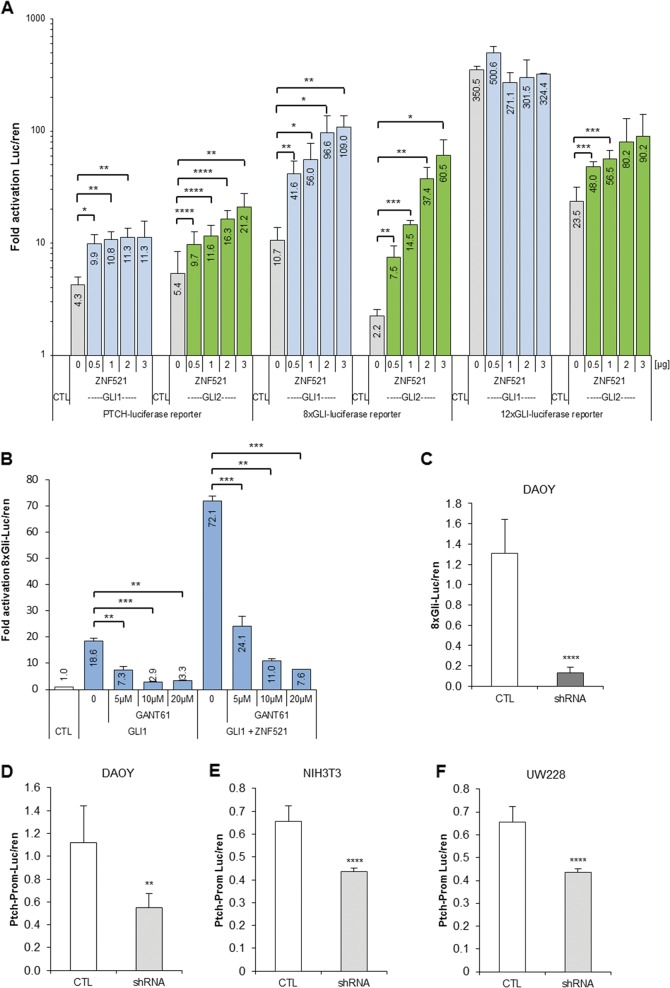
To assess the contribution of ZNF521 in modulating the activity of GLI factors, transactivation assays were performed using reporter constructs containing GBSs fused to the cDNA of firefly luciferase (luc). To this end, cells were co-transfected with a reporter construct and expression vectors carrying the cDNAs for ZNF521, GLI1 or GLI2 as indicated in Fig. 4a. The PTCH-luc construct contains a 4.3 kb fragment of the PTCH1 promoter region with one GBS at −704/−696 bp upstream of the transcription start site, while the engineered constructs 8xGli-luc and 12xGLI-luc have multiple GBSs (Fig. S2). As expected (Fig. 4a), both GLI1 and GLI2 alone (grey bars) activated all the reporter constructs. Instead, the co-expression of ZNF521 with GLI proteins (GLI1, blue and GLI2, green bars) significantly enhanced the transactivation, almost invariably in a ZNF521 dose-dependent fashion.
Fig. 4. ZNF521 acts with GLI proteins to co-transactivate GLI-responsive reporter constructs.
a The GLI-responsive reporters: PTCH promoter luciferase, 8xGli (GBS) luciferase and the 12xGLI-(GBS) luciferase were transfected in HEK293T cells with constant amounts of either GLI1 or GLI2 alone or together with increasing amounts (0–3 μg/well) of ZNF521 plasmid. The luciferase values were expressed as a ratio of the co-transfected Renilla activity (luc/ren). The results are expressed in a log scale as a fold activation in the absence of GLI1 or GLI2. b Transactivation of the 8x Gli reporter by GLI1 alone as well as with ZNF521 can be inhibited by 5–20 µM of the GLI1 inhibitor GANT61. c–f The cell line DAOY silenced for ZNF521 with shRNA had a reduced ability to transactivate the 8xGli-luciferase construct (c). DAOY (d), NIH3T3 (e), and UW228 (f) cells silenced for ZNF521 with shRNA had a reduced ability to transactivate the PTCH promoter-luciferase construct. *p < 0.05, **p < 0.01, ***p < 0.001, ****p < 0.0001
This phenomenon was particularly striking with 8xGli-luc, where ZNF521 expression resulted in a super-activation up to 10–30-fold greater than that induced by GLI1 or GLI2 alone. Instead, in the case of 12xGLI-luc, owing to the high responsiveness of this construct to GLI1 alone (350.5-fold activation), the additional effect of ZNF521 was masked by the already maximal activation level. Instead, the transactivation of 12xGLI-luc by GLI2 alone was relatively modest (23-fold activation) and the effect of ZNF521 resulted in a substantial up-regulation (up to 90-fold greater than that obtained by GLI2 alone).
The super-induction experiments described above were repeated in the presence of the GLI inhibitor GANT61, which specifically inhibits GLI1 binding to target promoters, thereby abrogating transactivation44,45. GANT61 treatment significantly inhibited, in a dose-dependent manner, 8xGli-luc transactivation by GLI1 alone and also, to a large extent, the super-activation by the combination of GLI1 and ZNF521 (Fig. 4b).
Cell lines (DAOY, NIH3T3 and UW228) silenced for ZNF521 and transfected with 8xGli-luc or the PTCH promoter luc reporter, which have a basal level of activity, were found to has a reduced transcriptional activation, indicating that the reduction of endogenous ZNF521 in these cells, which also express SHH pathway components, has attenuated the response (Fig. 4c–f).
These results delineate the existence of a cooperative action between ZNF521 and GLI factors in the transcriptional activation of GLI-binding regulatory sequences, which is sensitive to the GLI antagonist GANT61.
The ZNF521-GLI1 cooperation requires the recruitment of the HDAC-NuRD complex by ZNF521
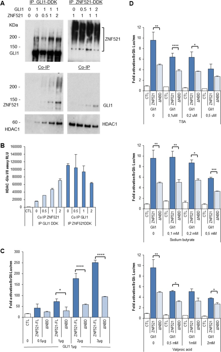
ZNF521 possesses a 12-amino-acid-long motif at its N-terminal end21–23,46–48, which is needed to bind the NuRD complex, and is required for the activity of ZNF521 in MB cells39. To assess the relevance of this interaction in the cooperation between ZNF521 and HH signalling, we performed experiments in which increasing amounts of expression vector carrying the cDNA for HA-ZNF521 were co-transfected with a constant amount of GLI1-DDK-Flag or, vice versa, increasing amounts of cDNA for 3xHA-GLI1 were co-transfected with a constant amount of ZNF521-DDK-Flag (Fig. 5a). The extracts obtained were then subjected to IP with an anti-DDK-Flag antibody (Fig. 5a, top panel). The precipitates were analysed with antibodies to ZNF521, GLI1 or histone deacetylase 1 (HDAC1) (Fig. 5a, middle and bottom panels). As expected, ZNF521 was found associated with GLI1, and, reciprocally, GLI1 co-precipitated with ZNF521. As shown in Fig. 5a (lane 2), IP of GLI1 was accompanied by detectable co-IP of endogenous HDAC1, even in the absence of transfected ZNF521. However, in the presence of increasing amounts of transfected ZNF521, the co-precipitation of HDAC1 was increased proportionally. Instead, when ZNF521 was immunoprecipitated the amounts of HDAC1 that were pulled down were higher than that co-precipitated with GLI1, and when additional GLI1 was co-transfected, no increase in HDAC1 co-precipitation was observed. These data were confirmed by the measurement of HDACI/II activity in the precipitates (Fig. 5b), and indicate that the interaction between HDAC1 (or HDAC1-containing complexes, such as NuRD) and ZNF521 is preferential compared to that with GLI1.
Fig. 5. The activity of the combination of ZNF521 and GLI1 is promoted by the NuRD-interacting N-terminal motif in ZNF521.
a Cells (HEK293T) were transfected either with GLI1-DDK (Flag) and increasing amounts of HA-ZNF521 (top left panel) or with ZNF521-DDK (Flag) and increasing amounts of 3xHA-GLI1 (top right panel). The IPs pulled down with anti-Flag-protein G complex were analysed with rabbit anti-DDK (Flag) antibody for GLI1 and ZNF521 and the Co-IPs with anti-ZNF521 or with anti-GLI1 antibodies, as well as for the presence of Co-IP HDAC1. b An aliquot of the IP beads were incubated with the 3xFlag peptide to release bound proteins and assayed with the HDAC-Glo™ I/II substrate and developer. c Cells were all transfected with GLI1, either with control vector (CTL) or with increasing concentrations of full-length (FL) ZNF521 or a construct lacking the first 12 amino acids (ZNF521-ΔNBD) and the 8xGli-luciferase reporter. d Cells were transfected with GLI1 either with control vector (CTL) or together with ZNF521 or ZNF521-ΔNBD (used as control for HDAC inhibitors), and after 24 h, HDAC inhibitors, TSA, NaBt or VPA were added. The reporter activity was measured at 48 h and calculated as fold activation of the 8xGLI-luc. *p < 0.05, **p < 0.01, ***p < 0.001, ****p < 0.0001
In additional experiments, we tested whether the binding of ZNF521 to the NuRD complex is required for the ZNF521-GLI transcriptional co-operational activity. These assays demonstrated that co-transfection of GLI1 (Fig. 5c) or GLI2 (Fig. S2D) together with a deletion mutant of ZNF521 lacking the NuRD-binding domain (ΔNBD)—which is able to bind GLI1 (Fig. S2E) but unable to recruit components of the NuRD complex23,24,46—resulted in a lower enhancement of the 8xGli-luc reporter activity than that achieved by full-length ZNF521. Furthermore, in a complementary set of experiments, treatment of transfected cells with three distinct HDAC inhibitors (trichostatin A (TSA); sodium butyrate (NaBt); valproic acid (VPA)) resulted in an aberration of the cooperative effect between ZNF521-FL and GLI1 (Fig. 5d) or GLI2 (Fig. S2F) on the 8xGli-luc reporter, such that once the maximum amount of HDAC inhibitor was used, the difference between the full length and the construct lacking the NuRD-binding motif was minimized. Taken together, the data described above indicate that the association between ZNF521 and the HDAC-NuRD complex takes part in the transcriptional synergism between ZNF521 and the HH pathway effectors.
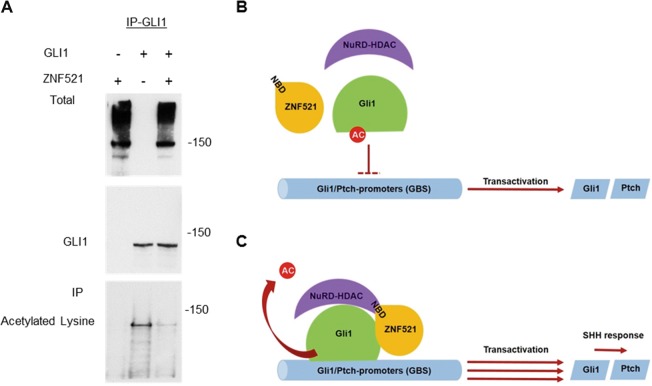
It is known that post-translational modifications of GLI proteins contribute to the fine control of the HH pathway. Specifically, it has been documented that deacetylation of GLI factors by HDAC1 induces an increase in their transcriptional activity by facilitating their access to the chromatin49–51 and enhancing transactivation of target genes. To test whether the interaction with ZNF521 modifies the GLI1 acetylation status, GLI1-DDK-Flag and ZNF521 were co-transfected and GLI1 was precipitated with anti-DDK-Flag antibody. Western blot analyses with an anti-acetylated lysine antibody showed a lower level of the acetylation of GLI1 in the presence of ZNF521 (Fig. 6a). This is likely to account for the increase in its activity induced by ZNF521.
Fig. 6. Activation of GLI1 is promoted by ZNF521 throught deacetylation of GLI1.
a The presence of ZNF521 results in a reduced acetylation of GLI1. Cells were transfected either with GLI1-DDK (Flag) and 3xHA-ZNF521 or with both together and IP was performed for GLI1 pulldown with anti-Flag-M2 agarose beads. Total input is shown for ZNF521 and IPs were hybridized with anti-GLI1 or with an anti-acetyl-lysine antibody. b, c Hypothesis for the mechanism of action for ZNF521 collaborating with GLI1 together with HDAC1-NuRD complex resulting in an increased transactivation of GLI-responsive genes. Diagrams for the action of GLI1 on the GLI1 target promoters in the absence (a) or presence (b) of ZNF521
Discussion
ZNF521 displays features compatible with a function as a transcriptional regulator, with widely acknowledged roles in stem cells of diverse organs and tissues23–30. A notable feature being, a short amino acid sequence located at its N-terminal end, which is shared with a family of transcriptional regulators (friend of GATA-1/2, SALL1–4, BCL11a/b and Zfp423) and has been shown to recruit the NuRD complex21,23,46,47,52–54.
In the present approach, regarding neural neoplasias, ZNF521 was shown to be a regulator of immature MB cells, where its enforced expression stimulates growth, clonogenicity, migration ability and tumorigenicity, and this action requires the integrity of the NuRD-binding motif39. Among the MB subgroups, particularly high expression of ZNF521 is present in the subgroup characterized by dysregulation of the SHH pathway. Additional analyses of a larger cohort of patients, conducted in the framework of the present study, revealed a striking positive association between levels of ZNF521 transcript and the expression of SHH pathway components, GLI1, GLI2 and PTCH. It was observed that high expression of ZNF521 was also detected in the WNT subgroup. However, when WNT genes were examined for correlations with ZNF521 in the MB cases, only two genes WNT5A and LEF1 showed a significant R value (Fig. S3); thus, ZNF521 was not further examined in the context of having a role in the WNT pathway.
ZNF521 being present at high levels in all SHH subgroup samples, it is likely to be crucial for this pathway, considering that low levels of ZNF521 are not found in this SHH subgroup. The functional and expression data for the SHH pathway39 encouraged us to explore the possibility that ZNF521 could physically and/or functionally interact with mediators of the SHH signalling. These results indicate that: (i) ZNF521 binds to GLI1 and GLI2; (ii) this interaction occurs in regulatory regions of SHH target genes that contain GLI-binding consensus sequences; (iii) enforced expression of ZNF521 in DAOY MB and NIH3T3 cells enhances the expression of SHH target genes both in the absence and in the presence of SAG; (iv) conversely, silencing of endogenous ZNF521 reduced GLI1 and PTCH1 mRNA; (v) RNA-Seq data showed that ZNF521 widely impacts the SHH signalling by modulating a large set of genes, whose expression is also regulated by the addition of SAG; (vi) ZNF521 synergistically cooperates with GLI1 and GLI2 in the transcriptional activation of SHH-responsive elements; and (vii) this synergism is more evident in the presence of the N-terminal NuRD-binding motif of ZNF521.
The SHH pathway is subjected to fine control, and the activity of GLI1 and GLI2 is known to be post-translationally modified by ubiquitination50,55 and sumoylation56 resulting in degradation. GLI1 can instead be activated by phosphorylation by the tyrosine kinase HCK, which disrupts the interaction with its inhibitor SUFU at the primary cilium57. An additional regulatory mechanism is mediated by acetylation/deacetylation of the GLI factors. Specifically, it has been determined that deacetylation of lysine 518 by HDAC1 is critical for the activation of GLI150 and that the HDAC1/2 inhibitor, vismodegib, could inhibit tumour growth in a mouse MB model51.
Consistent with the notion that the HDAC-rich NuRD complex is necessary for the transcriptional cooperation between ZNF521 and GLI, three HDAC class I inhibitors were able to counteract the super-induction of the GLI reporter by full-length ZNF521. This evidence underlies the importance of the interaction between ZNF521 and HDAC1-NuRD in the enhancement of GLI transactivation. It also fits well both with our previous finding that the NBD is essential for the activity of ZNF521 in MB, and with those of Coni et al.49,51 and Canettieri et al.50 showing that deacetylation of conserved lysines in GLI1 and GLI2 augments the GLI transcriptional activity by permitting easier access to chromatin. This is further supported by our experiments in vitro, in which the co-expression of GLI1 and ZNF521 resulted in a significant deacetylation of GLI1 (Fig. 6a) and is exemplified in Fig. 6b as a mechanism whereby GLI activity is enhanced by ZNF521 through deacetylation dependent on NuRD-associated HDAC1.
The classification of MB into subgroups with distinct molecular, demographic and clinical characteristics has offered the possibility to determine prognostic differences between the groups. The current therapeutic strategies in MB treatment include surgical resection, cranium–spinal irradiation and adjuvant chemotherapy. The cure rates of average and high-risk patients are fairly high (85 and 70%, respectively)58; however, they are associated with serious treatment-induced morbidity. Inter-tumoral heterogeneity has been detected within the four different subgroups, such that a further classification identified a total of 12 MB subtypes40, which may permit an increased level of stratification by providing more specific prognostic information and thus allowing a greater degree of “therapeutic tailoring” to minimize treatment-induced damage. Molecular biomarkers associated with specific phenotypes of these tumours are potentially important in identifying and categorizing specific types of MBs. High expression of ZNF521 is found not only in the SHH group but also in the WNT group and in a substantial fraction of cases in group 4. ZNF521 expression could thus help define a novel sub-category classification. In this context, and in the light of the fact that activation of the HH pathway is critical for the maintenance of the stem cell compartment in tumours derived from dysregulation of different signalling mechanisms (ref. 3 and references therein), the NuRD-dependent synergism between ZNF521 and GLI might represent a valuable biomarker for the identification of patients with MB—and possibly other SHH-driven malignancies—who may potentially benefit from a combination of SHH and HDAC inhibitors.
Materials and methods
Cell lines and culture conditions
HEK293T, DAOY, NIH3T3 and UW228 cells were cultured in Dulbecco’s modified Eagle’s medium supplemented with 10% foetal bovine serum, 50 U of penicillin and 50 μg of streptomycin/mL, at 37 °C in 5% CO2. All tissue culture reagents were from Life Technologies. For stimulation of the SHH pathway, SAG SMO ligand (ALX-270–426, ENZO Life Sciences, Italy) was solubilized in dimethyl sulfoxide and used at 2.5–200 nM for 48 h, after the cells had been starved for 24 h.
Plasmids and lentiviral vectors
The ZNF521 lentiviral vectors UBC promoter-transgene-IRES-EGFP (FUIGW), FUIGW-flag-ZNF521 and FUIGW-flag-ZNF521ΔNBD (lacking the first 12 amino acids that constitute the NuRD-binding motif) were used24,39 as well as TrueORF-Gold human NM_015461.2 pCMV6-ZNF521-Myc-DDK (Origene) and the mouse cDNA clones pCMV-3xHA-Zfp52137, Image Clone pCMV-Sport6-Zfp521 (Evi3) NM_145492.4 was sub-cloned in the lentiviral vector FUIGW as well as the corresponding Zfp521ΔNBD. Human and mouse hZNF521/mZfp521 are 97% identical in protein sequence and both or either constructs were used in the experiments and are indicated as ZNF521. shRNA lentiviral vectors for specific silencing of ZNF521 were as described in ref. 39.
GLI1 and GLI2 was transfected using the pCDNA-Flag-GLI1, pCDNA-3xHA-GLI1 and pCDNA-Flag-GLI2, as well as TrueORF-Gold human NM_005269 pCMV6-GLI1-Myc-DDK (Origene) plasmids. Promoter reporter assays were performed with PTCH-luc (4.3 b promoter fragment)59, 8xGli-luc, 12xGLI-luc (Cellogenetics) and transfections were normalized with the control pRL-TK Renilla plasmid (Promega).
Transfection and transduction of cell lines
Plasmids were transfected using polyethylenimine (PEI) (Polysciences) 1 µg/µl (3 µg for 1 µg plasmid DNA) or the calcium phosphate method in HEK293T cells using 10 μg plasmid/100 mm tissue culture plate. The medium was changed after 16 h and cells were harvested for IP or luc activity after 48 h. Cells were transduced60 with the control vector FUIGW or FUIGW-ZNF521 using the lentiviral packaging plasmids pCMV-VSVG and pCMV-deltaR8-91 and were 70–80% positive for the transgene EGFP (enhanced green fluorescent protein) by fluorescence-activated cell sorting analysis and stably expressed the ZNF521 or mutant ΔNBD at similar high levels. Cells were sorted for EGFP and were over 90% positive for EGFP.
Expression analysis by RT-qPCR
RNA was prepared using the TRIzol reagent (Life Technologies), quantified with the NanoDrop 2000/2000c Spectrophotometer (Thermo Fisher Scientific) and the quality was monitored using 1.5% agarose gels run in MOPS buffer, pH 7.1 (0.4 M MOPS (3-(N morpholino)propanesulfonic acid), 0.1 M NaAc, 20 mM EDTA), and 10% formaldehyde. cDNA was synthesized from 1 µg RNA using SuperScript III reverse transcriptase at 42 °C and 2.5 µM random hexamers (Life Technologies). Quantitative reverse transcription-PCR (RT-qPCR) reactions were carried out with the iQ™ SYBR® Green Supermix (Bio-Rad) with the qPCR amplifier QuantStudio3 (Applied Biosystems). One cycle of 3 min at 95 °C was followed by 45 cycles of 10 s at 95 °C, 10 s at 60 °C and 10 s at 72 °C, finishing with a melting curve. Relative gene expression was determined using the comparative threshold cycle Ct method, normalizing for housekeeping genes (GAPDH and UBC) such that the average the expression ratio was calculated as 2−ddCt. Primers used in this study were designed to have high stringency for qPCR and span exon–intron sites and the sequences were as follows (5′–3′): h-GLI1 (F) ACAGCCAGTGTCCTCGACTT, (R) ATAGGGGCCTGACTGGAGAT; h-PTCH (F) CTTCGCTCTGGAGCAGATTT, (R) CAGGACATTAGCACCTTCT; h-GAPDH (F) CACCATCTTCCAGGAGCGAG, (R) TCACGCCACAGTTTCCCGGA; h-UBC (F) ATTTGGGTCGCGGTTCTTG, (R) TGCCTTGACATTCTCGATGGT; m-Gli1 (F) ACCCACTCCAATGAGAAGCC, (R) CAGTTTGAGACCCCGAGACC; m-Ptch (F) TACCTCAACGGCCTACGAGA, (R) GCTGTCAGAAAGGCCAAAGC; m-Gapdh (F) TGACGTGCCGCCTGGAGAA, (R) AGTGTAGCCCAAGATGCCCTTCAG; m-Ubc (F) GCCCAGTGTTACCACCAAGA, (R) CCCATCACACCCAAGAACA.
Transcriptome sequencing and analysis
Poly-A-enriched strand-specific ribosomal RNA-depleted libraries were generated with the Illumina TruSeq Stranded Total RNA Library Prep Gold, according to the manufacturer’s instructions. Libraries were sequenced by Illumina HiSeq2000 resulting in paired 50 nt reads. Fastq files were aligned to the hg38 genome assembly using STAR61. STAR was also used to quantify gene expression for each gene. STAR gene counts were normalized applying the median of ratios method implemented in DESeq2 R package62. Briefly, the normalization process implies different steps: (i) for each gene, a pseudo-reference sample is created and is equal to the geometric mean across all samples; (ii) for every gene in a sample and for each sample, the ratios sample/ref are calculated; (iii) the median value of all ratios for a given sample is taken as the normalization factor (size factor) for that sample; (iv) for each gene in each sample the normalized count values is calculated dividing each raw count value by the sample’s normalization factor. BRB-ArrayTools (v4.6; https://brb.nci.nih.gov/BRB-ArrayTools/) were used to perform statistical tests, briefly: (i) normalized count values were thresholded if the intensity at the minimum value was below 1; (ii) genes were excluded if <25% of expression data have at least a 1.5-fold change in either direction from gene’s median value; (iii) paired t test (with random variance model) were used to select features with a significant gene expression change (p value < 0.05) in the selected conditions. Clustering analysis was performed by using Cluster 3.0 and Java TreeView (http://bonsai.hgc.jp/~mdehoon/software/cluster/software.htm). The uncentred correlation and centroid linkage were used to aggregate genes and conditions. Pathways overrepresentation analysis was performed by using the Molecular Signature Database v6.2 (MSIgDB)63.
Nuclear and cytoplasmic extracts
DAOY cells treated with or without 200 nM SAG were processed for nuclear and cytoplasmic extracts64. Cells were scraped, re-suspended in hypotonic lysis buffer (10 mM HEPES, pH 7.9, 10 mM KCl, 0.1 mM EDTA, protease inhibitors (P8849, Sigma) and phosphatase inhibitor cocktails 2 and 3 (P0044, P5726, Sigma) and incubated on ice for 20 min. After the addition of 0.25% Igepal-630 (NP40), samples were centrifuged at 3600 r.p.m for 5 min and supernatants containing the cytoplasmic extracts were recovered. Nuclear pellets were re-suspended in 20 mM HEPES, pH 7.9, 0.4 M NaCl, 1 mM EDTA with protease and phosphatase inhibitors. After three cycles of vortex and ice, samples were centrifuged at 12,000 r.p.m. for 20 min and the supernatants containing the nuclear extracts were collected. Proteins (50 μg) were denatured, reduced, separated on 4–12% NuPAGE Novex Bis-Tris gradient polyacrylamide gels (Life Technologies) and blotted onto nitrocellulose membranes. Membranes were quenched with 5% blotto (Bio-Rad), ZNF521 was detected with rabbit anti-ZNF521 (EHZF S15 sc-84808, Santa Cruz, Biotechnology; predicted molecular weight (MW): 148 kDa) at 1:2000, GLI1 with rabbit anti-GLI1 (C68H3, Cell Signalling; predicted MW: 118 kDa) at 1:5000, for nuclear extracts with rabbit anti-H1.2 at 1:10,000 (ab-4086, Abcam; predicted MW: 21.3 kDa) and cytoplasmic extracts with anti-GAPDH at 1:1000 (sc-166574, Santa Cruz Biotechnology; predicted MW: 36 kDa). Secondary rabbit and mouse horse radish peroxidase (HRP) antibodies were detected by the ImmunoCruz Western blotting luminal reagent (sc-2004, sc-2005, Santa Cruz, Biotechnology) and exposure to auto-radiographic film (GE Healthcare).
Co-IP interaction assays
Cells, HEK293T cultured to 60–70% confluence, were transfected using PEI and were used for IP after 48 h. After washing with 1× phosphate-buffered saline (PBS), cells were pelleted at 1000 r.p.m. for 5 min at 4 °C and re-suspended in 1 ml of IP buffer (50 mM Tris/HCl, pH 7.5, 250 mM NaCl, 0.1% Triton X-100, 0.1 nM ZnCl2) supplemented with protease and phosphatase inhibitor cocktails 2 and 3. Cell lysates were sonicated on ice for three times for 10 s at 100% amplitude (UP50H ultrasonic processor Hielscher, Ultrasound technology) and centrifuged twice at 13,000 r.p.m. for 20 min at 4 °C to remove debris.
IP was performed with 10 μg monoclonal anti-Flag-M2 (F3165, Sigma-Aldrich) and 20 μl Protein G Sepharose (P3296, Sigma-Aldrich). Cell lysates were added to the beads and incubated overnight at 4 °C with rotation. After four washes with IP buffer and then with PBS, bound proteins were released at 95 °C for 5 min in NuPAGE® LDS Sample Buffer plus reducing agent and loaded onto a NuPAGE Novex Bis-Tris 4–12% gel. Antibodies used for the detection of immunoprecipitated proteins were anti-Flag-M2-HRP (A8592), DYKDDDDK Tag (Flag) rabbit antibody (#2368 Cell Signalling), rabbit anti-HA (ab9110, Abcam) and rabbit anti-GLI1 (C68H3, Cell Signalling).
HDACI/II activity assays were performed on Flag-M2-agarose IPs after releasing proteins with 3xFlag peptide at 200 μg/ml for 30 min at 4 °C, serial dilutions were made for the assays and extracts incubated with the HDAC-Glo™ I/II substrate and developer (G6420, Promega) for luminescence reading with the GloMax Explorer luminometer in white 96-well plates. The anti-acetyl lysine antibody (ab21623, Abcam) was used to control the degree of acetylation in IPs of GLI1-myc-DDK in the presence of ZNF521.
Chromatin immunoprecipitation
ChIP was performed using ZNF521-myc-DDK- and/or GLI1-Flag-transfected 293T cells; 2 × 10⁶ cells in 500 μl PBS were cross-linked with 1% formaldehyde for 8 min and blocked with 1 mM glycine for 5 min, then washed twice with PBS and finally the cell pellets were frozen on dry ice. Cells were extracted in 200 μl of 20 mM HEPES, pH 7.9, 25% glycerol, 420 mM NaCl, 1.5 mM MgCl2, 0.2 mM EDTA and 1 nM ZnCl2, and then nuclei were recovered after centrifugation at 13,000 × g for 10 min. Nuclei were lysed and sonicated for 40 cycles of high voltage 30 s on/30 s off in a Bioruptor bath sonicator (Diagenode) at 4 °C in 300 μl of 50 mM Tris/HCl, pH 8.0, 1 mM EDTA, 150 mM NaCl, 0.2% SDS, 1% Triton X-100 and 1 nM ZnCl2. After centrifugation, it was verified that the sonication had resulted in fragments of 500–1000 base pairs (bp). Extracts were diluted with 1 ml of 50 mM Tris/HCl, pH 8.0, 1 mM EDTA, 150 mM NaCl, 0.1% Triton X-100 and 1 nM ZnCl2, and then centrifuged at 13,000 × g for 20 min at 4 °C. Soluble extracts were divided for specific IP with anti-Flag-M2 antibody and non-specific (control mouse IgG). Antibody complexes were recovered with Protein G Sepharose. After 16 h of mixing by rotation at 4 °C, the beads were washed three times with Triton buffer and twice with PBS and then de-cross-linked by incubation in 2.5 mM Tris/HCl, pH 6.8, 200 mM NaCl, 2% SDS and 10 mM dithiothreitol at 65 °C for 16 h. All buffers contained the protease inhibitor mix for His-tagged proteins from Sigma. DNA was extracted with phenol/chloroform, and precipitated with 0.3 M NaAc, pH 5.4, and 2.5 volumes of ethanol with 1 μg glycogen carrier. Pellets were washed with 70% ethanol and re-suspended in 30 μl H2O for qPCR analysis.
Primers used for amplification of the GLI1 and PTCH promoters were: GLI1 promoter distal ~2000 bp upstream, GLI1 promoter proximal ~200 bp upstream covering the GBS, PTCH ∼700 bp upstream covering the proximal GBS, and were as follows (5′–3′): GLI1 promoter distal (F) TAAGTGGGCTTTAGTGAGGGGCT, (R) TCTACGTCTCGAAGTTCTGGAGG; GLI1 promoter proximal (F) CGTAAGCAGTATAGGGTCCCTCA, (R) ACCCGCGAGAAGCGCAAACTT; PTCH promoter proximal (F) GTATTGCTGCGAGAAGGTGG, (R) TTTCTGCGACGCGATTGGCTCG.
Transactivation reporter assays
Cells were transfected with plasmids for GLI proteins and ZNF521 together with the reporter PTCH-luc, 8xGli-luc or 12xGLI-luc normalizing with pRT-TK Renilla using the PEI transfecting reagent. After 40 h, cells were washed with PBS and total soluble extracts were prepared by freezing and thawing in 250 mM Tris/HCl, pH 7.5, containing protease inhibitors. Proteins were quantified using the Bio-Rad reagent and 10 μg was used for the Dual-Glo luc assay system (E2920, Promega) performed in white 96-well plates and detected using the GloMax Explorer Luminometer (Promega). Ratios of luc/Renilla luminescence were calculated and presented as fold activation for each reporter.
The GLI1 inhibitor GANT61 (S8075, Selleckchem) was prepared in ethanol and used at 20–100 nM added 4 h after transfection. HDAC class I inhibitors, TSA (T8552, Sigma-Aldrich) used at 0.1–0.5 μm in DMSO, NaBt (B5887, Sigma-Aldrich) used at 0.1–0.5 mM prepared in PBS and VPA (ALX-550-304 from Enzo Life Sciences) used at 0.5–2mM in DMSO were added 24 h after transfection before assaying at 48 h.
Gene expression analysis of R2 platform database
The public data set of 763 tumour MB40 samples (Tumour Medulloblastoma - Cavalli - 763 - rma_sketch - hugene11t) (https://hgserver1.amc.nl/cgi-bin/r2/main.cgi) was interrogated for ZNF521 (8022612) vs. GLI1 (7956430), GLI2 (8044993) or PTCH (8162533) reporter probes, as well as for WNT genes. For graphical representation and statistical comparison between the subgroups and subtypes, data were transferred into Excel and the GraphPad prism version 5.03 program for analysis (t test of unpaired two-tailed analysis)
Statistical analysis
The Student’s t test assuming unequal variances between two samples was used to determine the significant differences. Groups were judged to differ significantly at p values lower than 0.05.
Supplementary information
Figure S3. Correlation between ZNF521 and WNT pathway genes in all Mb subgroups
Acknowledgements
The work was supported by funds AIRC 9204, PON03PE_00009_2 ICaRe and PON01_02834 PROMETEO, POR Calabria 2014-2020 DEMOCEDE. V.L., Y.M. and A.A. were supported by the Ph.D. Program in Molecular and Translation Oncology and Advanced Medical-Surgical Technologies. E.C. and S.S. were supported by fellowship from fund PON03PE_00009_2 ICaRe. M.G. was supported by fellowships from EMSAs and Fondazione IEO-CCM. F.B. was supported by the Italian Ministry of Health (Ricerca Finalizzata, GR-2016-02363975 and TRASCAN-2, CLEARLY). We are also particularly grateful to Ugo Cavallaro for his decisive support.
Authors’ contributions
S.S., M.G. and V.L. performed the experiments illustrated; Y.M., E.C., A.A. and B.C. carried out preliminary work; P.Z., F.B. and V.M. performed informatic analyses; E.De.S. provided essential reagents and advice for the experimental strategy; M.M., H.M.B. and G.M. devised the experimental design, supervised the research and wrote the manuscript.
Conflict of interest
The authors declare that they have no conflict of interest.
Footnotes
This paper is in memory of a dear friend and exceptional scientist, Prof. Giovanni Morrone
Edited by R. Mantovani
Publisher’s note Springer Nature remains neutral with regard to jurisdictional claims in published maps and institutional affiliations.
These authors contributed equally: Stefania Scicchitano, Marco Giordano, Valeria Lucchino
Contributor Information
Maria Mesuraca, Phone: +0961/3694081, Email: mes@unicz.it.
Heather M. Bond, Phone: +0961/3694081, Email: bond@unicz.it
Supplementary information
Supplementary Information accompanies this paper at (10.1038/s41419-019-1946-x).
References
- 1.Hui CC, Angers S. Gli proteins in development and disease. Annu. Rev. Cell Dev. Biol. 2011;27:513–537. doi: 10.1146/annurev-cellbio-092910-154048. [DOI] [PubMed] [Google Scholar]
- 2.Briscoe J, Thérond PP. The mechanisms of Hedgehog signalling and its roles in development and disease. Nat. Rev. Mol. Cell. Biol. 2013;14:416–429. doi: 10.1038/nrm3598. [DOI] [PubMed] [Google Scholar]
- 3.Aberger F, Ruiz I, Altaba A. Context-dependent signal integration by the GLI code: the oncogenic load, pathways, modifiers and implications for cancer therapy. Semin. Cell Dev. Biol. 2014;33:93–104. doi: 10.1016/j.semcdb.2014.05.003. [DOI] [PMC free article] [PubMed] [Google Scholar]
- 4.Pandolfi S, Stecca B. Cooperative integration between HEDGEHOG-GLI signalling and other oncogenic pathways: implications for cancer therapy. Expert Rev. Mol. Med. 2015;17:e5. doi: 10.1017/erm.2015.3. [DOI] [PMC free article] [PubMed] [Google Scholar]
- 5.Carballo GB, Honorato JR, de Lopes GPF, Spohr TCLSE. A highlight on Sonic hedgehog pathway. Cell Commun. Signal. 2018;16:11. doi: 10.1186/s12964-018-0220-7. [DOI] [PMC free article] [PubMed] [Google Scholar]
- 6.Fattahi S, Pilehchian Langroudi M, Akhavan-Niaki H. Hedgehog signalling pathway: epigenetic regulation and role in disease and cancer development. J. Cell. Physiol. 2018;233:5726–5735. doi: 10.1002/jcp.26506. [DOI] [PubMed] [Google Scholar]
- 7.Kinzler KW, Vogelstein B. The GLI gene encodes a nuclear protein which binds specific sequences in the human genome. Mol. Cell. Biol. 1990;10:634–642. doi: 10.1128/MCB.10.2.634. [DOI] [PMC free article] [PubMed] [Google Scholar]
- 8.Ruiz i Altaba A, Mas C, Stecca B. The Gli code: an information nexus regulating cell fate, stemness and cancer. Trends Cell Biol. 2007;17:438–447. doi: 10.1016/j.tcb.2007.06.007. [DOI] [PMC free article] [PubMed] [Google Scholar]
- 9.Northcott PA, et al. Subgroup-specific structural variation across 1,000 medulloblastoma genomes. Nature. 2012;488:49–56. doi: 10.1038/nature11327. [DOI] [PMC free article] [PubMed] [Google Scholar]
- 10.Northcott PA, et al. The whole-genome landscape of medulloblastoma subtypes. Nature. 2017;547:311–317. doi: 10.1038/nature22973. [DOI] [PMC free article] [PubMed] [Google Scholar]
- 11.Gibson P, et al. Subtypes of medulloblastoma have distinct developmental origins. Nature. 2010;468:1095–1109. doi: 10.1038/nature09587. [DOI] [PMC free article] [PubMed] [Google Scholar]
- 12.Dahmane N, Ruiz i Altaba A. Sonic hedgehog regulates the growth and patterning of the cerebellum. Development. 1999;126:3089–3100. doi: 10.1242/dev.126.14.3089. [DOI] [PubMed] [Google Scholar]
- 13.Wechsler-Reya RJ, Scott MP. Control of neuronal precursor proliferation in the cerebellum by Sonic Hedgehog. Neuron. 1999;22:103–114. doi: 10.1016/S0896-6273(00)80682-0. [DOI] [PubMed] [Google Scholar]
- 14.Wallace VA. Purkinje-cell-derived Sonic hedgehog regulates granule neuron precursor cell proliferation in the developing mouse cerebellum. Curr. Biol. 1999;9:445–448. doi: 10.1016/S0960-9822(99)80195-X. [DOI] [PubMed] [Google Scholar]
- 15.Rudin CM, et al. Treatment of medulloblastoma with hedgehog pathway inhibitor GDC-0449. N. Engl. J. Med. 2009;361:1173–1178. doi: 10.1056/NEJMoa0902903. [DOI] [PMC free article] [PubMed] [Google Scholar]
- 16.Robinson G, et al. Novel mutations target distinct subgroups of medulloblastoma. Nature. 2012;488:43–48. doi: 10.1038/nature11213. [DOI] [PMC free article] [PubMed] [Google Scholar]
- 17.Gonnissen A, Isebaert S, Haustermans K. Targeting the Hedgehog signalling pathway in cancer: beyond Smoothened. Oncotarget. 2015;6:13899–13913. doi: 10.18632/oncotarget.4224. [DOI] [PMC free article] [PubMed] [Google Scholar]
- 18.Kieran MW. Targeted treatment for sonic hedgehog-dependent medulloblastoma. Neuro Oncol. 2014;16:1037–1047. doi: 10.1093/neuonc/nou109. [DOI] [PMC free article] [PubMed] [Google Scholar]
- 19.Infante P, et al. Gli1/DNA interaction is a druggable target for Hedgehog-dependent tumors. EMBO J. 2015;34:200–217. doi: 10.15252/embj.201489213. [DOI] [PMC free article] [PubMed] [Google Scholar]
- 20.Rimkus TK, Carpenter RL, Qasem S, Chan M, Lo HW. Targeting the Sonic Hedgehog signalling pathway: review of smoothened and GLI inhibitors. Cancers. 2016;8:22. doi: 10.3390/cancers8020022. [DOI] [PMC free article] [PubMed] [Google Scholar]
- 21.Bond HM, et al. Early hematopoietic zinc finger protein (EHZF), the human homolog to mouse Evi3, is highly expressed in primitive human hematopoietic cells. Blood. 2004;103:2062–2070. doi: 10.1182/blood-2003-07-2388. [DOI] [PubMed] [Google Scholar]
- 22.Warming S, Suzuki T, Yamaguchi TP, Jenkins NA, Copeland NG. Early cell factor-associated zinc-finger gene is a frequent target of retroviral integration in murine B-cell lymphomas. Oncogene. 2004;23:2727–2731. doi: 10.1038/sj.onc.1207452. [DOI] [PubMed] [Google Scholar]
- 23.Bond HM, et al. Early hematopoietic zinc finger protein-zinc finger protein 521: a candidate regulator of diverse immature cells. Int. J. Biochem. Cell Biol. 2008;40:848–854. doi: 10.1016/j.biocel.2007.04.006. [DOI] [PubMed] [Google Scholar]
- 24.Mega T, et al. Zinc finger protein 521 antagonizes early B-cell factor 1 and modulates the B-lymphoid differentiation of primary hematopoietic progenitors. Cell Cycle. 2011;10:2129–2139. doi: 10.4161/cc.10.13.16045. [DOI] [PubMed] [Google Scholar]
- 25.Mesuraca M, et al. ZNF423 and ZNF521: EBF1 antagonists of potential relevance in B-lymphoid malignancies. Biomed. Res. Int. 2015;2015:165238. doi: 10.1155/2015/165238. [DOI] [PMC free article] [PubMed] [Google Scholar]
- 26.Holmfeldt P, et al. Functional screen identifies regulators of murine hematopoietic stem cell repopulation. J. Exp. Med. 2016;213:433–449. doi: 10.1084/jem.20150806. [DOI] [PMC free article] [PubMed] [Google Scholar]
- 27.Salerno L, Cosentino C, Morrone G, Amato F. Computational modeling of a transcriptional switch underlying B-lymphocyte lineage commitment of hematopoietic multipotent cells. PLoS ONE. 2015;10:e0132208. doi: 10.1371/journal.pone.0132208. [DOI] [PMC free article] [PubMed] [Google Scholar]
- 28.Kamiya D, et al. Intrinsic transition of embryonic stem-cell differentiation into neural progenitors. Nature. 2011;470:503–509. doi: 10.1038/nature09726. [DOI] [PubMed] [Google Scholar]
- 29.Lou S, et al. Incoherent feed-forward regulatory loops control segregation of C-mechanoreceptors, nociceptors, and pruriceptors. J. Neurosci. 2015;35:5317–5329. doi: 10.1523/JNEUROSCI.0122-15.2015. [DOI] [PMC free article] [PubMed] [Google Scholar]
- 30.Shahbazi E, et al. Conversion of human fibroblasts to stably self-renewing neural stem cells with a single zinc-finger transcription factor. Stem Cell Rep. 2016;6:539–551. doi: 10.1016/j.stemcr.2016.02.013. [DOI] [PMC free article] [PubMed] [Google Scholar]
- 31.Ohkubo N, et al. Abnormal behaviors and developmental disorder of hippocampus in zinc finger protein 521 (ZFP521) mutant mice. PLoS ONE. 2014;9:e92848. doi: 10.1371/journal.pone.0092848. [DOI] [PMC free article] [PubMed] [Google Scholar]
- 32.Han R, et al. MiR-9 promotes the neural differentiation of mouse bone marrow mesenchymal stem cells via targeting zinc finger protein 521. Neurosci. Lett. 2012;515:147–152. doi: 10.1016/j.neulet.2012.03.032. [DOI] [PubMed] [Google Scholar]
- 33.Kang S, et al. Regulation of early adipose commitment by Zfp521. PLoS Biol. 2012;10:e1001433. doi: 10.1371/journal.pbio.1001433. [DOI] [PMC free article] [PubMed] [Google Scholar]
- 34.Addison WN, et al. Direct transcriptional repression of Zfp423 by Zfp521 mediates a bone morphogenic protein-dependent osteoblast versus adipocyte lineage commitment switch. Mol. Cell. Biol. 2014;34:3076–3085. doi: 10.1128/MCB.00185-14. [DOI] [PMC free article] [PubMed] [Google Scholar]
- 35.Chiarella Emanuela, Aloisio Annamaria, Codispoti Bruna, Nappo Giovanna, Scicchitano Stefania, Lucchino Valeria, Montalcini Ylenia, Camarotti Augusto, Galasso Olimpio, Greco Manfredi, Gasparini Giorgio, Mesuraca Maria, Bond Heather Mandy, Morrone Giovanni. ZNF521 Has an Inhibitory Effect on the Adipogenic Differentiation of Human Adipose-Derived Mesenchymal Stem Cells. Stem Cell Reviews and Reports. 2018;14(6):901–914. doi: 10.1007/s12015-018-9830-0. [DOI] [PubMed] [Google Scholar]
- 36.Mesuraca M, et al. Expression profiling and functional implications of a set of zinc finger proteins, ZNF423, ZNF470, ZNF521, and ZNF780B, in primary osteoarthritic articular chondrocytes. Mediat. Inflamm. 2014;2014:318793. doi: 10.1155/2014/318793. [DOI] [PMC free article] [PubMed] [Google Scholar]
- 37.Hesse E, et al. Zinc finger protein 521, a new player in bone formation. Ann. NY Acad. Sci. 2010;1192:32–37. doi: 10.1111/j.1749-6632.2009.05347.x. [DOI] [PubMed] [Google Scholar]
- 38.Chiarella E, et al. ZNF521 represses osteoblastic differentiation in human adipose-derived stem cells. Int. J. Mol. Sci. 2018;19:E4095. doi: 10.3390/ijms19124095. [DOI] [PMC free article] [PubMed] [Google Scholar]
- 39.Spina R, et al. Critical role of zinc finger protein 521 in the control of growth, clonogenicity and tumorigenic potential of medulloblastoma cells. Oncotarget. 2013;4:1280–1292. doi: 10.18632/oncotarget.1176. [DOI] [PMC free article] [PubMed] [Google Scholar]
- 40.Cavalli FMG, et al. Intertumoral heterogeneity within medulloblastoma subgroups. Cancer Cell. 2017;31:737–754.e6. doi: 10.1016/j.ccell.2017.05.005. [DOI] [PMC free article] [PubMed] [Google Scholar]
- 41.Chen JK, Taipale J, Young KE, Maiti T, Beachy PA. Small molecule modulation of smoothened activity. Proc. Natl Acad. Sci. USA. 2002;99:14071–14076. doi: 10.1073/pnas.182542899. [DOI] [PMC free article] [PubMed] [Google Scholar]
- 42.Sasaki H, Hui C, Nakafuku M, Kondoh H. A binding site for Gli proteins is essential for HNF-3beta floor plate enhancer activity in transgenics and can respond to Shh in vitro. Development. 1997;124:1313–1322. doi: 10.1242/dev.124.7.1313. [DOI] [PubMed] [Google Scholar]
- 43.De Smaele E, et al. An integrated approach identifies Nhlh1 and Insm1 as Sonic Hedgehog-regulated genes in developing cerebellum and medulloblastoma. Neoplasia. 2008;10:89–98. doi: 10.1593/neo.07891. [DOI] [PMC free article] [PubMed] [Google Scholar]
- 44.Lauth M, Bergström A, Shimokawa T, Toftgård R. Inhibition of GLI-mediated transcription and tumor cell growth by small-molecule antagonists. Proc. Natl Acad. Sci. USA. 2007;104:8455–8460. doi: 10.1073/pnas.0609699104. [DOI] [PMC free article] [PubMed] [Google Scholar]
- 45.Agyeman A, Jha BK, Mazumdar T, Houghton JA. Mode and specificity of binding of the small molecule GANT61 to GLI determines inhibition of GLI-DNA binding. Oncotarget. 2014;5:4492–4503. doi: 10.18632/oncotarget.2046. [DOI] [PMC free article] [PubMed] [Google Scholar]
- 46.Bernaudo F, et al. Validation of a novel shotgun proteomic workflow for the discovery of protein–protein interactions: focus on ZNF521. J. Proteome Res. 2015;14:1888–1899. doi: 10.1021/pr501288h. [DOI] [PubMed] [Google Scholar]
- 47.Joshi P, et al. The functional interactome landscape of the human histone deacetylase family. Mol. Syst. Biol. 2013;9:672. doi: 10.1038/msb.2013.26. [DOI] [PMC free article] [PubMed] [Google Scholar]
- 48.Torchy MP, Hamiche A, Klaholz BP. Structure and function insights into the NuRD chromatin remodeling complex. Cell. Mol. Life Sci. 2015;72:2491–2507. doi: 10.1007/s00018-015-1880-8. [DOI] [PMC free article] [PubMed] [Google Scholar]
- 49.Coni S, et al. Gli2 acetylation at lysine 757 regulates hedgehog-dependent transcriptional output by preventing its promoter occupancy. PLoS ONE. 2013;8:e65718. doi: 10.1371/journal.pone.0065718. [DOI] [PMC free article] [PubMed] [Google Scholar]
- 50.Canettieri G, et al. Histone deacetylase and Cullin3-REN(KCTD11) ubiquitin ligase interplay regulates Hedgehog signalling through Gli acetylation. Nat. Cell Biol. 2010;12:132–142. doi: 10.1038/ncb2013. [DOI] [PubMed] [Google Scholar]
- 51.Coni S, et al. Selective targeting of HDAC1/2 elicits anticancer effects through Gli1 acetylation in preclinical models of SHH medulloblastoma. Sci. Rep. 2017;7:44079. doi: 10.1038/srep44079. [DOI] [PMC free article] [PubMed] [Google Scholar]
- 52.Harder L, Puller AC, Horstmann MA. ZNF423: transcriptional modulation in development and cancer. Mol. Cell. Oncol. 2014;1:e969655. doi: 10.4161/23723548.2014.969655. [DOI] [PMC free article] [PubMed] [Google Scholar]
- 53.Bond HM, et al. ZNF423: a new player in estrogen receptor-positive breast cancer. Front. Endocrinol. 2018;9:255. doi: 10.3389/fendo.2018.00255. [DOI] [PMC free article] [PubMed] [Google Scholar]
- 54.Miccio A, et al. NuRD mediates activating and repressive functions of GATA-1 and FOG-1 during blood development. EMBO J. 2010;29:442–456. doi: 10.1038/emboj.2009.336. [DOI] [PMC free article] [PubMed] [Google Scholar]
- 55.Bhatia N, et al. Gli2 is targeted for ubiquitination and degradation by beta-TrCP ubiquitin ligase. J. Biol. Chem. 2006;281:19320–19326. doi: 10.1074/jbc.M513203200. [DOI] [PubMed] [Google Scholar]
- 56.Cox B, Briscoe J, Ulloa F. SUMOylation by Pias1 regulates the activity of the Hedgehog dependent Gli transcription factors. PLoS ONE. 2010;5:e11996. doi: 10.1371/journal.pone.0011996. [DOI] [PMC free article] [PubMed] [Google Scholar]
- 57.Shi X, Zhan X, Wu J. A positive feedback loop between Gli1 and tyrosine kinase Hck amplifies shh signalling activities in medulloblastoma. Oncogenesis. 2015;4:e176. doi: 10.1038/oncsis.2015.38. [DOI] [PMC free article] [PubMed] [Google Scholar]
- 58.Kijima N, Kanemura Y. Molecular classification of medulloblastoma. Neurol. Med. Chir. 2016;56:687–697. doi: 10.2176/nmc.ra.2016-0016. [DOI] [PMC free article] [PubMed] [Google Scholar]
- 59.Agren M, Kogerman P, Kleman MI, Wessling M, Toftgård R. Expression of the PTCH1 tumor suppressor gene is regulated by alternative promoters and a single functional Gli-binding site. Gene. 2004;330:101–114. doi: 10.1016/j.gene.2004.01.010. [DOI] [PubMed] [Google Scholar]
- 60.Chiarella E, et al. UMG Lenti: novel lentiviral vectors for efficient transgene- and reporter gene expression in human early hematopoietic progenitors. PLoS ONE. 2014;9:e114795. doi: 10.1371/journal.pone.0114795. [DOI] [PMC free article] [PubMed] [Google Scholar]
- 61.Dobin A, et al. STAR: ultrafast universal RNA-seq aligner. Bioinformatics. 2013;29:15–21. doi: 10.1093/bioinformatics/bts635. [DOI] [PMC free article] [PubMed] [Google Scholar]
- 62.Love MI, Huber W, Anders S. Moderated estimation of fold change and dispersion for RNA-seq data with DESeq2. Genome Biol. 2014;15:550. doi: 10.1186/s13059-014-0550-8. [DOI] [PMC free article] [PubMed] [Google Scholar]
- 63.Subramanian A, et al. Gene set enrichment analysis: a knowledge-based approach for interpreting genome-wide expression profiles. Proc. Natl Acad. Sci. USA. 2005;102:15545–15550. doi: 10.1073/pnas.0506580102. [DOI] [PMC free article] [PubMed] [Google Scholar]
- 64.Codispoti B, et al. Recombinant TAT-BMI-1 fusion protein induces ex vivo expansion of human umbilical cord blood-derived hematopoietic stem cells. Oncotarget. 2017;8:43782–43798. doi: 10.18632/oncotarget.15156. [DOI] [PMC free article] [PubMed] [Google Scholar]
Associated Data
This section collects any data citations, data availability statements, or supplementary materials included in this article.
Supplementary Materials
Figure S3. Correlation between ZNF521 and WNT pathway genes in all Mb subgroups


