Abstract
Multiple system atrophy (MSA) is a sporadic neurodegenerative disease clinically characterized by cerebellar signs, parkinsonism, and autonomic dysfunction. Pathologically, MSA is an α-synucleinopathy affecting striatonigral and olivopontocerebellar systems, while neocortical and limbic involvement is usually minimal. In this study, we describe four patients with atypical MSA with clinical features consistent with frontotemporal dementia (FTD), including two with corticobasal syndrome, one with progressive non-fluent aphasia, and one with behavioral variant FTD. None had autonomic dysfunction. All had frontotemporal atrophy and severe limbic α-synuclein neuronal pathology. The neuronal inclusions were heterogeneous, but included Pick body-like inclusions. The latter were strongly associated with neuronal loss in the hippocampus and amygdala. Unlike typical Pick bodies, the neuronal inclusions were positive on Gallyas silver stain and negative on tau immunohistochemistry. In comparison to 34 typical MSA cases, atypical MSA had significantly more neuronal inclusions in anteromedial temporal lobe and limbic structures. While uncommon, our findings suggest that MSA may present clinically and pathologically as a frontotemporal lobar degeneration (FTLD). We suggest that this may represent a novel subtype of FTLD associated with α-synuclein (FTLD-synuclein).
Keywords: Multiple system atrophy, Frontotemporal lobar degeneration, Neuropathology, Pick body-like inclusions, α-Synuclein
Introduction
Multiple system atrophy (MSA) is a sporadic, adult-onset, neurodegenerative disease characterized clinically by cerebellar ataxia, parkinsonism and autonomic dysfunction with variable pyramidal signs [6, 21]. MSA encompasses three disorders that were at one time considered separate entities based on differing pathologic and clinical features: olivopontocerebellar atrophy (OPCA), striatonigral degeneration (SND), and Shy–Drager syndrome (SDS) [10]. In 1989, Papp and Lantos [30] described argyrophilic glial cytoplasmic inclusions (GCI) in oligodendrocytes in MSA, and GCI are now considered the pathognomonic lesions of MSA. The discovery that GCI were composed of α-synuclein led to classification of MSA as an α-synucleinopathy, a designation shared with Lewy body disease (LBD) presenting clinically as Parkinson’s disease or dementia with Lewy bodies [8, 36, 38, 43].
Glial cytoplasmic inclusions can be found throughout the central nervous system, being most numerous in striatonigral and olivopontocerebellar systems, areas that also have variable neuronal loss. Ozawa and co-workers [29] showed that density of GCI was correlated with both disease duration and severity of neuronal loss, indicating that GCI are likely an important factor in neurodegeneration in MSA. In addition to GCI, α-synuclein accumulates in cytoplasm, nuclei, and cell processes of neurons as neuronal cytoplasmic inclusions (NCI), neuronal intranuclear inclusions (NII), and dystrophic neurites [1, 23]. In typical MSA, NCI and dystrophic neurites are most often detected in the putamen, pontine nuclei, and inferior olivary nucleus [17, 27, 45], while limbic structures (e.g., hippocampus and amygdala) are affected to a lesser degree. The relationship between NCI and neurodegeneration is not clearly understood.
Neurodegeneration in neocortex and limbic structures is usually minimal in MSA. While motor cortex and supplementary motor cortex often have many GCI, neuronal loss is minimal [42]. There are a few reports of MSA with severe frontal or temporal lobe atrophy and numerous NCI [13, 16, 19, 31, 32, 46], but this is better recognized in Japan and is under recognized in Europe and North America. In the present study, we describe clinical and pathologic features of four MSA patients who presented with FTD clinical syndromes associated with frontotemporal and limbic system degeneration. We also compare the frequency and severity of NCI in neocortical and limbic areas of these four cases to those found in 34 typical MSA cases.
Materials and methods
Case material
Four atypical MSA patients were included in this study. Two were from a series of brains submitted for review or consultation to the neuropathology laboratory at the Mayo Clinic in Jacksonville, Florida (MCJ), between 1997 and 2014 (Cases 1 and 2). Out of 124 MSA cases accessioned during that time period, only these two had frontotemporal atrophy with numerous limbic α-synuclein NCI. The other two patients were from the University of Rochester in New York (Case 3) and the University of Texas Southwestern Medical Center (Case 4). Paraffin-embedded blocks from these two brains were available for histological examination and semi-quantitative evaluation. The four atypical MSA cases were compared to 34 typical MSA cases using semiquantitative methods.
Neuropathologic assessment
Formalin-fixed and paraffin-embedded sections from cortical, subcortical, brainstem, and cerebellar regions were stained with hematoxylin and eosin (H&E), a silver stain (Gallyas), and thioflavin-S for fluorescent microscopy. The density of senile plaques and neurofibrillary tangles (NFT) were assessed with thioflavin-S fluorescent microscopy and used to assign Thal amyloid phase [37] and Braak NFT stage [4], respectively. Luxol fast blue stain was used to assess white matter integrity on sections of anteromedial temporal lobe. Immunohistochemistry was performed on a DAKO Autostainer (Universal Staining System, Carpinteria, CA, USA) with the following antibodies:α-synuclein (NACP, 1:3000, rabbit polyclonal, Mayo Clinic [2, 12]), phosphorylated tau (CP13, 1:1000, mouse monoclonal, gift from Dr. Peter Davies, Feinstein Institute for Medical Research, North Shore/ Long Island), and TDP-43 (pS409/410, 1:5000 mouse monoclonal, Cosmo Bio USA, Carlsbad, CA or MC2085 [47], 1:2500 rabbit polyclonal, from Leonard Petrucelli, Mayo Clinic).
Sections of hippocampus were studied with double-labeling immunohistochemistry by combining α-synuclein polyclonal antibody (NACP) and phosphorylated tau monoclonal antibody (CP13 or PHF-1, 1:1000, mouse monoclonal, gift from Dr. Peter Davies, Feinstein Institute for Medical Research, North Shore/Long Island) or Aβ monoclonal antibody (4G8, 1:50,000, mouse monoclonal, BioSource International/Invitrogen Corp., Carlsbad, CA).
Semi-quantitative assessment of α-synuclein immunohistochemistry
A semi-quantitative assessment of α-synuclein pathology was performed in the following regions: cerebral cortex—middle frontal gyrus, superior temporal gyrus, parahippocampal gyrus, and cingulate gyrus; subcortical areas—hippocampus, amygdala, hypothalamus, and putamen; brainstem—substantia nigra, pontine nuclei, inferior olivary nucleus, medullary tegmentum, and pyramidal tract; and cerebellar white matter. The densities of NCI and GCI were scored separately using a 4-point scale: 0 = none, 1= sparse to mild, 2 moderate, 3 = severe. In addition, the density of NCI in select brain regions (hippocampus, hypothalamus, and inferior olivary nucleus) was assessed in 34 typical MSA cases for comparison to atypical MSA.
Immunoelectron microscopy
Formalin-fixed tissues from subiculum and amygdala from case 1 and from hippocampus and cerebellum from case 2 were processed for post-embedding immunogold electron microscopy as previously reported [23, 41]. Rabbit polyclonal antibody to α-synuclein and a mouse monoclonal antibody to phosphorylated tau (CP13 or PHF-1) were used for immuno-EM. Stained ultrathin sections were examined on a Philips 208S electron microscope (Phillips, Amsterdam, NL) fitted with a Gatan 831 Orius digital camera (Gatan, Inc., Pleasanton, CA, USA).
Genetic analysis
Genomic DNA was extracted from frozen brain tissue. For direct sequence analysis, each of the coding exons of SNCA (exons 2 through 6) was amplified by polymerase chain reaction (PCR) using exon-specific primers for SNCA (available upon request). Sequencing reactions were performed using BigDye Terminator chemistry. Genomic sequences were generated on an ABI 3730XL DNA Analyzer and read with SeqScape Software v2.5 (Life Technologies, Carlsbad, CA, USA). For SNCA genomic dosage analysis, TaqMan Copy Number assay targeted to exon 6 was used (details available on request). TaqMan Copy Number Reference Assay RNase P (human) was used as an endogenous control. Quantitative PCR was carried out using TaqMan expression chemistry protocol. All assays were performed in triplicate on the ABI 7900HT Fast Real-Time PCR System and analyzed with ABI SDS 2.2.2 Software (Life Technologies).
Statistical analysis
SigmaPlot 12.0 (Systat Software, Inc., San Jose, CA, USA) statistical software was used to analyze data. Due to the small sample size, non-parametric methods (Mann–Whitney rank-sum test) were used for group comparisons of continuous variables. Fisher’s exact test was used for group comparison of categorical data. Statistical significance was considered P < 0.05.
Results
Clinical features
Clinical features of atypical MSA cases are summarized in Table 1. The antemortem clinical diagnosis for Cases 1 and 3 was corticobasal syndrome (CBS) [3, 20], while case 2 was progressive non-fluent aphasia (PNFA) [11], and Case 4 was behavioral variant frontotemporal dementia (bvFTD) [26].
Table 1.
Clinical features of atypical MSA cases
| Case 1 | Case 2 | Case 3 | Case 4 | |
|---|---|---|---|---|
| Sex | Female | Female | Female | Female |
| Age at death, years | 91 | 88 | 70 | 73 |
| Age at onset, years | 88 | 70 | 67 | 66 |
| Duration of illness, years | 3 | 18 | 3 | 7 |
| Family history | None | None | None | n.a. |
| Initial symptoms | Dystonia | Aphasia | Abnormal tongue movement | Memory impairment, depression, personality change |
| Clinical diagnosis | CBS | PNFA | CBS | bvFTD |
| Dysarthria | + | + | + | |
| Dysphagia | + | + | ||
| Urinary problems | + | + | ||
| Parkinsonism | + | + | + | |
| Pyramids sign | + | + | ||
| Dystonia | + | |||
| Myoclonus | + | |||
| Apraxia | + | |||
| Involuntary movements | + | |||
| Memory impairment | + | + | + | |
| Depression | + | |||
| Personality change | + | + | ||
| Overeating | + | |||
| Aphasia | + | |||
| Cerebellar signs | + |
A particular clinical symptom or sign is displayed as (+) if specifically stated in the clinical records. Otherwise, they are displayed as blank space since it is difficult to conclude that a symptom or sign was absent from retrospective medical records
CBS corticobasal syndrome, PNFA progressive non-fluent aphasia, bvFTD behavior variant frontotemporal dementia, n.a. not available
Case 1
This 91-year-old right-handed woman had a 3-year history of progressive motor dysfunction. She was living independently and was asymptomatic until the age of 87 years. At age 88 years, she noticed her left hand curling up, which was thought to be dystonic posturing. The symptoms led to decreased ability to use her left arm. This progressed to her left leg. Eventually, she had left-side contractures. In the last year of her life, she developed similar problems on her right side. She had stiffness, difficulty walking, and tremors. She had dysphagia and recurrent bladder infections. On examination, she had marked weakness in all her extremities, and she had increased tone and rigidity, as well as dystonia. She had mild dysarthria and unintelligible speech. Her reflexes were brisk in both upper extremities, but they could not be elicited in either of the lower extremities. Her toes were mute on testing for Babinski sign, and there was no clonus. There was no upward or downward gaze palsy. She had no documented autonomic dysfunction. Her neurological diagnosis was CBS.
Case 2
This 88-year-old, right-handed woman had an 18-year history of progressive aphasia. When she initially presented at the age of 70 years, she had difficulty distinguishing the pronouns “he” and “she”. For the first 5 years, her illness remained mild. At age 75 years, her husband died, and she worsened. By age 79, she lost use of most nouns and adjectives. She was unable to write, although she could occasionally copy. Her verbal output was mildly decreased, but what she produced was generally garbled and incomprehensible. Her understanding of written language was better preserved. She was disinhibited and had some behavioral changes. She began to point out people inappropriately in public. She also started to collect sugar packets. At the age of 80 years, she developed a number of urinary tract infections. On examination at age 81 years, she was unable to give her name in response to a simple command, although she could remove her gloves and shoes without difficulty. Cognitive testing was compromised by her language deficits. On cranial nerve testing, there were no abnormalities, except mild dysarthria and unintelligible speech. No parkinsonism or cerebellar signs were noted, and she had no documented autonomic dysfunction. An MRI of the brain showed unilateral cortical atrophy, left-greater-than-right, anterior-greater-than-posterior, and temporal-greater-than-frontal atrophy. After this exam, her symptoms further progressed to terminal dementia. Her neurological diagnosis was PNFA.
Case 3
This 70-year-old woman had a 3-year history of progressive motor and cognitive dysfunction. At age 67, her daughter noticed that she would move her tongue abnormally. Six months later, she began to notice symptoms that affected her eating and speech. She began to stutter, especially when she was nervous. She developed twitching on the left side of her face. She had difficulty controlling her tongue and closing her eyes when she opened her mouth. She also had tremors in her left upper extremity and decreased fine motor movements in both upper extremities, with the left more affected than the right. Examination at the age of 68 years revealed orobuccal lingual apraxia, myoclonic jerks, involuntary movements, and cogwheel rigidity of both upper extremities, with the left more affected than the right. She also demonstrated frontal release signs, dysphasia, and intentional tremor. Her gait was slow, and she had decreased arm swing on the left side. In the final stage of her illness, her gait became unsteady. Her right extremities developed myoclonus. She had severe oral apraxia and bilateral facial myoclonus. She also had increased tone, throughout, with adventitious movements in all extremities, but mostly on the right side of her body. She had gait apraxia and severe instability while walking. She suffered from terminal dementia. She had no documented autonomic dysfunction. Her neurological diagnosis was CBS.
Case 4
This 73-year-old woman presented with a slowly- progressive memory decline that started when she was 66 years old. It was accompanied by depression and changes in her personality. Another notable feature was a change in her appetite; she developed significant overeating and weight gain. Toward the end of her life, she had abnormal gait and posture, as well as bradykinesia and cogwheel rigidity. Her symptoms progressed slowly over the years. She progressed to a point where she required assistance with activities of daily living, necessitating placement in an assisted-living facility. Ultimately, she needed skilled nursing. She had no documented autonomic dysfunction. Her neurological diagnosis was bvFTD.
Pathological findings
Neuronal loss and gliosis
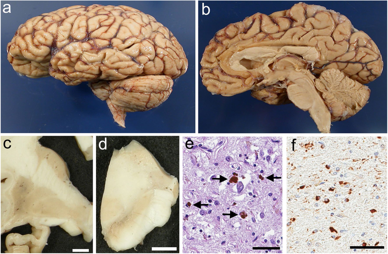
The distribution of cerebral atrophy and neuronal loss are shown in Table 2. In Case 1, cerebral atrophy was most marked in the frontal pole and the superior frontal gyrus, with less atrophy in the precentral gyrus. There was also peri-Sylvian atrophy affecting the inferior frontal lobe and the superior temporal lobe. In Cases 2 and 4, cerebral atrophy was most marked in the anterior frontal and temporal lobes (Fig. 1a, b). In Case 3, bilateral frontoparietal atrophy and softening of the right temporal lobe were remarkable. Marked medial temporal lobe atrophy, including the hippocampus and amygdala, was noted in three cases (Cases 2, 3, and 4) (Fig. 1c), but not in Case 1. Severe myelin pallor in the temporal lobe was noted in Case 2, but it was mild in Cases 3 and 4. No myelin pallor was noted in Case 1. Marked atrophy and dark gray-brown discoloration of the putamen was noted in three cases (Cases 1, 2, and 3) (Fig. 1c), but it was mild in Case 4. Neuronal loss and gliosis were most marked in the lateral and posterior putamen, and it was accompanied by iron pigment (Fig. 1e). Marked decreased neuromelanin pigmentation was noted in the substantia nigra of all cases (Fig. 1d). Neuronal loss and gliosis with extraneuronal neuromelanin was most marked in ventrolateral cell groups. Marked atrophy of the pontine base and cerebellum was noted in Case 2, but not in the other three cases (Fig. 1b). Mild neuronal loss and Bergmann gliosis were noted in internal granule cell and Purkinje cell layers in Case 2, but not in the other three cases.
Table 2.
Pathological features of atypical MSA cases
| Case 1 | Case 2 | Case 3 | Case 4 | |
|---|---|---|---|---|
| Brain weight, grams | 1220 | 560 | 1170 | 1090 |
| Cerebral atrophy | F > T | F < T | F, T, P | F, T |
| Braak NFT stage | III-IV | VI | II | I |
| Thal Aβ phase | 3 | 5 | 0 | 0 |
| TDP-43 pathology | None | None | None | None |
| Neuronal loss and gliosis | ||||
| Hippocampal dentate fascia | − | +++ | + | +++ |
| Hippocampal CA1/subiculum | − | +++ | ++ | +++ |
| Amygdala | + | +++ | n.a | +++ |
| Putamen | ++ | +++ | +++ | + |
| Substantia nigra | + | +++ | ++ | +++ |
| Pontine nuclei | − | +++ | − | − |
| Inferior olivary nucleus | + | +++ | − | − |
| Cerebellar granule cells | − | + | − | − |
| Purkinje cells | − | ++ | − | − |
| α-Synuclein-positive NCI | ||||
| Middle frontal cortex | + | +++ | +++ | +++ |
| Superior temporal cortex | + | ++ | n.a. | n.a. |
| Cingulate cortex | + | +++ | n.a. | n.a. |
| Hippocampal dentate fascia | + | +++ | +++ | +++ |
| Hippocampal CA1/subiculum | +++ | +++ | +++ | +++ |
| Parahippocampal gyrus | +++ | +++ | +++ | +++ |
| Amygdala | +++ | +++ | n.a. | +++ |
| Hypothalamus | +++ | +++ | +++ | +++ |
| Putamen | +++ | +++ | +++ | +++ |
| Substantia nigra | + | + | +++ | ++ |
| Pontine nuclei | +++ | +++ | ++ | +++ |
| Inferior olivary nucleus | +++ | +++ | − | + |
| α-Synuclein-positive GCI | ||||
| Frontal white matter | + | + | ++ | ++ |
| Hippocampus (alveus) | + | ++ | +++ | +++ |
| Amygdala | + | + | n.a. | ++ |
| Hypothalamus | + | + | + | + |
| Putamen | +++ | +++ | +++ | +++ |
| Pontine base | +++ | +++ | + | +++ |
| Medullary tegmentum | +++ | +++ | + | +++ |
| Pyramids | +++ | ++ | + | ++ |
| Cerebellar white matter | +++ | +++ | + | +++ |
F frontal lobe, T temporal lobe, P parietal lobe, n.a. not available
Fig. 1.
Macroscopic and microscopic findings in atypical MSA. Cerebral atrophy is marked in anterior frontal and temporal lobes (a). No atrophy is evident in the pons and cerebellum (b). Severe atrophy in the medial temporal lobe, including the hippocampus (c). Severe atrophy and dark gray-brown discoloration of the putamen (c). Marked decreased pigmentation of the substantia nigra (d). Severe neuronal loss and gliosis in the putamen, accompanied by iron pigment (arrows) (e). Many α-synuclein-positive glial cytoplasmic inclusions in the cerebellar white matter. Case 4 (a, b). Case 2 (c–f). H&E stain (e). α-synuclein immunohistochemistry (f). Scale bars c; 5 cm, d; 500 mm, e, f; 50 μm
α-Synuclein neuronal pathology
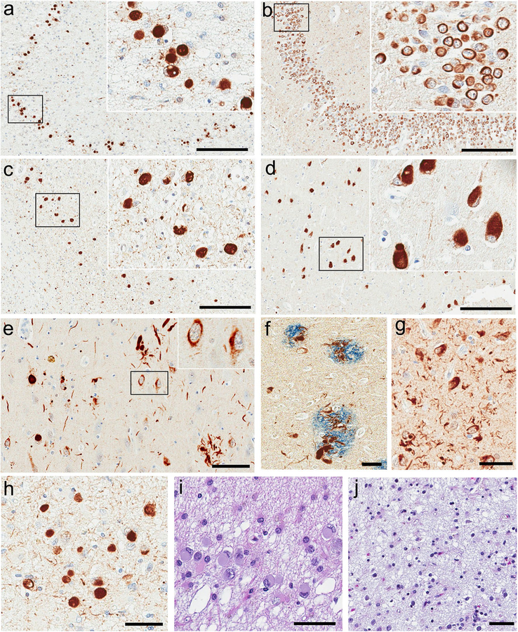
The distribution and severity of α-synuclein-positive NCI are shown in Table 2. In many areas, NCI were accompanied by variable numbers of dystrophic neurites. In the frontotemporal cortex, all cases had NCI and dystrophic neurites (Supplemental Fig. 1). The NCI were more frequent in the superficial and deep layers of the cortex. Some were visible on H&E stains as eosinophilic round inclusions, similar to Pick bodies.
In the limbic region, all cases had numerous NCI that were morphologically heterogeneous and included Pick body-like inclusions, as well as NFT-like and ring-shaped inclusions. The Pick body-like inclusions were most strongly associated with neuronal loss and gliosis. In Case 4 numerous Pick body-like inclusions were noted in the hippocampal dentate fascia (Fig. 2a). Similar to genuine Pick bodies some of these inclusions were visible on H&E stains (Fig. 2i). Numerous ring-shaped inclusions were noted in the dentate fascia in Case 3 (Fig. 2b). In Cases 3 and 4, numerous Pick body-like inclusions were noted in the hippocampal CA1 sector and subiculum (Fig. 2c, d). NFT-like inclusions were the predominant type of NCI in Case 1 (Fig. 2e). In addition, Case 1 had α-synuclein immunopositive dystrophic neurites in the corona of senile plaques in the subiculum (Fig. 2f) and the amygdala. Case 2 had heterogeneous NCI, including Pick body-like inclusions, as well as many dystrophic neurites in the dentate fascia and pyramidal cell layer of the hippocampus. All cases had abundant NCI in the entorhinal cortex (Fig. 3g), amygdala (Fig. 3h), and hypothalamus. NCI were frequent in the posterior hypothalamus of three cases (Cases 2, 3, and 4) and mild in one case (Case 1). Severe neuronal pathology was noted in the anterior hypothalamus in Cases 1 and 2, but this region was not available for study in Cases 3 and 4.
Fig. 2.
Limbic α-synuclein neuronal pathology in atypical MSA. Many Pick body-like (a) and ring-shaped (b) inclusions in the hippocampal dentate fascia. Many Pick body-like inclusions in the subiculum (c, d). Many NFT-like inclusions and neurites in the subiculum (e). Inset in (e) shows NFT-like inclusions. Thick short neurites in senile plaques in the subiculum (f). Many NFT-like inclusions and neurites in the entorhinal cortex (g). Many Pick body-like inclusions in the amygdala (h). Pick body-like inclusions in the dentate fascia are also visible on H&E stains (i). Severe neuronal loss and gliosis in the entorhinal cortex (j). Case 4 (a, c, h–j). Case 3 (b, d). Case 1 (e–g). α-Synuclein immunohistochemistry (a–e, g, h). Double immunohistochemistry with α-synuclein (brown) and amyloid-β (blue) (f). H&E stain (i, j). Scale bars a–d; 400 μm, e-j; 50 μm
Fig. 3.
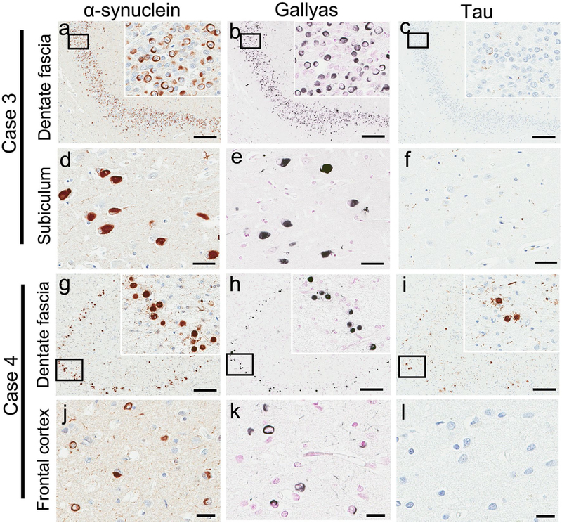
Neuronal inclusions are intensely positive on α-synuclein (a, d, g, j) and Gallyas silver stains (b, e, h, k), but tau immunoreactivity is variable among each case and region. In Case 3, almost no neuronal inclusions are positive on tau stains in the hippocampal dentate fascia (c) or subiculum (f). In Case 4, some of neuronal inclusions are positive on tau stains in the hippocampal dentate fascia (i), while no neuronal inclusions are positive on tau stains in the frontal cortex (l). Scale bars a–c, g–i; 200 μm, d–f, j–l; 50 μm
In the putamen, all cases had abundant NCI and variable dystrophic neurites. Case 3 had abundant NCI in the substantia nigra, while they were less frequent in the other three cases. In the inferior olivary nucleus, abundant NCI were noted in Cases 1 and 2, but they were sparse in Cases 3 and 4.
α-Synuclein glial pathology
The distribution and severity of GCI are shown in Table 2. All cases had many GCI that were morphologically identical those in MSA. The regions with the highest density of GCI were the striatonigral and olivopontocerebellar systems. All cases had many GCI in the putamen. Many GCI were present in pontine base, medullary tegmentum, and cerebellar white matter (Fig. 1f), in Cases 1, 2 and 4, while they were sparse in Case 3. GCI were present in the neocortical and limbic structures, but they were generally sparse.
Tau immunoreactivity for neuronal inclusions
The α-synuclein-positive NCI were intensely positive on Gallyas silver stains, while tau immunoreactivity was minimal. NCI that had tau immunoreactivity were variable with respect to number that were positive and the regions in which they were found (Fig. 3). In Case 3, numerous ringshaped (Fig. 3a–c) and Pick body-like inclusions (Fig. 3d–f) in the hippocampus were mostly negative for tau. In Case 4, some of the Pick body-like inclusions in the hippocampus were positive for tau, although the number was much less than those positive for α-synuclein and Gallyas stains (Fig. 3g–i). Double immunohistochemistry using sections of the hippocampus revealed co-localization of α-synuclein and tau in a subset of Pick body-like inclusions or NFT-like inclusions in three cases (Cases 1, 2 and 4) (Supplemental Fig. 2).
In the frontal cortex, almost no NCI were detected with tau immunohistochemistry in three cases (Cases 1, 3, and 4) (Fig. 4j–l). Case 2 had abundant neurofibrillary pathology in the frontal cortex that was a feature of concomitant AD-type pathology. In the putamen, pontine nuclei, and inferior olivary nucleus, almost no NCI were positive for tau in any of the cases.
Fig. 4.
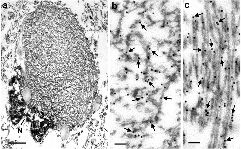
Electron micrograph of a Pick body-like inclusion that displaces the nucleus (N) to the periphery. The nucleus is highly indented and has condensed chromatin (a). Enlargement of central region shows randomly orientated granule-coated filaments heavily labeled with α-synuclein antibody. Arrows point to some of the 18 nm gold particles (b). Enlargement of peripheral region shows parallel granule-coated filaments. Arrows point to some gold particles (c). Case 2, hippocampus. Scale bars a; 1 μm, b, c; 100 nm
Immunoelectron microscopy
Pick body-like inclusions were composed of granule-coated filaments randomly oriented. The filaments were immunolabeled with α-synuclein antibody. The nucleus was often pushed to the side and indented (Fig. 4a–c). Dystrophic neurites, including dystrophic neurites in senile plaques (Supplemental Fig. 3), contained granule-coated α-synuclein-positive filaments. When α-synuclein and tau were found in the same neuron, as in Case 1, α-synuclein filaments tended to be perinuclear, while tau formed discrete cytoplasmic filamentous aggregates (Supplemental Fig. 4). Tau-positive filaments were almost all straight filaments; only a few paired helical filaments were detected.
Additional pathologies
The severity of AD-type pathology is shown in Table 2. Case 2 had severe AD-type pathology (Braak NFT stage VI, Thal Aβ phase 5), and Case 1 had mild AD-type pathology (Braak NFT stage III-IV, Thal Aβ phase 3). Minimal NFT pathology and no SP were observed in Case 3 and Case 4. No TDP-43 pathology was observed in any of the cases.
Genetic findings
For the two cases in which frozen tissue was available (Cases 1 and 2), whole gene sequencing of SNCA for point mutations and copy number variation was performed. Both cases were negative for SNCA point mutations and genomic multiplications.
Comparison of atypical MSA to typical MSA
The four atypical MSA cases were compared to 34 typical MSA cases with respect to frequency and density of NCI in limbic areas affected in atypical MSA (hippocampal dentate fascia, CA1/subiculum, and hypothalamus) and a region vulnerable to NCI in typical MSA (inferior olivary nucleus) (Table 3). All the atypical MSA cases were women, while women made up 44 % of the typical MSA group. The atypical MSA group had a significantly older median age at onset than the typical MSA group. There was no significant difference in the median brain weight, disease duration, Braak NFT stage, or Thal amyloid phase. NCI were often observed in the limbic regions in typical MSA, but they were very sparse compared to atypical MSA. In contrast, the frequency and severity of NCI in the inferior olivary nucleus were not different between atypical and typical MSA.
Table 3.
Comparison of atypical MSA to typical MSA
| Atypical MSA (n = 4) | Typical MSA (n = 34) | P value | |
|---|---|---|---|
| Female | 4/4 (100 %) | 15/34 (44 %) | 0.105 |
| Brain weight, grams | 1130 (693, 1208) | 1180 (1100, 1325) | 0.295 |
| Age at death, years | 81 (71, 90) | 66 (62, 71) | 0.015 |
| Age at onset, years | 69 (66, 84) | 58 (53, 62) | 0.010 |
| Duration of illness, years | 5 (3, 15) | 8 (6, 10) | 0.319 |
| Braak NFT stage | III (I, V) | I (I, II) | 0.085 |
| Thal Aβ phase | 1.5 (0, 4.5) | 2 (0, 2) | 0.590 |
| α-Synuclein-positive NCI | |||
| Hippocampal dentate fascia | |||
| Frequency | 4/4 (100 %) | 18/33 (55 %) | 0.131 |
| Scores | 3 (1.5, 3) | 1 (0, 1) | 0.005 |
| Hippocampal CA1/subiculum | |||
| Frequency | 4/4 (100 %) | 22/33 (67 %) | 0.296 |
| Scores | 3 (3, 3) | 1 (0, 1) | <0.001 |
| Hypothalamus | |||
| Frequency | 4/4 (100 %) | 21/32 (66 %) | 0.290 |
| Scores | 3 (3, 3) | 1 (0, 2) | 0.003 |
| Inferior olivary nucleus | |||
| Frequency | 3/4 (75 %) | 32/32 (100 %) | 0.111 |
| Scores | 2 (0.25, 3) | 2 (1, 3) | 0.873 |
All data are displayed as median (25th, 75th, range), unless otherwise noted. Neuronal cytoplasmic inclusion (NCI) scores are assessed semi-quantitatively using a 4-point grading scale (0 = none, 1 = sparse to mild, 2 = moderate, 3 = severe)
Discussion
In this study, we describe four patients with atypical MSA who presented clinically as FTD, but without documented autonomic dysfunction, a requirement for diagnosis of clinically probable MSA [9]. All cases met pathological criteria for MSA, with striatonigral degeneration and variable olivopontocerebellar degeneration; all had pathognomonic α-synuclein-positive GCI [21]. On the other hand, macroscopic and microscopic findings were suggestive of FTLD, except that neuronal inclusions were not composed of TDP-43 or tau, but rather α-synuclein. The clinical syndromes included CBS, PNFA, and bvFTD, which are clinical phenotypes of FTLD [15, 33, 35]. The association of Pick body-like NCI with neuronal loss suggests that cortical and limbic α-synuclein neuronal pathology contributed to the atypical clinical presentations. There are limited reports of MSA presenting with FTD clinical syndromes [33].
Neuronal inclusions detected with α-synuclein immunohistochemistry were most numerous in limbic and cortical regions and included Pick body-like, NFT-like, and ringshaped inclusions. Of these various morphologic subtypes, Pick -body-like inclusions were most strongly associated with neuronal loss and gliosis, particularly in the hippocampus and amygdala. The neuronal inclusions in atypical MSA were easily distinguished from Lewy bodies in that they were intensely positive on Gallyas stains, while Lewy bodies are negative on Gallyas stains [39]. In addition, tau immunohistochemistry was helpful in distinguishing the Pick body-like and NFT-like inclusions from genuine Pick bodies [48] and NFT because most of the NCI in atypical MSA were negative. Furthermore, Pick bodies are usually negative on Gallyas stains [40]. These findings support the notion that atypical MSA is not MSA with coexistent Pick’s disease, AD or LBD, but rather a variant of MSA [31, 32].
It is worth noting a previous case report of a 51-year-old woman with progressive dementia, psychosis and muscular rigidity with neuropathological features of both MSA and LBD [34]. This patient had frontotemporal and hippocampal atrophy with many α-synuclein-positive, ring-shaped NCI in the hippocampal dentate fascia, similar to those in one of our patients (Case 3). These findings lead us to conclude that this case might be another example of atypical MSA. Unfortunately, clinical details were sparse making it difficult to know if she had features of FTD.
Co-localization of α-synuclein and tau in a subset of Pick body-like and NFT-like inclusions was observed, which was consistent with a previous report of atypical MSA [31]. Co-localization of tau and α-synuclein has been reported in sporadic LBD [14] and familial LBD due to mutations in SNCA [5, 7, 18, 25, 28]. Although the mechanism of abnormal tau accumulation in α-synuclein inclusions is unknown, it may be noteworthy that α-synuclein oligomers can seed tau aggregation in vitro [22].
In addition to the possible case noted above [34], six other atypical MSA cases have been reported—one from the United States (US) [13] and five from Japan [16, 19, 31, 32, 46] (Table 4). Pathologically, all cases had similar features, including frontotemporal atrophy and numerous limbic NCI, including Pick body-like inclusions. Clinically, the course of the disease was different between US and Japanese cases. The median age of symptomatic onset was older in US than Japanese patients (70 vs. 53 years, P = 0.008). The disease duration tended to be shorter in US than Japanese patients (3 vs. 15 = years, P = 0.056). Unlike the US cases, at least three Japanese cases had antemortem clinical diagnosis of MSA, although marked cerebral atrophy noted in the early stages of the disease process indicated that they were atypical MSA. It is possible that greater severity of cerebellar and brainstem pathology in the Japanese cases may have led to recognition of antemortem MSA. In the US cases, only one had cerebellar atrophy; while two cases had cerebellar signs, they occurred later in the disease. Memory impairment, which is not a characteristic feature of MSA [9], was frequent in US (3/5) and most Japanese cases (4/5), including cases with an antemortem diagnosis of MSA.
Table 4.
Summary of previously reported atypical MSA cases
| Horoupian et al. [13] | Shibuya et al. [32] | Piao et al. [31] | Konagaya et al. [19] | Katayama et al. [16] | Yuasa et al. [46] | |
|---|---|---|---|---|---|---|
| Clinical features | ||||||
| Nationality | US | JPN | JPN | JPN | JPN | JPN |
| Sex | Female | Female | Female | Male | Female | Female |
| Age at death, years | 78 | 65 | 73 | 66 | 70 | 77 |
| Age at onset, years | 75 | 53 | 54 | 51 | 45 | 63 |
| Duration of illness, years | 3 | 12 | 19 | 15 | 25 | 14 |
| Clinical diagnosis | Parkinsonism plus | MSA | MSA | MSA | n.a. | SCD |
| Cerebellar signs | + | + | + | + | + | + |
| Parkinsonism | + | + | + | + | + | |
| Memory impairment | + | + | + | + | ||
| Pathologic features | ||||||
| Brain weight, grams | 1140 | 920 | 687 | 1115 | n.a | 1065 |
| Cerebral atrophy | F | T | F,T | F,T | T | T |
| SND/OPCA | SND > OPCA | SND < OPCA | SND = OPCA | SND = OPCA | SND < OPCA | SND < OPCA |
| Numerous GCI | Yes | Yes | Yes | Yes | Yes | Yes |
| Many limbic NCI | Yes | Yes | Yes | Yes | Yes | Yes |
| Morphology | PiB-like | PiB-like | PiB-like | n.a. | PiB-like | n.a. |
| α-Synuclein stain | n.a. | n.a. | Positive | Positive | Positive | Positive |
| Silver stain | Positive | Positive | Positive | Positive | Positive | Positive |
| Tau stain | Negative | Negative | Positive (partially) | Negative | Negative | Negative |
| AD-type pathology | Minimal | Minimal | Minimal | Minimal | n.a. | Minimal |
F frontal lobe, T temporal lobe, SND striatonigral degeneration, OPCA olivopontocerebellar degeneration, SCD spinocerebellar degeneration, US United states, JPN Japan, n.a. not available, PiB-like Pick body-like
Autonomic dysfunction, such as orthostatic hypotension and syncope, were not described in the clinical records of any of our four cases, although they all had severe α-synuclein neuronal pathology in the hypothalamus. The lack of autonomic dysfunction is at variance with a clinical diagnosis of MSA [9].
In comparison to typical MSA, limbic NCI were significantly more frequent and severe in atypical MSA. Only sparse NCI were detected in limbic structures in typical MSA [44]. Wang et al. reported that 55 % (17/31) of MSA cases had NCI in the hippocampus, including the dentate fascia. The density of limbic NCI in most of our typical MSA cases was very low; however, two cases had more abundant lesions. These two cases were not considered to be atypical MSA since they did not have cerebral atrophy or an FTD clinical syndrome. It will be important to examine the clinical impact of limbic α-synuclein neuronal pathology in larger autopsy cohorts of pathologically confirmed MSA. The variable severity of limbic NCI in typical MSA cases suggests that atypical MSA is an extreme phenotype. We consider that numerous Pick body-like inclusions in hippocampus and amygdala associated with neuronal loss and atrophy are the most specific features of atypical MSA, although exceptional cases (Case 1) may have NFT-like inclusions, rather than Pick body-like inclusions.
The nosology of the disorder shared by the four patients we report is worth discussion. While pathologically they had the hallmark GCI of MSA, clinically they presented with FTD syndromes and they had frontotemporal degeneration with severe limbic and cortical α-synuclein neuronal pathology. FTLD is currently classified based on the protein that accumulates within neuronal and glial lesions (e.g., FTLD-tau, FTLD-TDP, and FTLD-FUS). Atypical MSA does not fit into any of the current categories [24]. We propose a new category of FTLD for atypical MSA—FTLD-synuclein. Alternatively, atypical MSA could be considered a subtype of MSA in addition to MSA-P and MSA-C, namely MSA-FTD. From a clinical perspective, this is a more challenging diagnosis, since none of our patients carried an antemortem diagnosis of MSA; rather, the clinical picture was that of FTD.
Supplementary Material
Acknowledgments
We are grateful to all patients, family members, and caregivers who agreed to brain donation; without their donation these studies would have been impossible. We also acknowledge expert technical assistance of Linda Rousseau and Virginia Phillips for histology and Monica Castanedes-Casey for immunohistochemistry. This research was supported in part by a research grant from the Uehara Memorial Foundation, as well as NIH P50 NS072187, P50 AG16574 and P30 AG12300, as well as the CurePSP: Foundation for PSP|CBD and Related Disorders, The Mangurian Foundation, and The Robert E Jacoby Professorship in Alzheimer’s Disease Research.
Footnotes
Electronic supplementary material The online version of this article (doi:10.1007/s00401-015-1442-z) contains supplementary material, which is available to authorized users.
References
- 1.Ahmed Z, Asi YT, Sailer A, Lees AJ, Houlden H, Revesz T, Holton JL (2012) The neuropathology, pathophysiology and genetics of multiple system atrophy. Neuropathol Appl Neurobiol 38:4–24 [DOI] [PubMed] [Google Scholar]
- 2.Beach TG, White CL, Hamilton RL, Duda JE, Iwatsubo T, Dickson DW, Leverenz JB, Roncaroli F, Buttini M, Hladik CL, Sue LI, Noorigian JV, Adler CH (2008) Evaluation of alpha-synuclein immunohistochemical methods used by invited experts. Acta Neuropathol 116:277–288 [DOI] [PMC free article] [PubMed] [Google Scholar]
- 3.Boeve BF, Lang AE, Litvan I (2003) Corticobasal degeneration and its relationship to progressive supranuclear palsy and frontotemporal dementia. Ann Neurol 54(Suppl 5):S15–S19 [DOI] [PubMed] [Google Scholar]
- 4.Braak H, Braak E (1991) Neuropathological stageing of Alzheimer-related changes. Acta Neuropathol 82:239–259 [DOI] [PubMed] [Google Scholar]
- 5.Duda JE, Giasson BI, Mabon ME, Miller DC, Golbe LI, Lee VM, Trojanowski JQ (2002) Concurrence of alpha-synuclein and tau brain pathology in the Contursi kindred. Acta Neuropathol 104:7–11 [DOI] [PubMed] [Google Scholar]
- 6.Fanciulli A, Wenning GK (2015) Multiple-system atrophy. N Engl J Med 372:249–263 [DOI] [PubMed] [Google Scholar]
- 7.Fujishiro H, Imamura AY, Lin WL, Uchikado H, Mark MH, Golbe LI, Markopoulou K, Wszolek ZK, Dickson DW (2013) Diversity of pathological features other than Lewy bodies in familial Parkinson’s disease due to SNCA mutations. Am J Neurodegener Dis 2:266–275 [PMC free article] [PubMed] [Google Scholar]
- 8.Gai WP, Power JH, Blumbergs PC, Blessing WW (1998) Multiple-system atrophy: a new alpha-synuclein disease? Lancet 352:547–548 [DOI] [PubMed] [Google Scholar]
- 9.Gilman S, Wenning GK, Low PA, Brooks DJ, Mathias CJ, Trojanowski JQ, Wood NW, Colosimo C, Durr A, Fowler CJ, Kauf-mann H, Klockgether T, Lees A, Poewe W, Quinn N, Revesz T, Robertson D, Sandroni P, Seppi K, Vidailhet M (2008) Second consensus statement on the diagnosis of multiple system atrophy. Neurology 71:670–676 [DOI] [PMC free article] [PubMed] [Google Scholar]
- 10.Graham JG, Oppenheimer DR (1969) Orthostatic hypotension and nicotine sensitivity in a case of multiple system atrophy. J Neurol Neurosurg Psychiatry 32:28–34 [DOI] [PMC free article] [PubMed] [Google Scholar]
- 11.Grossman M (2010) Primary progressive aphasia: clinicopathological correlations. Nat Rev Neurol 6:88–97 [DOI] [PMC free article] [PubMed] [Google Scholar]
- 12.Gwinn-Hardy K, Mehta ND, Farrer M, Maraganore D, Muenter M, Yen SH, Hardy J, Dickson DW (2000) Distinctive neuropathology revealed by alpha-synuclein antibodies in hereditary parkinsonism and dementia linked to chromosome 4p. Acta Neuropathol 99:663–672 [DOI] [PubMed] [Google Scholar]
- 13.Horoupian DS, Dickson DW (1991) Striatonigral degeneration, olivopontocerebellar atrophy and “atypical” Pick disease. Acta Neuropathol 81:287–295 [DOI] [PubMed] [Google Scholar]
- 14.Ishizawa T, Mattila P, Davies P, Wang D, Dickson DW (2003) Colocalization of tau and alpha-synuclein epitopes in Lewy bodies. J Neuropathol Exp Neurol 62:389–397 [DOI] [PubMed] [Google Scholar]
- 15.Josephs KA, Hodges JR, Snowden JS, Mackenzie IR, Neumann M, Mann DM, Dickson DW (2011) Neuropathological background of phenotypical variability in frontotemporal dementia. Acta Neuropathol 122:137–153 [DOI] [PMC free article] [PubMed] [Google Scholar]
- 16.Katayama T, Yamaoka A, Yokokawa Y, Saito Y, Aiba L, Yasuda T, Yoshida M, Hashizume Y (2000) An autopsy case of multiple system atrophy (MSA) with marked temporal lobe atrophy and numerous Pick bodylike inclusions. Neuropathology 20:A29 [Google Scholar]
- 17.Kato S, Nakamura H (1990) Cytoplasmic argyrophilic inclusions in neurons of pontine nuclei in patients with olivopontocerebellar atrophy: immunohistochemical and ultrastructural studies. Acta Neuropathol 79:584–594 [DOI] [PubMed] [Google Scholar]
- 18.Kiely AP, Asi YT, Kara E, Limousin P, Ling H, Lewis P, Proukakis C, Quinn N, Lees AJ, Hardy J, Revesz T, Houlden H, Holton JL (2013) alpha-Synucleinopathy associated with G51D SNCA mutation: a link between Parkinson’s disease and multiple system atrophy? Acta Neuropathol 125:753–769 [DOI] [PMC free article] [PubMed] [Google Scholar]
- 19.Konagaya M, Sakai M, Yoshida M, Hashizume Y (2006) An autopsy case of long-course multiple system atrophy (MSA) with remarkable atrophy and numerous NCI in the temporal lobe. No To Shinkei 58:430–437 [PubMed] [Google Scholar]
- 20.Kouri N, Whitwell JL, Josephs KA, Rademakers R, Dickson DW (2011) Corticobasal degeneration: a pathologically distinct 4R tauopathy. Nat Rev Neurol 7:263–272 [DOI] [PMC free article] [PubMed] [Google Scholar]
- 21.Lantos PL (1998) The definition of multiple system atrophy: a review of recent developments. J Neuropathol Exp Neurol 57:1099–1111 [DOI] [PubMed] [Google Scholar]
- 22.Lasagna-Reeves CA, Castillo-Carranza DL, Guerrero-Muoz MJ, Jackson GR, Kayed R (2010) Preparation and characterization of neurotoxic tau oligomers. Biochemistry 49:10039–10041 [DOI] [PubMed] [Google Scholar]
- 23.Lin WL, DeLucia MW, Dickson DW (2004) Alpha-synuclein immunoreactivity in neuronal nuclear inclusions and neurites in multiple system atrophy. Neurosci Lett 354:99–102 [DOI] [PubMed] [Google Scholar]
- 24.Mackenzie IR, Neumann M, Baborie A, Sampathu DM, Du Plessis D, Jaros E, Perry RH, Trojanowski JQ, Mann DM, Lee VM (2011) A harmonized classification system for FTLD-TDP pathology. Acta Neuropathol 122:111–113 [DOI] [PMC free article] [PubMed] [Google Scholar]
- 25.Markopoulou K, Dickson DW, McComb RD, Wszolek ZK, Katechalidou L, Avery L, Stansbury MS, Chase BA (2008) Clinical, neuropathological and genotypic variability in SNCA A53T familial Parkinson’s disease. Variability in familial Parkinson’s disease. Acta Neuropathol 116:25–35 [DOI] [PMC free article] [PubMed] [Google Scholar]
- 26.Neary D, Snowden JS, Gustafson L, Passant U, Stuss D, Black S, Freedman M, Kertesz A, Robert PH, Albert M, Boone K, Miller BL, Cummings J, Benson DF (1998) Frontotemporal lobar degeneration: a consensus on clinical diagnostic criteria. Neurology 51:1546–1554 [DOI] [PubMed] [Google Scholar]
- 27.Nishie M, Mori F, Yoshimoto M, Takahashi H, Wakabayashi K (2004) A quantitative investigation of neuronal cytoplasmic and intranuclear inclusions in the pontine and inferior olivary nuclei in multiple system atrophy. Neuropathol Appl Neurobiol 30:546–554 [DOI] [PubMed] [Google Scholar]
- 28.Obi T, Nishioka K, Ross OA, Terada T, Yamazaki K, Sugiura A, Takanashi M, Mizoguchi K, Mori H, Mizuno Y, Hattori N (2008) Clinicopathologic study of a SNCA gene duplication patient with Parkinson disease and dementia. Neurology 70:238–241 [DOI] [PubMed] [Google Scholar]
- 29.Ozawa T, Paviour D, Quinn NP, Josephs KA, Sangha H, Kilford L, Healy DG, Wood NW, Lees AJ, Holton JL, Revesz T (2004) The spectrum of pathological involvement of the striatonigral and olivopontocerebellar systems in multiple system atrophy: clinicopathological correlations. Brain 127:2657–2671 [DOI] [PubMed] [Google Scholar]
- 30.Papp MI, Kahn JE, Lantos PL (1989) Glial cytoplasmic inclusions in the CNS of patients with multiple system atrophy (striatonigral degeneration, olivopontocerebellar atrophy and Shy– Drager syndrome). J Neurol Sci 94:79–100 [DOI] [PubMed] [Google Scholar]
- 31.Piao YS, Hayashi S, Hasegawa M, Wakabayashi K, Yamada M, Yoshimoto M, Ishikawa A, Iwatsubo T, Takahashi H (2001) Co-localization of alpha-synuclein and phosphorylated tau in neuronal and glial cytoplasmic inclusions in a patient with multiple system atrophy of long duration. Acta Neuropathol 101:285–293 [DOI] [PubMed] [Google Scholar]
- 32.Shibuya K, Nagatomo H, Iwabuchi K, Inoue M, Yagishita S, Itoh Y (2000) Asymmetrical temporal lobe atrophy with massive neuronal inclusions in multiple system atrophy. J Neurol Sci 179:50–58 [DOI] [PubMed] [Google Scholar]
- 33.Shinagawa S, Miller B (2015) Frontotemporal dementia In: Rosenberg RN, Padcual JM (eds) Rosenberg’s molecular and genetic basis of neurological and psychiatric disease, vol 69, 5th edn Elsevier, London, pp 779–791 [Google Scholar]
- 34.Sikorska B, Papierz W, Preusser M, Liberski PP, Budka H (2007) Synucleinopathy with features of both multiple system atrophy and dementia with Lewy bodies. Neuropathol Appl Neurobiol 33:126–129 [DOI] [PubMed] [Google Scholar]
- 35.Snowden J, Neary D, Mann D (2007) Frontotemporal lobar degeneration: clinical and pathological relationships. Acta Neuropathol 114:31–38 [DOI] [PubMed] [Google Scholar]
- 36.Spillantini MG, Crowther RA, Jakes R, Cairns NJ, Lantos PL, Goedert M (1998) Filamentous alpha-synuclein inclusions link multiple system atrophy with Parkinson’s disease and dementia with Lewy bodies. Neurosci Lett 251:205–208 [DOI] [PubMed] [Google Scholar]
- 37.Thal DR, Rub U, Orantes M, Braak H (2002) Phases of A beta-deposition in the human brain and its relevance for the development of AD. Neurology 58:1791–1800 [DOI] [PubMed] [Google Scholar]
- 38.Tu PH, Galvin JE, Baba M, Giasson B, Tomita T, Leight S, Nakajo S, Iwatsubo T, Trojanowski JQ, Lee VM (1998) Glial cytoplasmic inclusions in white matter oligodendrocytes of multiple system atrophy brains contain insoluble alpha-synuclein. Ann Neurol 44:415–422 [DOI] [PubMed] [Google Scholar]
- 39.Uchihara T, Nakamura A, Mochizuki Y, Hayashi M, Orimo S, Isozaki E, Mizutani T (2005) Silver stainings distinguish Lewy bodies and glial cytoplasmic inclusions: comparison between Gallyas-Braak and Campbell-Switzer methods. Acta Neuropathol 110:255–260 [DOI] [PubMed] [Google Scholar]
- 40.Uchihara T, Tsuchiya K, Nakamura A, Akiyama H (2005) Silver staining profiles distinguish Pick bodies from neurofibrillary tangles of Alzheimer type: comparison between Gallyas and Campbell-Switzer methods. Acta Neuropathol 109:483–489 [DOI] [PubMed] [Google Scholar]
- 41.Uchikado H, Lin WL, DeLucia MW, Dickson DW (2006) Alzheimer disease with amygdala Lewy bodies: a distinct form of alpha-synucleinopathy. J Neuropathol Exp Neurol 65:685–697 [DOI] [PMC free article] [PubMed] [Google Scholar]
- 42.Wakabayashi K, Takahashi H (2006) Cellular pathology in multiple system atrophy. Neuropathology 26:338–345 [DOI] [PubMed] [Google Scholar]
- 43.Wakabayashi K, Yoshimoto M, Tsuji S, Takahashi H (1998) Alpha-synuclein immunoreactivity in glial cytoplasmic inclusions in multiple system atrophy. Neurosci Lett 249:180–182 [DOI] [PubMed] [Google Scholar]
- 44.Wang L, Hashizume Y, Yoshdia M, Hishikawa N (2002) Temporal lobe lesions in multiple system atrophy. Neuropathology 22:A18 [Google Scholar]
- 45.Yokoyama T, Kusunoki JI, Hasegawa K, Sakai H, Yagishita S (2001) Distribution and dynamic process of neuronal cytoplasmic inclusion (NCI) in MSA: correlation of the density of NCI and the degree of involvement of the pontine nuclei. Neuropathology 21:145–154 [DOI] [PubMed] [Google Scholar]
- 46.Yuasa H, Mitake S, Yoshdia M, Hashizume Y, Ojika K (2002) An autopsy case of multiple system atrophy (MSA) with dementia and marked atrophy of the temporal lobe. Neuropathology 22:A18 [Google Scholar]
- 47.Zhang YJ, Xu YF, Cook C, Gendron TF, Roettges P, Link CD, Lin WL, Tong J, Castanedes-Casey M, Ash P, Gass J, Rangachari V, Buratti E, Baralle F, Golde TE, Dickson DW, Petrucelli L (2009) Aberrant cleavage of TDP-43 enhances aggregation and cellular toxicity. Proc Natl Acad Sci 106:7607–7612 [DOI] [PMC free article] [PubMed] [Google Scholar]
- 48.Zhukareva V, Mann D, Pickering-Brown S, Uryu K, Shuck T, Shah K, Grossman M, Miller BL, Hulette CM, Feinstein SC, Trojanowski JQ, Lee VM (2002) Sporadic Pick’s disease: a tauopathy characterized by a spectrum of pathological tau isoforms in gray and white matter. Ann Neurol 51:730–739 [DOI] [PubMed] [Google Scholar]
Associated Data
This section collects any data citations, data availability statements, or supplementary materials included in this article.


