Abstract
Background:
Classically, arthroscopy has been considered one of the diagnostic gold standards for assessing intra-articular knee and shoulder abnormality.
Purpose:
To assess the risks associated with in-office needle arthroscopy.
Study Design:
Case series; Level of evidence, 4.
Methods:
A retrospective case series analysis was performed by evaluating consecutive diagnostic needle arthroscopies performed by 13 physicians at 13 independent institutions. The findings of both major and minor complications were reported by each of the 13 surgeons based on office documentation. The data were analyzed as a lump sum of both knee and shoulder cases and then subdivided and examined separately. The patients’ ages ranged from 14 to 78 years, and no statistical difference was noted between the numbers of men and women. A major complication was defined as infection, chondral toxicity, or the need for alternative treatment at an urgent care or emergency room secondary to the procedure. Minor complications were defined as a vasovagal event, pain that persisted after 24 hours, or the need for crutches or sling postprocedure.
Results:
Of the 1419 cases, no major complications were reported. The overall rate of vasovagal events was 1.9% for all procedures (1.6% in knees, 3% in shoulders). Persistent pain longer than 24 hours postprocedure was reported in 0.3% of cases. No patient required crutches or a sling. Postarthroscopy magnetic resonance imaging was needed in 1.4% of cases. No device failures were reported.
Conclusion:
Previous literature has evaluated the efficacy, sensitivity, and specificity of in-office diagnostic arthroscopy, and this study validates needle arthroscopy as safe in the office setting, with minimal risk of major or minor complications.
Keywords: Outpatient, needle arthroscopy, clinic, diagnostic arthroscopy
Classically, arthroscopy has been considered the diagnostic gold standard for assessing intra-articular knee abnormality.5,14,21 Unfortunately, this diagnostic tool requires a surgical procedure. Magnetic resonance imaging (MRI) has been proposed as a valid tool for the diagnosis of intra-articular knee abnormality, especially in elderly patient populations, in whom surgical anesthesia carries significant risk.25 Although MRI has been shown to be specific for meniscal and ligamentous injury, the sensitivity of MRI for identifying meniscal damage, or early stage chondral damage, is significantly less than that of traditional arthroscopy.2 Other studies have highlighted differences between MRI and arthroscopy, further stating that the reliability of MRI to diagnose a complete anterior cruciate ligament tear had a sensitivity, specificity, accuracy, and negative predictive value (NPV) of 90.9%, 84.6%, 88.6%, and 84.6%, respectively.15,22 The sensitivity, specificity, accuracy, and NPV of MRI to detect medial meniscal abnormality were 100%, 52.6%, 64%, and 100%, respectively, and to detect lateral meniscal abnormality they were 55.6%, 83.3%, 75.8%, and 83.3%, respectively.15,22 The objective measures of test performance for MRI are not perfect by any means, leading experts to question its overall reliability while also seeking a superior method.11
In-office arthroscopy has existed since the early 1990s; however, its use has been limited. Historically, the challenges associated with this technique included capital cost, procedural pain, time, lack of standardized surgical technique, and unclear literature concerning complications. All of these factors, combined with the relative ease of obtaining an MRI, have limited the growth of in-office arthroscopy as a diagnostic tool. However, improvements in technology, specifically the size and portability of the equipment and the quality of the images, has led to an insurgence in the use of in-office arthroscopy for diagnostic purposes, and recent work using in-office arthroscopy has been promising.17,9
Although MRI is useful in the diagnosis of many intra-articular lesions, many patients have a contraindication to this imaging modality. For some patients, an MRI is contraindicated because of metallic implants, obesity, or claustrophobia. Additional drawbacks include the increased time and cost required for an MRI, including an additional visit to the MRI facility, a follow-up visit to the prescribing physician, and the risk of incidental findings.16
An in-office diagnostic system enables clinicians to provide clinical solutions in an office-based setting. The ability to obtain intra-articular images for diagnostic purposes offers a statistically significant benefit compared with traditional MRI for the evaluation of intra-articular abnormality.6,9 The potential cost savings associated with in-office arthroscopy are also worth noting. Voigt et al26 demonstrated in 2014 that in-office arthroscopy procedures are responsible for a net saving of US$151 million per year compared with traditional use of MRI.
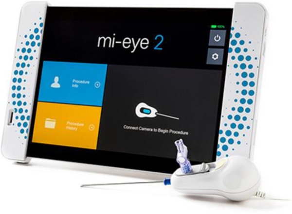
The system used for the current study was the Trice Medical Mi-Eye2 (Figure 1). The Mi-Eye2 consists of a disposable 14-gauge needle arthroscope and a handpiece that connects to a reusable portable tablet. This needle scope uses a “single-stick” mode of joint entry and allows for the 0° camera and light source to provide a 120° field of visualization, secondary to a retractable needle lumen after entry.
Figure 1.

The Mi-Eye2 system.
The purpose of this retrospective series was to assess the risks and complications associated with in-office needle arthroscopy immediately during or after the procedure. These risks include but are not limited to infection, need for further imaging, systemic symptoms, and pain.
Methods
A retrospective case series analysis was performed by evaluating consecutive diagnostic needle arthroscopies performed at 13 independent institutions by 13 physicians, all with experience in needle arthroscopy and selected by the senior author (N.H.A.). Procedures took place in an office setting during scheduled office hours. Although specific sterilization techniques differed among the surgeons included in this study, all of them performed sterile needle arthroscopy through a single needle stick using the Mi-Eye2 system. Data were collected from April 2016 through June 2018 for all diagnostic needle arthroscopies performed on the knee and shoulder. Although needle arthroscopy can be performed across all large joints, patients who had ankle, elbow, and wrist needle arthroscopies were excluded from this study. The patients’ ages ranged from 14 to 78 years, and no statistical difference was noted between the numbers of men and women.
Major and minor complications associated with the procedure over a 2-year period were reviewed. A major complication was defined as an infection, chondral toxicity (rapid destruction of cartilage surfaces),7,13,24 or the need for alternative treatment at an urgent care or emergency room secondary to the procedure. Minor complications included a vasovagal event, pain that persisted after 24 hours, and the need for crutches or sling after the procedure secondary to apprehension or pain. Additionally, the treating physicians documented the rationale for advanced imaging after the procedure (Table 1).
Table 1.
Major and Minor Complications as Defined by the 13 Participating Physicians
| Major Complication | Minor Complication |
|---|---|
| Infection | Vasovagal episode |
| Chondrotoxicitya | Pain lasting >24 h or requiring narcotics |
| Need for emergency department or urgent care evaluation | Need for crutches or sling postprocedure Need for MRI postprocedure |
All the patients underwent a problem-based focused history and physical examination by the treating physician. Patients who had corroborating findings on history that pointed to intra-articular abnormality such as joint swelling, pain, mechanical symptoms, and positive provocative physical examination tests were indicated for the diagnostic needle arthroscopy using the Mi-Eye2 system. Signed consent was obtained from each patient prior to the procedure.
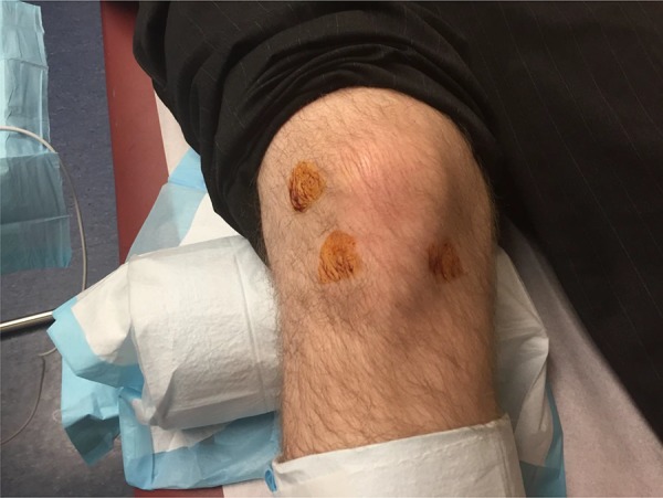
The aseptic preparation technique was documented for each individual surgeon (Figure 2). The local anesthetic agent used on the skin and capsule, the length of time between numbing and the start of the procedure, and whether the numbing agent was intentionally placed in the joint were recorded. Additionally, patient positioning for the procedure was recorded. Also recorded was the method for entry into the joint (ie, whether a scalpel blade or trocar was initially used to create access to the joint, or whether the needle-scope was introduced into the joint with a single-stick method) (Table 2).
Figure 2.

Example of aseptic preparation of a right knee by use of superolateral, medial, and lateral portal sites with alcohol and Betadine (Avrio Health).
Table 2.
Breakdown of Each Physician’s Mode of Entry Into the Joint, Technique for Closure, and Pre- and Postprocedure Antibiotic Regimensa
| Physician No. | Scalpel for Entry | Suture or Glue for Closure | Preprocedure Antibiotics | Postprocedure Antibiotics |
|---|---|---|---|---|
| 1 | N | N | N | N |
| 2 | N | N | N | N |
| 3 | N | N | N | N |
| 4 | N | N | N | N |
| 5 | N | N | N | N |
| 6 | N | N | N | N |
| 7 | N | N | N | N |
| 8 | N | N | N | N |
| 9 | N | N | N | N |
| 10 | N | N | N | N |
| 11 | N | N | N | N |
| 12 | N | N | N | N |
| 13 | N | N | N | N |
aN, none given or used.
After the patient was anesthetized, the treating physician began the procedure via medial or lateral portal entry into the knee joint based upon the location of presumed abnormality, determined via prior physical examination, history, and clinical concern. Similarly, for the shoulder, entry into the joint was gained primarily through a posterior approach. However, entry through the rotator interval was used when the suspected abnormality was located along the posterior aspect of the glenohumeral joint. In some cases, the treating physician elected to use a probe to inspect the abnormality in greater detail, in which a separate entry was made after a sterile preparation, independent of the needle portal. Each physician performed a thorough investigation of the joint in question and documented the findings per his or her own medical record. At the conclusion of the procedure, a bandage and/or a small occlusive dressing was applied, and the patients were instructed to use ice and their choice of either a nonsteroidal anti-inflammatory drug or acetaminophen based upon the individual physician’s postinjection protocol. Major and minor complications were noted and treated.
Results
For this study, 1419 consecutive cases of diagnostic needle arthroscopy of the knee or shoulder were reviewed. Statistical analyses were performed on the outcome data from these cases. The findings of both major and minor complications were reported by each of the 13 surgeons based upon office documentation. The aggregate data were deidentified and collated in a secure encryption by the lead author (S.M.). The data were analyzed as a lump sum of both knee and shoulder cases and then subdivided and examined separately (Table 3).
Table 3.
Sterile Technique, Number of Cases, Anesthetic Preparation, and Infections, Broken Down by Physician and Jointa
| Physician No. | Joint | No. of Cases | Standard Sterile Preparation Protocol | Standard Anesthetic Preparation Protocol | Infections |
|---|---|---|---|---|---|
| 1 | Knee | 224 | Betadine swab sticks, then alcohol on sterile gauze for anesthetic, then repeat same for Mi-Eye2. | 10-15 mL of 1% lidocaine for skin and capsule | 0 |
| Shoulder | 116 | 0 | |||
| 2 | Knee | 137 | ChloraPrep | 15 mL total of 1% lidocaine/0.25% Marcaine | 0 |
| Shoulder | 7 | ChloraPrep | 30 mL total of 1% lidocaine/0.25% Marcaine | 0 | |
| 3 | Knee | 7 | ChloraPrep | 1% lidocaine: 5 mL anterolateral, 5 mL anteromedial, 10 mL suprapatellar per joint | 0 |
| Shoulder | 4 | ChloraPrep | 10 mL of 1% lidocaine, posterior approach | 0 | |
| 4 | Knee | 187 | Betadine swab | 10 mL of 1% lidocaine with epinephrine in each portal | 0 |
| Shoulder | 4 | 10 mL of 1% lidocaine with epinephrine in each portal | 0 | ||
| 5 | Knee | 172 | Alcohol and ChloraPrep first (then infiltration tract). Next, cover the site and repeat preparation with ChloraPrep stick (no drapes). | 20-30 mL of 1% lidocaine with 2 mL of bicarbonate | 0 |
| Shoulder | 51 | 20-30 mL of 1% lidocaine with 2 mL of bicarbonate | 0 | ||
| 6 | Knee | 22 | ChloraPrep | 5 mL of 1% lidocaine with epinephrine in each portal | 0 |
| Shoulder | 1 | 5 mL of 1% lidocaine with epinephrine in each portal | 0 | ||
| 7 | Knee | 5 | Gowned first and then preparation with hydrogen peroxide, followed by Betadine. Anesthetic given and allowed to set up for 10 min. Then another Betadine preparation with ethyl chloride for procedure. | 10-15 mL of 1% lidocaine | 0 |
| Shoulder | 82 | 0 | |||
| 8 | Knee | 39 | Chlorhexidine, sterile gloves, no drape. | 10 mL of 1% lidocaine | 0 |
| 9 | Knee | 19 | Betadine swab sticks, then alcohol for Mi-Eye2 needle. | Anesthetic: cold spray, 20 mL of 1% lidocaine without epinephrine in portals and joint | 0 |
| 10 | Knee | 127 | Betadine swab sticks, then alcohol on sterile gauze for anesthetic, then repeat same for Mi-Eye2. | 20 mL total, half lidocaine 1%, half Marcaine 0.25% with epinephrine | 0 |
| Shoulder | 9 | Same as for knee | 0 | ||
| 11 | Knee | 50 | Iodine swab for injection of lidocaine-Marcaine mix followed by ChloraPrep and drape for the procedure. | 0 | |
| Shoulder | 6 | 0 | |||
| 12 | Knee | 56 | Alcohol followed by chlorhexidine preparation for injection, repeat chlorhexidine preparation for procedure, sterile gloves, no drapes. | Initial preparation with cold spray, then 15-20 mL of 1% lidocaine with epinephrine in skin and the track down to capsule; about 5 mL into the joint | 0 |
| Shoulder | 16 | 0 | |||
| 13 | Knee | 74 | ChloraPrep swab, no drape. | 8 mL of 1% lidocaine with epinephrine, 2-3 mL in capsule, minimal amount in joint | 0 |
| Shoulder | 4 | 0 |
aManufacturers: Betadine, Avrio Health; ChloraPrep, Becton Dickinson and Co; Marcaine, Pfizer.
Of the 1419 cases, no major complications were reported. In particular, no cases of joint infections or trips to the urgent care and/or emergency department due to the procedure were reported. Upon secondary query of the investigating surgeons, no cases of superficial cellulitis around the needle arthroscopy site were reported, nor were there any known cases of chondral toxicity.
Minor complications are noted in Table 4. The rate of overall vasovagal events was 1.9% for all procedures (1.6% for knees and 3% for shoulders). The incidence of persistent pain more than 24 hours postprocedure was 0.3%. The need for crutches or a sling postprocedure was 0%. The need for an MRI after diagnostic needle arthroscopy was overall 1.4%. The most common reason for ordering the MRI was inability to visualize the abnormality within the joint; per the physicians reporting, 55% of those cases occurred during the physicians’ initial learning curve, defined as their first 15 cases. Other reasons for ordering an MRI were persistent pain despite a negative needle arthroscopy, looking for the presence of subchondral bone marrow edema based upon the needle arthroscopy findings, inability of the patient to tolerate the procedure, and mandate from 1 insurance carrier who did not recognize diagnostic needle arthroscopy as an acceptable form of diagnosis at the time of the procedure. Additional miscellaneous reporting included 2 cases of superficial ecchymosis around the portal sites and 1 patient receiving Plavix (Bristol-Myers Squibb Co, Sanofi SA) who had persistent bleeding from the procedure site requiring the addition of topical glue. No device failures were reported for either the tablet or the disposable handpiece. No statistical difference in minor complications was found between sexes.
Table 4.
Minor Complications and Patient Positioning, Broken Down by Physician and Jointa
| Physician No. | Joint | Cases, n | Standard Patient Positioning | Vasovagal, n | Persistent Pain (>24 h), n | Need for Additional MRI, n |
|---|---|---|---|---|---|---|
| 1 | Knee | 224 | Supine | 3 | 0 | 4 |
| Shoulder | 116 | Lateral | 0 | 0 | 1 | |
| 2 | Knee | 137 | Sitting | 5 | 0 | 2 |
| Shoulder | 7 | Sitting | 0 | 0 | 0 | |
| 3 | Knee | 7 | Supine (knee flexed to 90°, extended, frog-leg standard) | 0 | 0 | 0 |
| Shoulder | 4 | Sitting upright in examination chair | 0 | 0 | 1 | |
| 4 | Knee | 187 | Seated with knee bent | 2 | 0 | 2 |
| Shoulder | 4 | Lateral, holding IV pole | 0 | 0 | 0 | |
| 5 | Knee | 172 | Supine | 0 | 2 | 2 |
| Shoulder | 51 | Beach-chair | 3 | |||
| 6 | Knee | 22 | Supine, then seated with knee bent | 1 | 0 | 0 |
| Shoulder | 1 | Seated | 0 | 0 | 0 | |
| 7 | Knee | 5 | Lateral | 1 | 0 | 0 |
| Shoulder | 82 | Beach-chair | 3 | 0 | 0 | |
| 8 | Knee | 39 | Supine | 1 | 0 | 2 |
| 9 | Knee | 19 | Supine | 1 | 0 | 0 |
| 10 | Knee | 127 | Lying supine with legs flexed at knees, off end of examination table | 2 | 3 | |
| Shoulder | 9 | Sitting upright at end of examination table | 0 | 0 | 0 | |
| 11 | Knee | 50 | Supine | 0 | 0 | 0 |
| Shoulder | 6 | Beach-chair | 2 | 0 | 0 | |
| 12 | Knee | 56 | Seated | 2 | 2, different from vasovagal patients | 0 |
| Shoulder | 16 | Seated | 0 | 0 | ||
| 13 | Knee | 74 | Supine | 0 | 1 | 2 |
| Shoulder | 4 | 2 seated, 2 lateral | 1 | 0 | 1 |
aIV, intravenous; MRI, magnetic resonance imaging.
Discussion
Over the past decade, the number of in-office diagnostic needle arthroscopy procedures has significantly increased, as the evolving technology allows physicians to visualize and accurately diagnose abnormalities. A multitude of factors have contributed to this increase, including patient demand, improved technologies, decreased delay in treatment, physician preference, and cost benefit to both the patient and the health care system. As the ability to diagnose intra-articular abnormality in the office setting becomes increasingly more common, the risks associated with diagnostic needle arthroscopy must be better understood. To our knowledge, this is the first study to critically evaluate the complications associated with in-office arthroscopy for the knee and shoulder joint.
Surgical arthroscopy has been considered the gold standard for intra-articular abnormalities associated with the knee and shoulder. However, it is not always prudent or possible to perform surgical diagnostic arthroscopy because of its inherent invasive nature, need for anesthesia, costliness, and other associated risks. In an effort to reproduce the information provided from surgical arthroscopy, diagnostic needle arthroscopy has increased in popularity.14 In particular, pre- and postsurgical cartilage evaluation and postmeniscal repair have been strong indications for the procedure.4 Chambers et al4 noted that if the physician is unsure whether to order an MRI, it appears the value of the MRI is more unreliable. Other literature suggests that degenerative knee abnormality can be difficult to determine solely by MRI, and although MRIs provide some degree of information, they often do not add diagnostic value.1 In addition to diagnostic difficulties noticed with MRI in this patient population, recent studies have shown a higher sensitivity and specificity in using needle-based arthroscopy versus MRI for the evaluation of meniscal abnormality.9
Although previous literature on needle arthroscopy has focused on efficacy, our study focused on complications and overall safety. The results of this study demonstrate the safety of diagnostic needle arthroscopy as it pertains to both major and minor complications. Previously reported rates of infection for in-office needle arthroscopy were hypothesized to be similar to rates of infection for arthrocentesis (<1 in 10,000).3 However, to our knowledge, no large series has documented rates of infection or complication.8–10
Our analysis demonstrated a 0% infection rate with a standard injection aseptic technique for a single-stick needle arthroscopy in 1419 patients. All closures were performed with simple bandages and/or a small compressive wrap. No scalpels were required for any incisions. The integrated system of the percutaneous camera and needle eliminates the need to make multiple passes within the joint, potentially reducing the risk of infections with a single-entry system.
The methods of anesthetizing the patients varied to some degree; however, a consensus was noted among the physicians that the use of lidocaine 1% with or without the addition of 0.25% Marcaine (Hospira) was sufficient to numb the skin and the capsule. Surgeons varied in their opinions regarding direct injection of a local anesthetic into the joint, and no cases of chondrotoxicity were reported followed during the 2-year study period. In addition, each surgeon attempted to remove all the residual fluid from the joint capsule at the end of the procedure to minimize nociceptor activation due to distention after the completion of the procedure. Each surgeon understood the potential risks associated with chondrotoxicity as reported by Kreuz et al.13 They reported that minimal amounts of these agents will not pose a significant risk to cartilage, especially if sterile saline is used as an irrigant.13
Understanding the potential risk for a vasovagal event is important. According to the physicians whose patients experienced a vasovagal event, only 3 of the 27 patients noted that the episode was due to pain. The majority of the patients reported that the episode occurred secondary to a phobia to needles or an “awkward sensation” during the procedure. The overall 1.9% rate of vasovagal events should be examined with the understanding that a subset of patients also cannot tolerate an MRI or MRI-arthrogram. The inability to tolerate an MRI for any reason has been reported to range between 0.7% and 20%.12,27,28 To avoid the risk of a vasovagal event, the lead author has advocated placing the patient in a lateral position for shoulder needle arthroscopies and allowing the patient to lie either 45° or flat for knee arthroscopies. In addition to creating a comfortable office environment, it may be ideal to turn the tablet away from the patient’s sight until the abnormality is identified. The terms used during the consultation with the patient are critical for patients with needle phobias; the term needle scope can be substituted with small probe or camera. This can help reduce anxiety in patients who may have a fear of needles.18–20
Finally, the need for additional imaging was extremely low within this cohort of patients; 1.4% of the patients who underwent the needle arthroscopy required an additional MRI for further evaluation. Within the cohort of patients analyzed, the majority of the MRIs were ordered during the surgeon’s early adaptation of the integrated system in the office. For investigation of shoulder labral injuries, knee or shoulder cartilage defects, or postsurgical reinjuries, a conventional MRI is often ordered and is often inconclusive.8,23 As such, either a repeat MRI with the addition of arthrography or a surgical diagnostic evaluation is necessary to determine the true diagnosis. The realization that no single diagnostic tool is perfect or “always” indicated allows for further imaging should the treating physician deem it necessary. Additionally, challenges exist in creating a comfortable environment not only for the patient but also for the surgeon. Training and repetition will allow the clinician to become familiar with the 0° arthroscope and to avoid pitfalls such as “becoming entrapped in the fat pad.”
Limitations
This was a multicenter retrospective study that evaluated in-office, needle-based diagnostic arthroscopy. Because of the retrospective nature of the study design, the results were dependent on the accuracy of the records kept as well as the patients’ self-reporting of any nonacute complications. We realize that additional data points could have been obtained in a prospective randomized controlled study. Nevertheless, we were satisfied that the primary endpoint—demonstrating the safety of needle-based diagnostic arthroscopy—was achieved.
Conclusion
Previous literature has evaluated the efficacy, sensitivity, and specificity of in-office diagnostic arthroscopy, and this study validates needle arthroscopy as safe in the office setting with minimal risk of major or minor complications.
Footnotes
One or more of the authors has declared the following potential conflict of interest or source of funding: S.M. has received hospitality payments from Stryker and consulting fees from Arthrex, C.R. Bard, Exactech, DePuy Mitek, Linvatec, Rotation Medical, Smith & Nephew, Trice Medical, and Zimmer Biomet. A.C. has received educational support from Arthrex and Stryker and consulting fees from Arthrex, Cayenne Medical, Trice Medical, and Zimmer Biomet. N.H.A. has received research support from Pacira, Smith & Nephew, and Trice Medical; consulting fees from Biom’Up, DePuy, Pacira, Smith & Nephew, and Trice; and hospitality payments from Novadaq Technologies. AOSSM checks author disclosures against the Open Payments Database (OPD). AOSSM has not conducted an independent investigation on the OPD and disclaims any liability or responsibility relating thereto.
Ethical approval for this study was obtained from the Mayo Clinic Institutional Review Board.
References
- 1. Altınel L, Er MS, Kaçar E, Erten RA. Diagnostic efficacy of standard knee magnetic resonance imaging and radiography in evaluating integrity of anterior cruciate ligament before unicompartmental knee arthroplasty. Acta Orthop Traumatol Turc. 2015;49(3):274–279. [DOI] [PubMed] [Google Scholar]
- 2. Behairy NH, Dorgham MA, Khaled SA. Accuracy of routine magnetic resonance imaging in meniscal and ligamentous injuries of the knee: comparison with arthroscopy. Int Orthop. 2009;33(4):961–967. [DOI] [PMC free article] [PubMed] [Google Scholar]
- 3. Bert JM, Bert TM. Management of infections after arthroscopy. Sports Med Arthrosc Rev. 2013;21(2):75–79. [DOI] [PubMed] [Google Scholar]
- 4. Chambers S, Jones M, Michla Y, Kader D. The accuracy of magnetic resonance imaging in detecting meniscal pathology. Orthop Proc. 2012;94-B(suppl XXIX):61. [Google Scholar]
- 5. Crawford R, Walley G, Bridgman S, Maffulli N. Magnetic resonance imaging versus arthroscopy in the diagnosis of knee pathology, concentrating on meniscal lesions and ACL tears: a systematic review. Br Med Bull. 2007;84(1):5–23. [DOI] [PubMed] [Google Scholar]
- 6. Deirmengian CA, Dines JS, Vernace JV, Schwartz MS, Creighton RA, Gladstone JN. Use of a small-bore needle arthroscope to diagnose intra-articular knee pathology: comparison with magnetic resonance imaging. Am J Orthop (Belle Mead NJ). 2018;47(2). [DOI] [PubMed] [Google Scholar]
- 7. Dragoo JL, Braun HJ, Kim HJ, Phan HD, Golish SR. The in vitro chondrotoxicity of single-dose local anesthetics. Am J Sports Med. 2012;40(4):794–799. [DOI] [PubMed] [Google Scholar]
- 8. El-Liethy N, Kamal H, Elsayed RF. Role of conventional MRI and MR arthrography in evaluating shoulder joint capsulolabral-ligamentous injuries in athletic versus non-athletic population. Egypt J Radiol Nucl Med. 2016;47(3):969–984. [Google Scholar]
- 9. Gill TJ, Safran M, Mandelbaum B, Huber B, Gambardella R, Xerogeanes J. A prospective, blinded, multicenter clinical trial to compare the efficacy, accuracy, and safety of in-office diagnostic arthroscopy with magnetic resonance imaging and surgical diagnostic arthroscopy. Arthroscopy. 2018;34(8):2429–2435. [DOI] [PubMed] [Google Scholar]
- 10. Halbrecht JL, Jackson DW. Office arthroscopy: a diagnostic alternative. Arthroscopy. 1992;8(3):320–326. [DOI] [PubMed] [Google Scholar]
- 11. Hoyt M, Goodemote P, Morton JR. How accurate is an MRI at diagnosing injured knee ligaments? J Fam Pract. 2010;59(2):118–120. [PubMed] [Google Scholar]
- 12. Kennedy DJ, Schneider BJ, Casey EK, Rittenberg JD, Lento PH, Smuck M. Vasovagal rates in fluoroscopically guided interventional procedures: a study of over 8,000 injections. Spine J. 2013;13(9):S23–S24. [DOI] [PMC free article] [PubMed] [Google Scholar]
- 13. Kreuz PC, Steinwachs M, Angele P. Single-dose local anesthetics exhibit a type-, dose-, and time-dependent chondrotoxic effect on chondrocytes and cartilage: a systematic review of the current literature. Knee Surg Sports Traumatol Arthrosc. 2018;26(3):819–830. [DOI] [PubMed] [Google Scholar]
- 14. Kuikka P-I, Kiuru MJ, Niva MH, Kröger H, Pihlajamäki HK. Sensitivity of routine 1.0-Tesla magnetic resonance imaging versus arthroscopy as gold standard in fresh traumatic chondral lesions of the knee in young adults. Arthroscopy. 2006;22(10):1033–1039. [DOI] [PubMed] [Google Scholar]
- 15. Laoruengthana A, Jarusriwanna A. Sensitivity and specificity of magnetic resonance imaging for knee injury and clinical application for the Naresuan University Hospital. J Med Assoc Thai. 2012;95(10):S151–S157. [PubMed] [Google Scholar]
- 16. McMillan S, Schwartz M, Jennings B, Faucett S, Owens T, Ford E. In-office diagnostic needle arthroscopy: understanding the potential value for the US healthcare system. Am J Orthop. 2017;46(5):252–256. [PubMed] [Google Scholar]
- 17. Meister K, Harris NL, Indelicato PA, Miller G. Comparison of an optical catheter office arthroscope with a standard rigid rod-lens arthroscope in the evaluation of the knee. Am J Sports Med. 1996;24(6):819–823. [DOI] [PubMed] [Google Scholar]
- 18. van Minde D, Klaming L, Weda H. Pinpointing moments of high anxiety during an MRI examination. Int J Behav Med. 2014;21(3):487–495. [DOI] [PubMed] [Google Scholar]
- 19. Munn Z, Jordan Z. Interventions to reduce anxiety, distress and the need for sedation in adult patients undergoing magnetic resonance imaging: a systematic review. Int J Evid Based Healthc. 2013;11(4):265–274. [DOI] [PubMed] [Google Scholar]
- 20. Murphy KJ, Brunberg JA. Adult claustrophobia, anxiety and sedation in MRI. Magn Reson Imaging. 1997;15(1):51–54. [DOI] [PubMed] [Google Scholar]
- 21. Nikolaou VS, Chronopoulos E, Savvidou C, et al. MRI efficacy in diagnosing internal lesions of the knee: a retrospective analysis. J Trauma Manag Outcomes. 2008;2(1):4. [DOI] [PMC free article] [PubMed] [Google Scholar]
- 22. Panigrahi R, Priyadarshi A, Palo N, Marandi H, Kumar Agrawalla D, Ranjan Biswal M. Correlation of clinical examination, MRI and arthroscopy findings in menisco-cruciate injuries of the knee: a prospective diagnostic study. Arch Trauma Res. 2017;6(1):e30364. [Google Scholar]
- 23. Pavic R, Margetic P, Bensic M, Brnadic RL. Diagnostic value of US, MR and MR arthrography in shoulder instability. Injury. 2013;44:S26–S32. [DOI] [PubMed] [Google Scholar]
- 24. Ravnihar K, Barlič A, Drobnič M. Effect of intra-articular local anesthesia on articular cartilage in the knee. Arthroscopy. 2014;30(5):607–612. [DOI] [PubMed] [Google Scholar]
- 25. Subhas N, Sakamoto FA, Mariscalco MW, Polster JM, Obuchowski NA, Jones MH. Accuracy of MRI in the diagnosis of meniscal tears in older patients. AJR Am J Roentgenol. 2012;198(6):W575–W580. [DOI] [PubMed] [Google Scholar]
- 26. Voigt JD, Mosier M, Huber B. In-office diagnostic arthroscopy for knee and shoulder intra-articular injuries: its potential impact on cost savings in the United States. BMC Health Serv Res. 2014;14(1):203. [DOI] [PMC free article] [PubMed] [Google Scholar]
- 27. Weinreb JC, Maravilla KR, Peshock R, Payne J. Magnetic resonance imaging: improving patient tolerance and safety. AJR Am J Roentgenol. 1984;143(6):1285–1287. [DOI] [PubMed] [Google Scholar]
- 28. Wollman DE, Beeri MS, Weinberger M, Cheng H, Silverman JM, Prohovnik I. Tolerance of MRI procedures by the oldest old. Magn Reson Imaging. 2004;22(9):1299–1304. [DOI] [PubMed] [Google Scholar]


