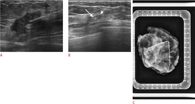
Fig. 4. Visible Cormark with ultrasound-guided localization (no residual carcinoma, fibrosis with collagen-like material deposition and giant cell reaction) in a 46-year-old woman with human epidermal growth factor receptor 2-positive breast cancer with residual ductal carcinoma in situ.
A. Initial tumor lesion was seen on ultrasound with the marker. B. Only the Cormark (marker itself [arrow], adjacent collagen [arrowhead]) is visible on ultrasound after neoadjuvant chemotherapy. C. Localization was confirmed using specimen mammogram.

