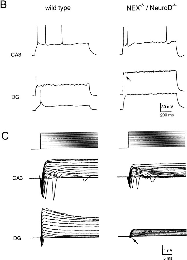
In the article “Neuronal Basic Helix-Loop-Helix Proteins (NEX and BETA2/Neuro D) Regulate Terminal Granule Cell Differentiation in the Hippocampus,” by Markus H. Schwab, Angelika Bartholomae, Bernd Heimrich, Dirk Feldmeyer, Silke Druffel-Augustin, Sandra Goebbels, Frank J. Naya, Shanting Zhao, Michael Frotscher, Ming-Jer Tsai, and Klaus-Armin Nave, which appeared on pages 3714–3724 of the May 15, 2000 issue, the lower left graph of Figure 5C [the IV curve of a control wild-type granule cell in the dentate gyrus (DG)] is a duplication of another curve just above it [a control wild-type pyramidal cell (CA3)]. The correct version of the figure, as well as the legend, is printed here.
B, Firing patterns of pyramidal cells (CA3, top) and dentate GC (DG, bottom) recorded in wild-type (WT, left) and NEX−/− *BETA2/NeuroD−/− mice (DKO, right). The membrane potential was set to −60 mV before action potentials were elicited by injection of 1 sec current pulses. Note that CA3 pyramidal cells of either genotype could fire regenerative action potentials with peak amplitudes of 70–100 mV. In differentiated GC, only nonregenerative action potentials (amplitude, 60 mV) could be elicited. Presumptive GC in NEX−/− *BETA2/NeuroD−/− mice never showed action potentials. Only a small current deflection on top the voltage response was observed (arrow). Firing patterns and current responses (C) are from the same neurons.C, Current responses of CA3 pyramidal cells (top) and dentate GC (bottom) in slices from wild-type (left) and NEX−/− *BETA2/NeuroD−/− mice (right). The membrane potential was held at −70 mV, and 10 mV voltage steps up to +100 mV were applied. Peak Na+current generally exceeded 1 nA in CA3 pyramidal cells, but was smaller in DG granule cells. Presumptive GC in NEX−/− *BETA2/NeuroD−/− mutants had peak Na+ currents <500 pA (arrow).


