Abstract
Chronic exposure to nicotine leads to long-term changes in both the abundance and activity of nicotinic acetylcholine receptors, processes thought to contribute to nicotine addiction. We have found that in Caenorhabditis elegans, prolonged nicotine treatment results in a long-lasting decrease in the abundance of nicotinic receptors that control egg-laying. In naive animals, acute exposure to cholinergic agonists led to the efficient stimulation of egg-laying, a response mediated by a nicotinic receptor functionally expressed in the vulval muscle cells. Overnight exposure to nicotine led to a specific and long-lasting change in egg-laying behavior, which rendered the nicotine-adapted animals insensitive to simulation of egg-laying by the nicotinic agonist and was accompanied by a promoter-independent reduction in receptor protein levels. Mutants defective in the gene tpa-1, which encodes a homolog of protein kinase C (PKC), failed to undergo adaptation to nicotine; after chronic nicotine exposure they remained sensitive to cholinergic agonists and retained high levels of receptor protein in the vulval muscles. These results suggest that PKC-dependent signaling pathways may promote nicotine adaptation via regulation of nicotinic receptor synthesis or degradation.
Keywords: nicotinic, acetylcholine, Caenorhabditis elegans, protein kinase C, adaptation, levamisole
After prolonged exposure to nicotine or other agonists, nicotinic acetylcholine receptors undergo changes in both activity and abundance. For example, in some cell types, long-term nicotine treatment has been shown to cause a long-lasting inactivation of nicotinic receptors that is distinct in both time course and persistence from the rapid, receptor-intrinsic desensitization induced by receptor agonists (Simasko et al., 1986; Lukas, 1991). In addition, chronic exposure to nicotinic agonists has been shown to cause changes in the abundance of nicotinic receptors in both neurons and muscle cells. For example, long-term exposure to nicotine leads to an increase in the density of α2β4 nicotinic receptors in the mammalian CNS (Wonnacott, 1990; Flores et al., 1992). Conversely, chronic exposure to nicotinic agonists leads to a decreased abundance of extrajunctional receptors in chick myotubes (Gardner and Fambrough, 1979). Depending on the context and cell type, α7-containing receptors have been shown to be either downregulated (Messing, 1982) or upregulated (Barrantes et al., 1995; Rogers and Wonnacott, 1995; Molinari et al., 1998) by chronic nicotine treatment. In some cases, agonist-induced changes in receptor abundance appear to be mediated at the transcriptional level (Changeaux, 1991), whereas in other cases, a variety of post-transcriptional processes, including changes in rates of receptor assembly, intracellular targeting, and protein turnover, have been implicated (Marks et al., 1992; Peng et al., 1994; Bencherif et al., 1995; Rothhut et al., 1996). Yet despite the importance of these processes to synaptic plasticity and their potential involvement in nicotine addiction (Dani and Heinemann, 1996), at present there is little information about the specific molecules and intracellular signaling pathways that regulate nicotinic receptor activity and abundance.
One way to investigate the mechanisms underlying nicotinic receptor regulation is to use a genetically tractable animal such asCaenorhabditis elegans. C. elegans has a simple, well characterized nervous system and is well suited for investigating how specific neurotransmitters, receptors, and signaling molecules function within the context of the nervous system to produce behavior. C. elegans contains a diverse family of nicotinic receptor genes (Ballivet et al., 1996; Bargmann, 1998; Mongan et al., 1998), including both neuromuscular and neuronal receptor subtypes (Squire et al., 1995;Treinin and Chalfie, 1995; Baylis et al., 1997). Several of these, including the unc-29 gene, have been shown to encode functional receptor subunits when expressed ectopically in oocytes (Fleming et al., 1997). Nicotinic receptor agonists have specific and easily assayed effects on several aspects of C. elegansbehavior, including locomotion, feeding, and egg-laying (Lewis et al., 1980; Trent et al., 1983; Avery and Horvitz, 1990). A number of paradigms for behavioral plasticity have been defined in C. elegans (Hedgecock and Russell, 1975; Rankin et al., 1990; Colbert and Bargmann, 1995; Schafer and Kenyon, 1995; Wen et al., 1997), demonstrating that these animals are capable of at least simple forms of learning. Thus, it seemed reasonable to ask whether chronic exposure to nicotine resulted in long-term changes in the abundance or activity of C. elegans nicotinic receptors and, if so, to identify molecules required for these processes.
MATERIALS AND METHODS
Egg-laying assays. Unless otherwise stated, nematodes were grown and assayed at room temperature on standard nematode growth medium (NGM) seeded with Escherichia coli strain OP50 as a food source. All animals tested were young adult (i.e., within 24 hr of the L4–adult molt) hermaphrodites. For dose–response experiments, individual animals were placed in microtiter wells containing liquid M9 and the indicated concentration of drug. In the acute-response experiments (see Fig. 1), the number of eggs laid in response to levamisole was assayed after 1 hr. For nicotine-adaptation experiments (see Figs. 2, 4, 6), worms were placed on 30 mmnicotine (Sigma, St. Louis, MO)-seeded NGM plates for varying lengths of time and tested for the response to levamisole in individual liquid M9 assays. To maximize the possibility that a levamisole-sensitive animal would lay eggs, eggs were counted after 4 hr in levamisole in these assays. Because of the relative impermeability of the nematode cuticle, internal concentrations of all drugs have been estimated to be several orders of magnitude lower than their concentrations in the growth medium (Lewis et al., 1980).
Fig. 1.
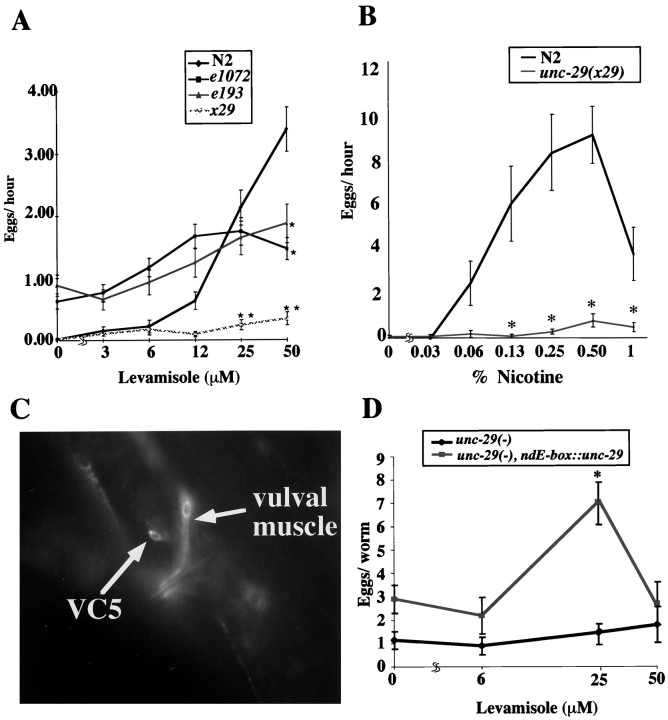
Requirement of the UNC-29 receptor protein for the acute response to nicotinic agonists. Egg-laying responses to levamisole or nicotine were assayed in liquid M9 after 1 hr of drug exposure. The unc-29(x29) allele contains a stop codon that interrupts the conserved fourth transmembrane region of the UNC-29 receptor protein (D. S. Poole, unpublished observations) and thus should result in a severe, if not complete, loss of receptor function. The N2 strain is the wild-type strain in these assays.A, Response of unc-29 mutants to levamisole is shown. Animals carrying any of threeunc-29 mutant alleles showed a dramatic reduction in levamisole-induced egg-laying. Individual points and error bars indicate the mean and SEM of the following numbers of trials: N2, 155; e193, 144; e1072, 96;x29, 96. Asterisks indicate significantly less egg-laying according to the Mann–Whitney rank-sum test (**p < 0.001; *p < 0.01). Other genes that were required for the levamisole response in this assay included unc-38, unc-63, unc-74, lev-1, lev-8, andlev-9 (Kim, Poole, and Schafer, unpublished observations). B, Response ofunc-29 mutants to nicotine is shown.unc-29 mutants showed a significant reduction in nicotine-induced egg-laying. Individual points and error bars indicate the mean and SEM of at least 12 trials.Asterisks indicate significantly less egg-laying according to the Mann–Whitney rank-sum test (*p < 0.01). C, Localization of GFP-tagged UNC-29 protein is shown on a digital image of a ZZ2001 adult hermaphrodite (genotype:unc-29(x29); Ex[ punc-29:: unc-29:: GFP, rol-6d]), which expresses unc-29:: GFPunder the control of the unc-29 promoter. In this ventral/lateral view, the VC5 neuronal cell body and one set of vulval muscles are strongly fluorescent. Not shown in this view are other fluorescent cells, including additional ventral cord motoneurons and various unidentified head and tail neurons. Punctate body fluorescence is caused by autofluorescence of gut granules. D, UNC-29 receptors function in the vulval muscles. The graph compares the levamisole response of an unc-29 mutant [ZZ29,unc-29(x29)] with that of a transgenic line that express a functional unc-29 allele under control of the vulval muscle-specific 18ndE-box enhancer (AQ497,unc-29(x29); dpy-20(e1282); ljEx8[18ndE-box:: unc-29, dpy-20(+)]). Individual points and error bars indicate the mean and SEM of 20 or more trials. At 25 μm, the AQ497 worms laid significantly more eggs according to the Mann–Whitney rank-sum test (*p < 0.001).
Fig. 2.
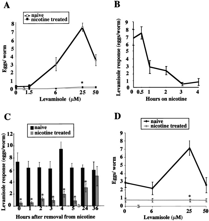
Long-term adaptation of levamisole receptors to nicotine. A, Effect of overnight nicotine treatment on the levamisole response. Shown are levamisole dose–response curves for wild-type animals cultured overnight in the presence of nicotine (30 mm); the dose–response curve of naive animals under the same conditions is shown as a control. Egg-laying was measured after 4 hr at the indicated condition. Points and error bars indicate the mean and SEM of 18 or more trials; at 25 μm, the nicotine-adapted animals laid significantly fewer eggs than did the naive animals according to the Mann–Whitney rank-sum test (*p < 0.001). These nicotine-adapted animals responded normally to serotonin; egg-laying rates in M9 salts containing 7.5 mm serotonin were as follows: naive, 2.7 ± 1.2 eggs/hr; nicotine-adapted, 2.5 ± 0.6 eggs/hr (n = 12 in both cases). B, Time course of nicotine adaptation. N2 hermaphrodites were placed on seeded NGM containing 30 mm nicotine for the indicated length of time and then assayed individually for egg-laying in M9 + 25 μm levamisole as described in A.Points and error bars indicate the mean and SEM of 18 trials. C, Long-term persistence of nicotine adaptation. The histogram shows the time course of recovery of levamisole responses in nicotine-adapted animals. N2 animals were grown overnight on NGM with 30 mm nicotine and then transferred to drug-free NGM plates for the indicated times; egg-laying in response to 25 μm levamisole was tested as described.Asterisks indicate time points in which the levamisole response was significantly lower in the nicotine-adapted animals than in the mock-treated animals grown on NGM without nicotine (*p < 0.001). Verticalbars and error bars indicate the mean and SEM of at least 18 trials. D, Nicotine adaptation in animals expressing only vulval muscle UNC-29 receptors. Shown are the levamisole dose–response curves for nicotine-treated animals expressing unc-29 only in the vulval muscles; naive animals are shown as a control. Animals of the strain AQ497 (genotypeunc-29(x29); dpy-20(e1282); ljEx8 [18ndE-box:: unc-29, dpy-20(+)]) were cultured overnight on NGM containing 30 mm nicotine and then assayed for egg-laying in response to levamisole as described. Points and error bars represent the mean and SEM of 20 or more trials. An asteriskindicates significantly less egg-laying according to the Mann–Whitney rank-sum test (*p < 0.001).
Fig. 4.
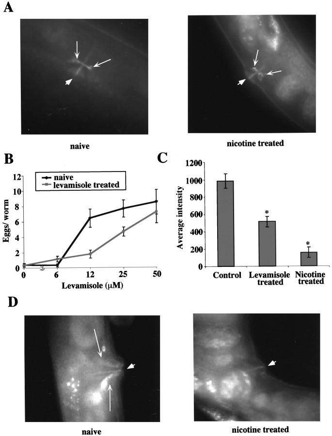
Specificity of the long-term effects of nicotine on egg-laying. A, Effect of long-term nicotine on VC–vulval muscle synapses. Shown here is the expression pattern of a synaptic marker (VAMP:: GFP) that illuminates the VC–vulval muscle synapse, in both nicotine-treated (right) and naive (left) adult animals (synapse identified bylongarrows; the vulva identified by ashortarrow). Animals of the strain NM670 (genotype, lin-15(n765); jsIs42x [punc-4:snb-1:: GFP, lin-15(+)]) were cultured on NGM containing 30 mmnicotine overnight, and images were captured as described in Materials and Methods. Naive animals cultured on NGM alone are shown for comparison. B, Effect of chronic levamisole treatment on the levamisole response. Shown is the levamisole dose–response curve of wild-type animals grown in the presence of levamisole; the dose–response curve of naive animals under the same conditions is shown as a control. N2 hermaphrodites were cultured overnight on NGM containing 50 μm levamisole and then assayed individually for egg-laying for 4 hr under the indicated condition as described in Figure 2. Points and error bars indicate the mean and SEM of 18 or more trials. C, Effect of chronic levamisole treatment on UNC-29 receptor abundance. The histogram compares the UNC-29:: GFP fluorescence intensity in the vulval muscles between naive ZZ2171 animals (see Fig. 3) and ZZ2171 animals treated overnight with levamisole (50 μm) or nicotine (30 mm). Verticalbars and error bars indicate the mean and SEM of five or more trials;asterisks indicate a statistically significant difference from naive animals according to the Mann–Whitney rank-sum test (*p < 0.02). D, Effect of nicotine on vulval muscle UNC-29 protein levels in lev-1mutants. Shown are digital images of naive (left) and nicotine-treated (right) lev-1hermaphrodites expressing UNC-29:: GFP in the vulval muscles. Animals of the strain AQ533 (genotype, unc-29(x29); lev-1(e211); Ex [punc-29:: unc-29:: GFP; rol-6d]) were cultured on NGM containing 30 mm nicotine overnight; UNC-29:: GFP fluorescence was measured as described previously (longarrows indicate vulval muscles, ashortarrow indicates the vulva). Average fluorescence intensities were 120 ± 20 (naive) and 0 ± 0 (nicotine-treated); the difference between these values was statistically significant (p < 0.001) according to the Mann–Whitney rank-sum test. Similar effects were seen in lev-8 and lev-9 mutant animals (J. Kim and W. R. Schafer, unpublished observations).
Fig. 6.
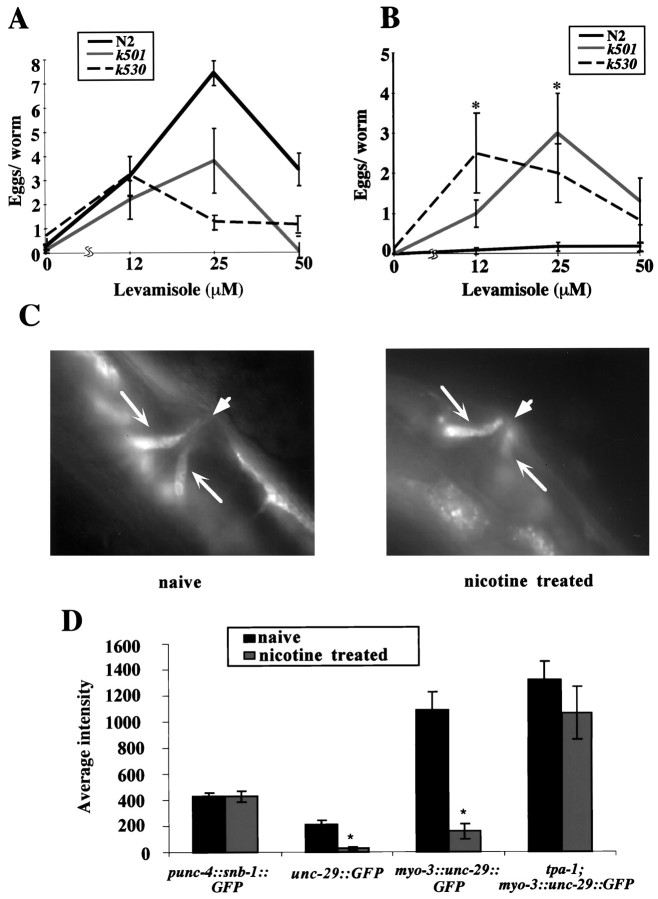
Dependence of nicotine adaptation on PKC.A, Response of tpa-1 mutants to levamisole. Levamisole dose–response curves for wild-type (N2) andtpa-1 mutant animals are shown. Animals were assayed for 4 hr under the indicated condition; points and error bars indicate the mean and SEM of at least 12 trials. B, Defect of tpa-1 mutants in nicotine adaptation. Levamisole dose–response curves (assayed at 4 hr) for wild-type andtpa-1 mutant strains after overnight treatment with 30 mm nicotine are shown. Points and error bars indicate the mean and SEM of at least 12 trials. The responses of bothtpa-1 mutants were significantly greater than that of the wild-type under this condition according to the Mann–Whitney rank-sum test (*p < 0.001). By the same test, thetpa-1 mutants showed no significant reduction in response to levamisole after overnight nicotine treatment (p > 0.5). C, Effect of nicotine on vulval muscle UNC-29 protein levels in tpa-1mutants. Shown here are digital images of naive (left) and nicotine-treated (right) tpa-1 mutant hermaphrodites expressing UNC-29:: GFP in the vulval muscles under the control of the myo-3 promoter (see Fig. 4).Long arrows indicate vulval muscles, and ashortarrow indicates the vulva. Animals of the strain AQ521 (genotype, unc-29(x29); tpa-1(k501); Ex [pmyo-3:: unc-29:: GFP; rol-6d]) were cultured on NGM containing 30 mm nicotine overnight; UNC-29:: GFP fluorescence was measured as described previously. D, Quantification of UNC-29:: GFP and SNB-1:: GFP fluorescence in nicotine-treated and untreated animals. The average intensity of fluorescence for nicotine-treated and naive animals expressing UNC-29:: GFP protein under the control of the unc-29 promoter (unc-29:: GFP) or themyo-3 promoter (myo-3:: unc-29:: GFP) or the synaptic marker VAMP:: GFP under the control of the VC-specific unc-4 promoter (punc-4:: snb-1:: GFP) are shown. Asterisks indicate significantly less fluorescence after nicotine treatment according to the Mann–Whitney rank-sum test (*p < 0.001). The histogram and error bars represent the mean and SEM of five trials.
Microscopy. The strain ZZ2001 (genotype,unc-29(x29); Ex[rol-6d, unc-29:: GFP]; kindly provided by Jim Lewis) was used for localization of UNC-29 receptors. For visualization of VC–vulval muscle synapses, we used the strain NM670 [genotype, lin-15(n765); jsIs42x [punc-4:: snb-1:: GFP, lin-15(+)]; kindly provided by Mike Nonet (1999)]. For visualization of thetpa-1 gene product protein kinase C (PKC), the strain TF6 (see below) was used. Worms were placed on agar pads and immobilized with 30 mm sodium azide. Green fluorescent protein (GFP) was visualized using standard immunofluorescence techniques at a magnification of 100×. Images were collected using a high-speed monochrome CCD camera (Hamamatsu) and analyzed using Metamorph image-processing software. To quantitate fluorescence intensity, the average background intensity was determined for each image and subtracted from the final fluorescent intensity. Then, the average, maximum, and minimum intensities were measured, along with the average area of fluorescence.unc-29:: GFP-expressing lines generated in our own laboratory using dpy-20(+) as a coinjection marker showed fluorescence in essentially the same set of cells as ZZ2001 (data not shown).
Generation of transgenic lines. Transgenic lines expressing the UNC-29 protein in specific cell types were constructed in the following manner. To express UNC-29 in the vulval muscles, a 2510 bp piece of unc-29 cDNA was cut from the plasmid pPD95.86. This piece was subcloned into the vector pSAK-10 (obtained from A. Fire) downstream of the region containing 18 repeats of the vulval muscle-specific ndE-box enhancer. This plasmid was injected at ∼100 ng/μl into unc-29(x29); dpy-20(e1282) worms using wild-type dpy-20(+) DNA as a coinjection marker (25 ng/μl). The F1 generation was scored for rescue of the dpyphenotype. Transgenic lines transmitting anndE-box:: unc-29; dpy-20(+) extrachromosomal array were identified by assaying the non-Dpy progeny of non-Dpy F1 transformants for egg-laying in the presence of 25 μm levamisole in M9.
tpa-1:: GFP reporter constructs. The expression pattern of tpa-1 was evaluated using the strain TF6 (genotype, unc-119(e2498); Is [tpa-1A:: GFP; unc-119(+)]). Two mRNA species, tpa-1A andtpa-1B, are transcribed from the tpa-1 gene, which consists of 11 exons. The tpa-1A mRNA contains all of the 11 exons, and the tpa-1B mRNA contains exons V–XI. Thetpa-1A:: GFP reporter construct was generated by inserting a 9 kb NaeI fragment, which contained thetpa-1A 5′ upstream region along with the first two exons of the tpa-1A-coding sequence, into the SmaI site of the GFP expression plasmid pGFP-N3 (Clontech, Cambridge, UK). The resulting plasmid tpa-1A:: GFP was injected into the germ lines of unc-119 mutant animals using wild-typeunc-119(+) DNA as a cotransformation marker; rescued transgenic lines expressing tpa-1A:: GFP were identified among the progeny. The [tpa-1A:: GFP; unc-119(+)] extrachromosomal array was integrated into the chromosome by UV irradiation.
Analysis of egg-laying patterns. Egg-laying in C. elegans occurs in a specific temporal pattern; egg-laying events are clustered, with periods of active egg-laying, or active phases, separated by long inactive phases during which eggs are retained. Both the duration of the inactive phases (“intercluster intervals”) and the duration of intervals between egg-laying events in a cluster (“intracluster intervals”) model as exponential random variables with different time constants (Waggoner et al., 1998). We found (Table1) that in wild-type animals treated overnight with nicotine, the duration of the inactive phase was significantly lengthened, while the rate of egg-laying within the active phase was actually increased (Table 1, rows 1, 2). Even after 24 hr in the absence of drug, nicotine-adapted animals exhibited significantly longer inactive egg-laying periods (Table 1, row 3).unc-29 mutant animals also showed a longer-than-normal inactive phase (Table 1, row 4). The egg-laying behavior of individual animals on solid media (NGM agar) was recorded for 4–8 hr as described using an automated tracking system (Waggoner et al., 1998).
Table 1.
Effect of long-term nicotine treatment on the temporal pattern of egg-laying
| Animal type (number, hours, intervals) | Intracluster time constant (1/λ1; sec) | Intercluster time constant (1/pλ2; sec) | p | λ1 (sec−1) | λ2 (sec−1× 10⁻³) |
|---|---|---|---|---|---|
| N2 (naive) (8, 46, 237) | 18 ± 2 | 1240 ± 160 | 0.545 ± 0.035 | 0.057 ± 0.008 | 1.5 ± 0.22 |
| Nicotine-adapted N2 (t = 0 hr) (6, 33, 50) | 5 ± 2 | 38401-160 ± 1080 | 0.380 ± 0.082 | 0.203 ± 0.152 | 0.7 ± 0.25 |
| Nicotine-adapted N2 (t = 24 hr) (5, 30, 105) | 11 ± 2 | 20401-160 ± 1080 | 0.490 ± 0.053 | 0.095 ± 0.021 | 1.0 ± 0.22 |
| unc-29(x29) (3, 19, 78) | 11 ± 2 | 19801-160 ± 480 | 0.673 ± 0.050 | 0.090 ± 0.015 | 0.75 ± 0.38 |
F1-160: The intercluster intervals (>300 sec in duration) were significantly longer than those in wild type as determined by the Mann-Whitney rank-sum test (p < 0.05). See Materials and Methods for parameter estimation and SD calculation.
Maximum likelihood estimates of intercluster and intracluster time constants were determined as described (Zhou et al., 1998). Briefly, the intracluster time constant is the reciprocal of the estimated parameter λ1, and the intercluster time constant is the reciprocal of the product of the parametersp and λ2. The expected variance of estimated time constants was determined by generating 100 independent sets of simulated egg-laying data using the coupled Poisson probability density function and computing the SD of the parameters estimated from these simulations.
RESULTS
Nicotinic receptors containing UNC-29 stimulate egg-laying inC. elegans
Both nicotine and the more specific nicotinic agonist levamisole have dramatic effects on nematode egg-laying behavior. For example, both nicotine and levamisole stimulate egg-laying in hypertonic liquid medium (M9), a condition that normally inhibits egg-laying (Trent et al., 1983; Weinshenker et al., 1995). To identify the receptor that mediates this response, we assayed the effect of these drugs on the egg-laying behavior of mutants known to be defective in specific nicotinic acetylcholine receptor (nAChR) subunit proteins. In the body muscle, levamisole specifically activates a nicotinic receptor subtype containing a non-α subunit encoded by the unc-29 gene (Fleming et al., 1997). We observed that whereas wild-type animals showed a robust dose-dependent stimulation of egg-laying by levamisole, mutants carrying recessive alleles of unc-29 showed little or no response to levamisole (Fig.1A).unc-29 mutants were also less responsive to stimulation of egg-laying by acute treatment with the more general agonist nicotine (Fig. 1B). These results indicated that a nicotinic receptor containing the UNC-29 subunit protein facilitated egg-laying in C. elegans and that stimulation of egg-laying by the agonist levamisole represents a functional assay for the activity of these receptors in vivo.
To understand how the UNC-29-containing receptor promotes egg-laying, we investigated the expression pattern of the UNC-29 protein. Recently published studies indicated that UNC-29 receptors are expressed in the body wall muscle, as well as some unidentified neurons (Fleming et al., 1997); however, UNC-29 receptor expression within the cells involved in egg-laying was not reported. To determine whether the egg-laying neurons or muscle cells contained UNC-29 receptors, we used a sensitive CCD imaging system to investigate the expression pattern of a chimeric UNC-29 protein tagged with GFP at its C terminal. This UNC-29:: GFP chimera was expressed under the control of its own promoter and could functionally rescue the movement (Fleming et al., 1997) and egg-laying (see Fig. 3A) phenotypes of unc-29 recessive mutants. In multiple independent transgenic lines, we observed expression of UNC-29:: GFP protein in the vulval muscles (Fig.1C). UNC-29:: GFP expression was also detected in one of the two classes of egg-laying motoneurons, the VCs. To determine whether activation of UNC-29 receptors in the vulval muscles functioned to promote egg-laying, we analyzed the egg-laying behavior of animals that expressed functional UNC-29 protein only in these cells. We analyzed a transgenic line that carried an unc-29loss-of-function allele on its chromosomes and expressed a functionalunc-29 gene under the control of the ndE-boxpromoter, which drives expression only in the vulval muscles (Harfe and Fire, 1998). For this transgenic line, levamisole induced robust, dose-dependent stimulation of egg-laying, indicating that the activity of vulval muscle UNC-29 receptors was sufficient to promote egg-laying (Fig. 1D). Thus, egg-laying in response to levamisole resulted at least in part from the activity of UNC-29 receptors in the vulval muscles.
Fig. 3.
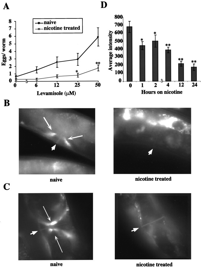
Effect of long-term nicotine on UNC-29 receptor abundance. A, Nicotine response and adaptation in animals expressing UNC-29:: GFP chimeric receptors. The levamisole dose–response curves for nicotine-adapted (i.e., cultured overnight on 30 mm nicotine) and naive animals of the strain ZZ2171 (genotype, unc-29(x29); Ex[pmyo-3:: unc-29:: GFP, rol-6d]), which expresses GFP-tagged UNC-29 protein in vulval and body muscles, are shown. Points and error bars represent the mean and SEM of 20 or more trials.Asterisks indicate significantly less egg-laying according to the Mann–Whitney rank-sum test (*p < 0.05; **p < 0.02). Similar results were obtained in animals expressing UNC-29:: GFP under the control of theunc-29 promoter (data not shown). B, Effect of chronic nicotine on vulval muscle UNC-29:: GFP levels. Shown is a digital image of a nicotine-treated adult hermaphrodite expressing UNC- 29:: GFP in the vulval muscles under the control of the unc-29 promoter (indicated by longarrows; the vulva indicated by a shortarrow). An image of a naive animal of the same transgenic line (strain ZZ2001; genotype,unc-29(x29); Ex[punc- 29:: unc-29:: GFP, rol-6d]) is shown for comparison. C, Effect of nicotine on vulval muscle UNC-29 levels inpmyo-3:: unc-29:: GFP animals. Shown are images of nicotine-adapted and naive ZZ2171 hermaphrodites, which express UNC-29:: GFP in the vulval muscles under the control of the muscle myosin promoter pmyo-3.Longarrows indicate vulval muscles; ashortarrow indicates the vulva.D, Time course of loss of UNC-29:: GFP expression in the vulval muscles. ZZ2171 hermaphrodites were placed on seeded NGM containing 30 mm nicotine for the indicated length of time; fluorescence intensity of the vulval muscles is indicated. Verticalbars and error bars indicate the mean and SEM of five or more trials.Asterisks indicate significantly less fluorescence according to the Mann–Whitney rank-sum test (*p < 0.05; **p < 0.001). Lost UNC-29:: GFP fluorescence was 60% recovered after 24 hr on drug-free medium and completely recovered after 36 hr (data not shown).
Effects of long-term nicotine exposure on egg-laying behavior
Using egg-laying as an assay, we investigated whether long-term exposure to nicotinic agonists led to adaptation ofunc-29-dependent nicotinic responses. We therefore cultured wild-type hermaphrodites for long (16 hr) periods in the presence of nicotine and then assayed their egg-laying behavior. We observed that chronic nicotine treatment did not prevent egg-laying muscle contraction per se. However, the nicotine-adapted animals were strongly resistant to stimulation of egg-laying by levamisole (Fig.2A). Because these animals still laid eggs in response to the neurotransmitter serotonin, long-term nicotine treatment appeared to downregulate specifically the egg-laying response to levamisole. This adaptive response to nicotine was induced slowly, because loss of the levamisole response occurred only after 1–3 hr of nicotine exposure (Fig. 2B). Adaptation to nicotine was also surprisingly long lasting; when nicotine-adapted animals were transferred to drug-free medium, even 24 hr after removal from nicotine most animals remained levamisole insensitive (Fig. 2C). Only at 36 hr after removal from nicotine did the nicotine-adapted animals recover their full responsiveness to levamisole. Thus, prolonged exposure to nicotine resulted in a persistent loss of sensitivity to nicotinic agonists with respect to egg-laying behavior. Because transgenic lines that expressed functional UNC-29 only in the vulval muscles also became levamisole insensitive after overnight nicotine treatment (Fig.2D), this adaptation to nicotine appeared to affect the nicotinic response of the vulval muscles themselves.
Long-term nicotine treatment also caused changes in egg-laying behavior in the absence of drug. Egg-laying in C. elegans occurs in a specific temporal pattern; egg-laying events are clustered, with periods of active egg-laying, or active phases, separated by long inactive phases during which eggs are retained. Both the duration of the inactive phases (intercluster intervals) and the duration of intervals between egg-laying events in a cluster (intracluster intervals) model as exponential random variables with different time constants (Waggoner et al., 1998). Although wild-type hermaphrodites that had been exposed to nicotine overnight could still lay eggs, their pattern of egg-laying behavior was abnormal; their overall rate of egg-laying was lower, and the inactive egg-laying phase was significantly longer than that of naive animals under the same condition. This temporal pattern was similar to (although more severely abnormal than) the pattern seen in animals carrying mutations inunc-29 (Table 1). These results were therefore consistent with the possibility that the alteration in egg-laying behavior induced by long-term nicotine exposure resulted at least in part from a loss ofunc-29 function in the vulval muscles.
Chronic nicotine treatment results in decreased UNC-29 abundance
In principle, inactivation of UNC-29 receptor activity could occur by a variety of mechanisms. One possibility we considered was that chronic nicotine exposure could cause changes in the expression or localization of the UNC-29 receptor protein, leading to a reduction in the number of receptors at the cell surface. To investigate this possibility, we used transgenic animals expressing GFP-tagged UNC-29 protein to examine the effect of long-term nicotine exposure on UNC-29 protein levels. The unc-29:: GFP chimera we used functionally rescued the behavioral abnormalities of unc-29loss-of-function mutations, including those associated with egg-laying, and the levamisole response mediated by the chimeric receptor protein was also subject to adaptation by long-term nicotine treatment (Fig.3A). Interestingly, we observed that chronic nicotine treatment also caused a dramatic reduction in the level of UNC-29:: GFP fluorescence in the vulval muscles (Fig. 3B). This nicotine-induced reduction in UNC-29:: GFP did not require the unc-29 promoter or 3′-untranslated region (3′-UTR), because transgenic animals that expressed UNC-29:: GFP in the vulval muscles under the control of the ectopic myo-3 promoter and unc-54 3′-UTR showed a qualitatively and quantitatively similar response (Fig.3C). The time course of downregulation of UNC-29:: GFP abundance was slow; 12–24 hr of nicotine exposure was required to observe the maximum effect (Fig.3D). The loss of UNC-29:: GFP fluorescence was not merely the result of a nonspecific effect of nicotine on GFP, because long-term nicotine exposure did not alter the fluorescent intensity of a VC-expressed VAMP:: GFP synaptic marker (Fig.4A) or of GFP itself expressed in the vulval muscles under the control of thetpa-1 promoter (see Fig. 5). Thus, the inhibitory effect of chronic nicotine on vulval muscle nAChR activity was apparently accompanied by a corresponding reduction in UNC-29 receptor abundance in the vulval muscle cells, an effect most likely mediated via a post-transcriptional regulatory mechanism.
Fig. 5.
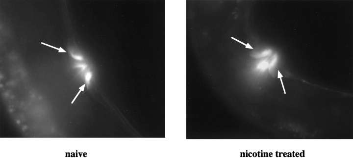
Expression of tpa-1 in the vulval muscles. Shown are digital images of adult hermaphrodites of thetpa-1A:: GFP-expressing strain TF6. In these ventral/lateral views, the vulval muscles are strongly fluorescent and are identified by arrows. Not shown in this view are other fluorescent cells, including vulval epidermal cells, the canal-associated neuron (CAN), and various unidentified head and tail neurons (Tabuse and Miwa, unpublished observations). Expression was never detected in the egg-laying motoneurons VC4, VC5, or the HSNs. Punctate fluorescence in the body is caused by autofluorescence of gut granules.
Interestingly, the more specific nicotinic agonist levamisole appeared to be less potent in inducing adaptation than was nicotine. Although animals treated with levamisole overnight were less sensitive to subsequent stimulation of egg-laying by levamisole, they retained significantly more responsiveness to levamisole than did nicotine-adapted animals (Fig. 4B). Moreover, long-term exposure to levamisole caused only a small (although significant) reduction in the abundance of GFP-tagged UNC-29 protein in the vulval muscles (Fig. 4C). One possible explanation of these results was that nicotine and levamisole might interact with the UNC-29 receptor molecule in different ways; alternatively, the long-term effect of nicotine on UNC-29 receptors might depend on a second, levamisole-insensitive nicotinic receptor in the vulval muscles. In agreement with the latter hypothesis, we found that the acute egg-laying response to nicotinic agonists was genetically separable from the long-term response to nicotine. Several genes, including lev-8, lev-9, and lev-1, are required for the acute stimulation of egg-laying by levamisole (J. Kim, D. S. Poole, and W. R. Schafer, unpublished observations), although they are not necessary for the assembly of levamisole-binding nAChRs (Lewis et al., 1987; Fleming et al., 1997). However, we observed that none of these genes was required for the downregulation of UNC-29:: GFP abundance in the vulval muscles in response to chronic nicotine treatment (Fig. 4D). Thus, although levamisole-sensitive UNC-29 receptors may be necessary for the acute effects of nicotine on egg-laying, a second nicotinic receptor may be at least partially responsible for nicotine's long-term effects on this behavior.
The PKC homolog TPA-1 is required for UNC-29 downregulation
What genes are required for nicotine adaptation in the egg-laying muscles? To investigate this question, we assayed C. elegansmutants with defects in genes encoding signaling molecules that function in the vulval muscles. One such gene was tpa-1,which encodes a C. elegans isoform of protein kinase C. Recessive mutations in tpa-1 confer resistance to phorbol esters but do not dramatically impair the health, fertility, or behavior of the worm (Tabuse et al., 1989). We had demonstrated previously that TPA-1 is required for the acute stimulation of egg-laying by serotonin (Waggoner et al., 1998). To determine whethertpa-1 was actually expressed in the vulval muscles, we generated transgenic lines expressing a GFP reporter under the control of one of the two tpa-1 promoters. We observed that one of these promoter fusions, tpa-1A:: GFP, was strongly expressed in the vulval muscles, as well as in vulval epidermal cells and a variety of neurons, including the canal-associated neuron and many unidentified cells in the head and tail (Fig. 5). The other promoter fusion, tpa-1B:: GFP, was expressed in the lateral epidermis and in a few head neurons (Y. Tabuse and J. Miwa, unpublished observations). Thus, tpa-1 indeed appeared to encode an isoform of protein kinase C that was expressed in the vulval muscles.
To assess the possible role of tpa-1 in nicotine adaptation, we first assayed the effect of tpa-1 mutations on the induction of egg-laying by levamisole and on the inactivation of this response by long-term nicotine treatment. We found that naivetpa-1 mutant animals laid eggs in response to levamisole over the same range of concentrations as did wild-type, although the magnitude of this stimulation was somewhat lower in thetpa-1 mutant (Fig.6A). Strikingly, however, we observed that tpa-1 mutants showed little or no reduction in levamisole-induced egg-laying after overnight exposure to nicotine (Fig. 6B), indicating that tpa-1mutant animals were completely defective in nicotine adaptation. We also tested the effect of tpa-1 on the ability of nicotine to decrease UNC-29 receptor abundance in the vulval muscles. We observed that tpa-1 mutant animals expressing the UNC-29:: GFP protein chimera in the vulval muscles retained high levels of fluorescence even after overnight nicotine treatment (Fig. 6C,D). Thus, the tpa-1-encoded isoform of PKC appeared to be necessary for the nicotine-induced downregulation of UNC-29 receptor abundance and biological activity in the vulval muscles.
DISCUSSION
Nicotinic agonists have both acute and chronic effects on egg-laying behavior in C. elegans. We have shown that the acute stimulation of egg-laying by nicotine and the nicotinic agonist levamisole is mediated by nAChRs containing the subunit protein UNC-29. We found that these UNC-29 receptors are expressed in the vulval muscles, and expression of functional UNC-29 protein in the vulval muscles alone is sufficient to confer egg-laying sensitivity to nicotinic agonists. Thus, nicotinic agonists appear to stimulate egg-laying at least in part via direct excitation of UNC-29-containing receptors in the vulval muscles. However, because unc-29mutants are still capable of laying eggs, UNC-29 receptors do not appear to be necessary for egg-laying muscle contraction. The dispensability of UNC-29 for muscle contraction could be caused by a second, levamisole-insensitive nAChR in the vulval muscles that displays some functional redundancy with the UNC-29 receptor, as has been observed in the C. elegans body muscle (Richmond and Jorgensen, 1999). Alternatively, the vulval muscles, like the C. elegans pharyngeal muscles, could possess intrinsic contractile activity and use nicotinic receptors only as a means to induce more rapid contraction (Raizen and Avery, 1994).
Chronic exposure to nicotinic agonists had dramatic effects on C. elegans egg-laying behavior that were long lasting and specific. Overnight treatment with nicotine induced a strong and persistent resistance to the stimulation of egg-laying by levamisole and resulted in an altered egg-laying pattern resembling that of nicotinic receptor-deficient mutants. Yet this adaptation to nicotine did not prevent egg-laying muscle contraction per se, nor did it affect the stimulation of egg-laying by noncholinergic agents such as serotonin. Chronic nicotine treatment also attenuated the levamisole responses of animals expressing functional UNC-29 protein only in the vulval muscles, indicating that the behavioral effects of long-term nicotine treatment result at least in part from effects on the vulval muscles themselves. Interestingly, chronic exposure to levamisole, an agonist that appears to be somewhat specific for UNC-29-containing receptors, downregulated UNC-29 receptor abundance only moderately. Moreover, the ability of nicotine to downregulate UNC-29 receptor levels was independent of several genes that are required for acute stimulation of egg-laying by levamisole, including another candidate subunit of the levamisole receptor (i.e., lev-1). Thus, the regulation by nicotine of UNC-29 receptor abundance in the vulval muscles may not be simply a consequence of prolonged activation of levamisole receptors in the vulval muscles but rather might depend on the activity of other levamisole-insensitive nicotinic receptors in the egg-laying muscles and/or neurons.
Interestingly, the loss of behavioral sensitivity to nicotinic agonists was accompanied by a concomitant reduction in UNC-29 protein abundance in the vulval muscle cells. This effect on UNC-29 protein level was independent of the unc-29 promoter, because downregulation still occurred in transgenic animals that expressed UNC-29:: GFP under the control of the myo-3 muscle myosin promoter. Downregulation was also independent of theunc-29 3′-UTR sequences, which in nearly all cases are the critical determinant of mRNA stability, export, and translatability inC. elegans (Ahringer and Kimble, 1991; Seydoux, 1996; Graves et al., 1999). Because the unc-29-coding region appeared to be sufficient to confer UNC-29 downregulation in the vulval muscles, this suggests that this downregulation most likely involves a post-translational mechanism. Post-translational regulation of nicotinic receptor levels has been observed in a number of vertebrate systems (Marks et al., 1992; Peng et al., 1994; Bencherif et al., 1995); however, the genetic and molecular requirements for these processes remain primarily uncharacterized. Because the chronic effect of nicotine on C. elegans egg-laying behavior depends on a process at least formally analogous to nAChR downregulation in vertebrates, genetic analysis of these processes in nematodes may provide important clues to the molecular basis of nAChR regulation in other organisms.
One gene that we found to be essential for the nicotine-induced downregulation of UNC-29 abundance was tpa-1, which encodes a homolog of protein kinase C. In contrast to wild-type animals,tpa-1 mutants maintained high levels of UNC-29 receptors in the vulval muscles and remained sensitive to the behavioral effects of levamisole after long-term exposure to nicotine. The mechanistic basis for why TPA-1/PKC is required for nicotine regulation of UNC-29 receptor abundance remains to be determined. Probably the simplest model for TPA-1 action is that direct phosphorylation of one or more subunits of the nicotinic receptors in the vulval muscles by TPA-1/PKC somehow targets them for increased degradation. The UNC-29 protein, like other candidate subunits of the levamisole receptor (e.g., LEV-1 and UNC-38), contains consensus sequences for PKC phosphorylation within the M3–M4 cytoplasmic loop (Pearson and Kemp, 1991; Fleming et al., 1997), and preliminary biochemical experiments indicate that these sequences can serve as substrates for the human ortholog of TPA-1in vitro (L. E. Waggoner, unpublished observations). Thus, direct phosphorylation of one or more subunits of the vulval muscle levamisole receptors is a reasonable possibility. It is well established that phosphorylation of the M3–M4 loop by protein kinases can modulate the activity of nAChRs by enhancing their rate of desensitization (Huganir and Greengard, 1983; Huganir et al., 1984,1986; Eusebi et al., 1985; Downing and Role, 1987; Safran et al., 1987;Hopfield et al., 1988). It is reasonable to suppose that receptor phosphorylation by PKC may also be linked to processes affecting nAChR abundance, such as protein targeting or degradation. In this regard, it is interesting to note that in rat muscle cells, a variety of intracellular signaling pathways have been shown to regulate the degradation rate of nicotinic receptors during formation of the neuromuscular junction (O'Malley et al., 1997). Alternatively, some or all of the effects of PKC could be indirect, and the actual substrates of PKC could be molecules that directly or indirectly regulate the stability or targeting of the receptor. For example, rapsyn, a peripheral membrane protein that acts to stabilize nicotinic receptors in skeletal muscle (Wang et al., 1999), contains a potential target sequence for PKC; thus, phosphorylation of a C. elegansrapsyn might inhibit its ability to stabilize UNC-29 receptors, leading to increased receptor turnover. It is also possible that PKC signaling in neighboring cells, for example, the vulval epidermis, could influence nicotinic receptor abundance in the vulval muscles via neuroendocrine signaling. The availability of simple and robust assays for nicotinic receptor activity and abundance in C. elegansshould make it possible to use genetic analysis to identify the PKC target(s) relevant to nicotine adaptation, along with other molecules that participate in long-term responses to nicotine.
Footnotes
This work was supported by the National Institute on Drug Abuse Grants DA 11556 and DA 12891 (W.R.S.); additional support for work in our lab was provided by National Science Foundation Award IBN-9723250 (W.R.S.), awards from the Beckman, Sloan, and Klingenstein Foundations (W.R.S.), and a National Institutes of Health predoctoral training grant (L.E.W). We express our appreciation to Jim Lewis for his generous assistance. We thank Jim Lewis, Mike Nonet, Kelly Liu, and theCaenorhabditis Genetics Center for strains; Jinah Kim for assistance in strain construction; and Cori Bargmann, Darwin Berg, Cynthia Kenyon, and Rachel Kindt for comments on this manuscript.
Correspondence should be addressed to Dr. William Schafer, Department of Biology, University of California, San Diego, 9500 Gilman Drive, La Jolla, California 92093-0349. E-mail: wschafer@ucsd.edu.
REFERENCES
- 1.Ahringer J, Kimble J. Control of the sperm-oocyte switch in Caenorhabditis elegans hermaphrodites by the fem-3 3′ untranslated region. Nature. 1991;349:346–348. doi: 10.1038/349346a0. [DOI] [PubMed] [Google Scholar]
- 2.Avery L, Horvitz HR. Effects of starvation and neuroactive drugs on feeding in Caenorhabditis elegans. J Exp Zool. 1990;253:263–270. doi: 10.1002/jez.1402530305. [DOI] [PubMed] [Google Scholar]
- 3.Ballivet M, Alliod C, Bertrand S, Bertrand D. Nicotinic acetylcholine receptors in the nematode Caenorhabditis elegans. J Mol Biol. 1996;258:261–269. doi: 10.1006/jmbi.1996.0248. [DOI] [PubMed] [Google Scholar]
- 4.Bargmann CI. Neurobiology of the Caenorhabditis elegans genome. Science. 1998;282:2028–2033. doi: 10.1126/science.282.5396.2028. [DOI] [PubMed] [Google Scholar]
- 5.Barrantes GE, Rogers AT, Lindstrom J, Wonnacott S. alpha-Bungarotoxin binding sites in rat hippocampal and cortical cultures: initial characterisation, colocalisation with alpha 7 subunits and up-regulation by chronic nicotine treatment. Brain Res. 1995;672:228–236. doi: 10.1016/0006-8993(94)01386-v. [DOI] [PubMed] [Google Scholar]
- 6.Baylis HA, Matsuda K, Squire MD, Fleming JT, Harvey RJ, Darlison MG, Barnard EA, Sattelle DB. ACR-3, a Caenorhabditis elegans nicotinic acetylcholine receptor subunit. Receptors Channels. 1997;5:149–158. [PubMed] [Google Scholar]
- 7.Bencherif M, Fowler K, Lukas RJ, Lippiello PM. Mechanisms of up-regulation of neuronal nicotinic acetylcholine receptors in clonal cell lines and primary cultures of fetal rat brain. J Pharmacol Exp Ther. 1995;275:987–994. [PubMed] [Google Scholar]
- 8.Changeaux JP. Compartmentalized transcription of acetylcholine receptor genes during motor endplate epigenesis. New Biol. 1991;3:413–429. [PubMed] [Google Scholar]
- 9.Colbert HA, Bargmann CI. Odorant-specific adaptation pathways generate olfactory plasticity in C. elegans. Neuron. 1995;14:803–812. doi: 10.1016/0896-6273(95)90224-4. [DOI] [PubMed] [Google Scholar]
- 10.Dani JA, Heinemann S. Molecular and cellular aspects of nicotine abuse. Neuron. 1996;16:905–908. doi: 10.1016/s0896-6273(00)80112-9. [DOI] [PubMed] [Google Scholar]
- 11.Downing JE, Role LW. Activators of protein kinase C enhance acetylcholine receptor desensitization in sympathetic ganglion neurons. Proc Natl Acad Sci USA. 1987;84:7739–7743. doi: 10.1073/pnas.84.21.7739. [DOI] [PMC free article] [PubMed] [Google Scholar]
- 12.Eusebi F, Molinaro M, Zani BM. Agents that activate protein kinase C reduced acetylcholine sensitivity in cultured myotubes. J Cell Biol. 1985;100:1339–1342. doi: 10.1083/jcb.100.4.1339. [DOI] [PMC free article] [PubMed] [Google Scholar]
- 13.Fleming JT, Squire MD, Barnes TM, Tornoe C, Matsuda K, Ahnn J, Fire A, Sulston JE, Barrard EA, Sattelle DB, Lewis JA. Caenorhabditis elegans levamisole resistance genes lev-1, unc-29, and unc-38 encode functional nicotinic acetylcholine receptor subunits. J Neurosci. 1997;17:5843–5857. doi: 10.1523/JNEUROSCI.17-15-05843.1997. [DOI] [PMC free article] [PubMed] [Google Scholar]
- 14.Flores CM, Rogers SW, Pabreza LA, Wolfe BB, Kellar KJ. A subtype of nicotinic cholinergic receptor in rat brain is composed of α4 and β2 subunits and is upregulated by chronic nicotine treatment. Mol Pharmacol. 1992;41:31–37. [PubMed] [Google Scholar]
- 15.Gardner JM, Fambrough DM. Acetylcholine receptor degradation measured by density labeling: effects of cholinergic ligands and evidence against recycling. Cell. 1979;16:661–674. doi: 10.1016/0092-8674(79)90039-4. [DOI] [PubMed] [Google Scholar]
- 16.Graves LE, Segal S, Goodwin EB. TRA-1 regulates the cellular distribution of the tra-2 mRNA in C. elegans. Nature. 1999;399:802–805. doi: 10.1038/21682. [DOI] [PubMed] [Google Scholar]
- 17.Harfe BD, Fire A. Muscle and nerve-specific regulation of a novel NK-2 class homeodomain factor in Caenorhabditis elegans. Development. 1998;125:421–429. doi: 10.1242/dev.125.3.421. [DOI] [PubMed] [Google Scholar]
- 18.Hedgecock EM, Russell RL. Normal and mutant thermotaxis in the nematode Caenorhabditis elegans. Proc Natl Acad Sci USA. 1975;72:4061–4065. doi: 10.1073/pnas.72.10.4061. [DOI] [PMC free article] [PubMed] [Google Scholar]
- 19.Hopfield JF, Tank DW, Greengard P, Huganir RL. Functional modulation of the nicotinic acetylcholine receptor by tyrosine phosphorylation. Nature. 1988;336:677–680. doi: 10.1038/336677a0. [DOI] [PubMed] [Google Scholar]
- 20.Huganir RL, Greengard P. cAMP-dependent protein kinase phosphorylates the nicotinic acetylcholine receptor. Proc Natl Acad Sci USA. 1983;80:1130–1134. doi: 10.1073/pnas.80.4.1130. [DOI] [PMC free article] [PubMed] [Google Scholar]
- 21.Huganir RL, Miles K, Greengard P. Phosphorylation of the nicotinic acetylcholine receptor by an endogenous tyrosine-specific protein kinase. Proc Natl Acad Sci USA. 1984;81:6968–6972. doi: 10.1073/pnas.81.22.6968. [DOI] [PMC free article] [PubMed] [Google Scholar]
- 22.Huganir RL, Delcour AH, Greengard P, Hess GP. Phosphorylation of the nicotinic acetylcholine receptor regulates its rate of desensitization. Nature. 1986;321:774–776. doi: 10.1038/321774a0. [DOI] [PubMed] [Google Scholar]
- 23.Lewis JA, Wu C-H, Levine JH, Berg H. Levamisole-resistant mutants of the nematode Caenorhabditis elegans appear to lack pharmacological acetylcholine receptors. Neuroscience. 1980;5:967–989. doi: 10.1016/0306-4522(80)90180-3. [DOI] [PubMed] [Google Scholar]
- 24.Lewis JA, Fleming JT, McLafferty S, Murphy H, Wu C. The levamisole receptor, a cholinergic receptor of the nematode Caenorhabditis elegans. Mol Pharmacol. 1987;31:185–193. [PubMed] [Google Scholar]
- 25.Lukas RJ. Effects of chronic nicotinic ligand exposure on functional activity of nicotinic acetylcholine receptors expressed by cells of the PC12 rat pheochromocytoma or the TE671/RD human clonal line. J Neurochem. 1991;56:1134–1145. doi: 10.1111/j.1471-4159.1991.tb11403.x. [DOI] [PubMed] [Google Scholar]
- 26.Marks MJ, Pauly JR, Gross SD, Deneris ES, Hermans-Borgmeyerr I, Heinemann SF, Collins AC. Nicotine binding and nicotinic receptor subunit RNA after chronic nicotine treatment. J Neurosci. 1992;12:2765–2784. doi: 10.1523/JNEUROSCI.12-07-02765.1992. [DOI] [PMC free article] [PubMed] [Google Scholar]
- 27.Messing A. Cholinergic agonist-induced down regulation of neuronal α-bungarotoxin receptors. Brain Res. 1982;232:479–484. doi: 10.1016/0006-8993(82)90292-x. [DOI] [PubMed] [Google Scholar]
- 28.Molinari EJ, Delbono O, Messi ML, Renganathan M, Arneric SP, Sullivan JP, Gopalakrishnan M. Up-regulation of human alpha7 nicotinic receptors by chronic treatment with activator and antagonist ligands. Eur J Pharmacol. 1998;347:131–139. doi: 10.1016/s0014-2999(98)00084-3. [DOI] [PubMed] [Google Scholar]
- 29.Mongan NP, Baylis HA, Adcock C, Smith GR, Sansom MS, Sattelle DB. An extensive and diverse gene family of nicotinic acetylcholine receptor α subunits in Caenorhabditis elegans. Receptors Channels. 1998;6:213–228. [PubMed] [Google Scholar]
- 30.Nonet M. Visualization of synaptic specializations in live C. elegans with synaptic vesicle protein-GFP fusions. J Neurosci Methods. 1999;89:33–40. doi: 10.1016/s0165-0270(99)00031-x. [DOI] [PubMed] [Google Scholar]
- 31.O'Malley JP, Moore CT, Salpeter MM. Stabilization of acetylcholine receptors by exogenous ATP and its reversal by cAMP and calcium. J Cell Biol. 1997;138:159–165. doi: 10.1083/jcb.138.1.159. [DOI] [PMC free article] [PubMed] [Google Scholar]
- 32.Pearson RB, Kemp BE. Protein kinase phosphorylation site sequences and consensus specificity motifs: tabulations. Methods Enzymol. 1991;200:62–81. doi: 10.1016/0076-6879(91)00127-i. [DOI] [PubMed] [Google Scholar]
- 33.Peng X, Gerzanich V, Anand R, Whiting PJ, Lindstrom J. Chronic nicotine treatment up-regulates alpha3 and alpha7 acetylcholine receptor subtypes expressed by the human neuroblastoma cell line SH-SY5Y. Mol Pharmacol. 1994;46:523–530. doi: 10.1124/mol.51.5.776. [DOI] [PubMed] [Google Scholar]
- 34.Raizen DM, Avery L. Electrical activity and behavior in the pharynx of Caenorhabditis elegans. Neuron. 1994;12:483–495. doi: 10.1016/0896-6273(94)90207-0. [DOI] [PMC free article] [PubMed] [Google Scholar]
- 35.Rankin CH, Chiba C, Beck C. Caenorhabditis elegans: a new model system for the study of learning and memory. Behav Brain Res. 1990;37:89–92. doi: 10.1016/0166-4328(90)90074-o. [DOI] [PubMed] [Google Scholar]
- 36.Richmond JE, Jorgensen EM. One GABA and two acetylcholine receptors function at the C. elegans neuromuscular junction. Nat Neurosci. 1999;2:1–7. doi: 10.1038/12160. [DOI] [PMC free article] [PubMed] [Google Scholar]
- 37.Rogers AT, Wonnacott S. Nicotine-induced upregulation of alpha bungarotoxin (alpha Bgt) binding sites in cultured rat hippocampal neurons. Biochem Soc Trans. 1995;23:48S. doi: 10.1042/bst023048s. [DOI] [PubMed] [Google Scholar]
- 38.Rothhut B, Romano SJ, Vijayaraghavan S, Berg DK. Post-translational regulation of neuronal acetylcholine receptors stably expressed in a mouse fibroblast cell line. J Neurobiol. 1996;29:115–125. doi: 10.1002/(SICI)1097-4695(199601)29:1<115::AID-NEU9>3.0.CO;2-E. [DOI] [PubMed] [Google Scholar]
- 39.Safran A, Sagi-Eisenberg R, Neumann D, Fuchs S. Phosphorylation of the acetylcholine receptor by protein kinase C and identification of the phosphorylation site within the receptor δ subunit. J Biol Chem. 1987;262:10506–10510. [PubMed] [Google Scholar]
- 40.Schafer WR, Kenyon CJ. A calcium channel homologue required for adaptation to dopamine and serotonin in Caenorhabditis elegans. Nature. 1995;375:73–78. doi: 10.1038/375073a0. [DOI] [PubMed] [Google Scholar]
- 41.Seydoux G. Mechanisms of translational control in early development. Curr Opin Genet Dev. 1996;6:555–561. doi: 10.1016/s0959-437x(96)80083-9. [DOI] [PubMed] [Google Scholar]
- 42.Simasko SM, Soares JR, Weiland GA. Two components of carbamylcholine-induced loss of nicotinic acetylcholine receptor function in the neuronal cell line PC12. Mol Pharmacol. 1986;30:6–12. [PubMed] [Google Scholar]
- 43.Squire MD, Tornoe C, Baylis HA, Fleming JT, Barnard EA, Sattelle DB. Molecular cloning and functional co-expression of a Caenorhabditis elegans nicotinic acetylcholine receptor subunit (acr-2). Receptors Channels. 1995;3:107–115. [PubMed] [Google Scholar]
- 44.Tabuse Y, Nishiwaki K, Miwa J. Mutations in a protein kinase C homolog confer phorbol resistance in Caenorhabditis elegans. Science. 1989;243:1713–1716. doi: 10.1126/science.2538925. [DOI] [PubMed] [Google Scholar]
- 45.Treinin M, Chalfie M. A mutated acetylcholine receptor subunit causes neuronal degeneration in C. elegans. Neuron. 1995;14:871–877. doi: 10.1016/0896-6273(95)90231-7. [DOI] [PubMed] [Google Scholar]
- 46.Trent C, Tsung N, Horvitz HR. Egg-laying defective mutants of the nematode Caenorhabditis elegans. Genetics. 1983;104:619–647. doi: 10.1093/genetics/104.4.619. [DOI] [PMC free article] [PubMed] [Google Scholar]
- 47.Waggoner L, Zhou GT, Schafer RW, Schafer WR. Control of behavioral states by serotonin in Caenorhabditis elegans. Neuron. 1998;21:203–214. doi: 10.1016/s0896-6273(00)80527-9. [DOI] [PubMed] [Google Scholar]
- 48.Wang Z-Z, Mathias A, Gautam M, Hall ZW. Metabolic stabilization of muscle nicotinic acetylcholine receptor by rapsyn. J Neurosci. 1999;19:1998–2007. doi: 10.1523/JNEUROSCI.19-06-01998.1999. [DOI] [PMC free article] [PubMed] [Google Scholar]
- 49.Weinshenker D, Garriga G, Thomas JH. Genetic and pharmacological analysis of neurotransmitters controlling egg-laying in C. elegans. J Neurosci. 1995;15:6975–6985. doi: 10.1523/JNEUROSCI.15-10-06975.1995. [DOI] [PMC free article] [PubMed] [Google Scholar]
- 50.Wen JY, Kumar N, Morrison G, Rambaldini G, Runciman S, Rousseau J, van der Kooy D. Mutations that prevent associative learning in C. elegans. Behav Neurosci. 1997;111:354–368. doi: 10.1037//0735-7044.111.2.354. [DOI] [PubMed] [Google Scholar]
- 51.Wonnacott S. The paradox of nicotinic acetylcholine receptor upregulation by nicotine. Trends Pharmacol Sci. 1990;11:216–218. doi: 10.1016/0165-6147(90)90242-z. [DOI] [PubMed] [Google Scholar]
- 52.Zhou GT, Schafer WR, Schafer RW. A three-state biological point process model and its parameter estimation. IEEE Trans Signal Process. 1998;46:2698–2707. [Google Scholar]


