Figure 2:
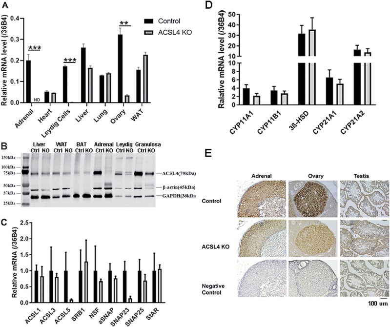
Generation of tissue-specific ACSL4 KO mice. (A) Levels of ACSL4 mRNA in control and ACSL4 KO mice. mRNA was isolated from adrenals and other tissues of 16- to 24-week-old mice (n = 3), and levels of ACSL4 mRNA were analyzed using RT-PCR. Experiments were repeated three times. (B). Western blot analysis of ACSL4 protein in various tissues and isolated cells from control and ACSL4 KO mice. Total cell extracts were prepared from tissues of 16- to 24-week-old control and ACSL4 KO mice (n = 3 to 5). Leydig and granulosa cells were isolated from male and female mice, respectively, following the protocol described in the Materials and Methods. Protein levels of ACSL4 were analyzed by immunoblotting with ACSL4 antibody. (C) Analysis of expression of various ACSL genes and selected genes that are involved in steroidogenesis in the adrenal using TaqMan quantitative PCR. (D) Analysis of expression of various genes involved in steroidogenesis in the adrenal using TaqMan quantitative PCR. (E) Histochemical analysis of ACSL4 in the adrenal, ovary, and testis of control and ACSL4 KO mice. Data are expressed as means ± SEM. **P < 0.01; ***P < 0.005. BAT, brown adipose tissue; Ctrl, control; ND, not detected; WAT, white adipose tissue.

