Abstract
Purpose
Our retrospective study addresses the largely unknown effect of fixation pin type on depth of skull penetration during Gamma Knife frame placement.
Methods
Absolute and relative depths were compared for 404 pins of aluminum and titanium in 101 patients who underwent Gamma Knife frame placement at 0.4 Nm (3.5 lb-in) torque.
Results
Effect of pin type was significant: penetration was less in aluminum than titanium pins, for absolute (0.27 mm vs. 0.83 mm) and relative (5.16% vs. 15.53%) depths (p < 0.001 each). Fifty percent of aluminum pins did not measurably penetrate the outer table of the skull whereas 95% of titanium pins penetrated into cancellous bone.
Conclusions
Although these pin penetration differences did not have a clear clinical correlate, we now recommend titanium pins for their consistent, deeper penetration, which may result in greater frame stability favorable for most clinical applications. Future studies may define optimal torque for aluminum pins.
Keywords: Radiosurgery, stereotaxic techniques, quality assurance, complications
Introduction
Gamma Knife radiosurgery utilizes a rigid frame to immobilize the patient during imaging and treatment. The frame is secured to the skull with either aluminum or titanium pins. If the depth of skull penetration is too superficial, then solid fixation may not be achieved and the frame could shift before radiation delivery [1]. In contrast, if skull penetration is too deep, violation of the inner table can occur, resulting in injury to the underlying parenchyma. Many neurosurgeons use a torque wrench to achieve a consistent level of force, however, the depth of penetration is not well understood.
In examining the relationship between torque and depth of skull penetration for the Fischer head fixation system (Leibinger, Freiberg, Germany), Toyota et al. correlated the pin depth seen on stereotactic CT scans with the torque level applied to the pins [2]. They reported that absolute depth, relative depth, and incidence of inner table violation decreased when torque was reduced from 0.9 Nm (8 lb-in) to 0.7 Nm (6.2 lb-in). However, whether depth of penetration is affected by pin composition, such as aluminum or titanium, or tip design is largely unknown.
The purpose of the present study is to evaluate the depth of pin penetration at a standard torque level utilizing the Leksell G Frame (Elekta Instrument AB, Stockholm, Sweden) and to determine if there are significant differences between the aluminum and titanium fixation pins. The results of this study may also guide the clinical application of each pin type.
Material and Methods
Inclusion/exclusion criteria
Data collection and analysis were approved by the Jewish Hospital institutional review board. In this retrospective study (July 2013-June 2018), 104 adult patients were identified who underwent single-fraction, framed Gamma Knife radiosurgery with a stereotactic CT scan for treatment planning in addition to the standard stereotactic MRI scan. The 101 patients included in the final analysis had fixation with a single type of pin, either aluminum or titanium; 3 patients each of whom had a combination of aluminum and titanium pins used for frame placement were excluded. Patients were grouped by pin type for analysis.
Fixation pins
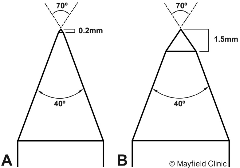
Two types of fixation pins were used based on availability on the day of treatment. The Titanium Fixation Screw (Elekta Instrument AB, Stockholm, Sweden) was composed of medical grade titanium characterized as high density (4.45 g/cm3) and non-magnetic. At its tip, the initial 0.2 mm had a 70o angle and the remaining cone had a 40o angle (Figure 1). The Reusable Fixation Screw (Elekta Instrument AB, Stockholm, Sweden), an anodized aluminum alloy pin of lower density (2.81 g/cm3) and non-magnetic materials, was designed to reduce artifact during CT and MRI scanning. At its tip, the initial 1.5 mm had a 70o angle and the remaining cone had a 40o angle (Figure 1).
Figure 1.
Comparison of two types of commonly used Elekta Fixation Screws, titanium (A) and aluminum (B). The distance of the initial tip angle (70o) is different (0.2 vs. 1.5 mm). (Reprinted with permission from the Mayfield Clinic).
Frame placement
All frames were placed by a single, experienced neurosurgeon. Briefly, with the patient under conscious sedation, four pin sites were prepped with chlorhexidine and injected with a mixture of lidocaine, bupivacaine hydrochloride, and sodium bicarbonate. The Leksell G Frame (Elekta Instrument AB) was secured to the skull using four fixation pins of various lengths. Diagonal pins were tightened in a sequential fashion using a torque wrench calibrated to 0.4 Nm (3.5 lb-in). This torque level had been used by our radiosurgery center for 25 years and is consistent with torque recommendations from other Gamma Knife centers (0.4-0.45 Nm) [3-5] The patient then underwent both stereotactic MRI and CT scans (Aquilion One, Toshiba Medical Systems, Japan) using the appropriate fiducial localizer. CT scanning parameters consisted of a 1-mm slice thickness and a matrix size of 512 x 512 (1 pixel = 0.588 mm).
Pin depth measurements
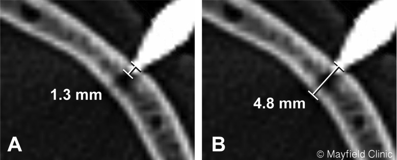
The stereotactic CT images were transferred to the Gamma Knife planning system (Leksell GammaPlan, versions 10 and 11). A bone window was selected with a level of 32,500 and width of 35,000 and magnified 8 times. At each of four pin sites, two measurements were made by the neurosurgeon defined as the following: A = distance from the surface of the bone to the tip of the pin, and B = thickness of the skull along the trajectory of the pin (Figure 2). Depth of penetration by percentage (%) was then calculated using the formula A/B x 100.
Figure 2.
Sample screenshot of stereotactic CT images depict the method used to measure depth of pin penetration (A) into bone and thickness of skull (B). (Reprinted with permission from the Mayfield Clinic).
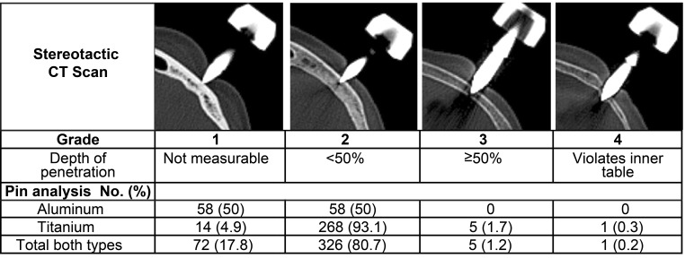
Next, the depth of penetration was graded using the 4-point scale that represented a modification of the grading system proposed by Toyota et al. [2]. Our scale defined: (1) No measurable penetration of the outer table, (2) < 50% penetration of the skull thickness, (3) > 50% penetration of the skull thickness, and (4) Penetration through the inner table (Figure 3). Age, sex, diagnosis, pin type (aluminum or titanium), and pin site complications were also documented.
Figure 3.
Four-point grading system based on relative depth of skull penetration. Distribution by grade for 404 pins in 101 patients and by pin type for 116 aluminum and 288 titanium pins. (Reprinted with permission from the Mayfield Clinic).
Statistical analysis
Descriptive statistics were generated for demographics and diagnosis at baseline. A linear mixed model analysis was used to manage correlated within-subject errors that resulted from repeated measures on four pin sites, using the mixed procedure in SPSS 22.0 (IBM Corp. Armonk, NY) [6]. For our primary analyses, we examined the effects of pin type and pin site, and the interaction between these two variables. The models were fitted using unstructured covariance structure and maximum likelihood estimation. After identification of the significant main effect, post-hoc analyses were also performed with Bonferroni correction and linear contrast tests were used to examine several other hypotheses.
Results
Clinical characteristics
From July 2013 to June 2018, 104 patients underwent single-fraction, framed Gamma Knife radiosurgery that included a stereotactic CT scan for treatment planning. Three patients were excluded because a combination of aluminum and titanium pins were used for frame placement within the same patient. Of the remaining 101 patients, the median age was 61 years (range 31-91) and 61% were female. The most common diagnoses were trigeminal neuralgia (41%), meningioma (25%), and vestibular schwannoma (16%). Aluminum pins were used in 29 patients (29%) and titanium pins in 72 patients (71%). Table 1 summarizes demographic and clinical characteristics of this study cohort.
Table 1.
Demographic and clinical Information for 101 patients who underwent Gamma Knife radiosurgery. The 62 women and 39 men ranged in age from 31 to 91 years old (mean 61 years).
| Characteristic | No. (%) |
| Diagnosis | |
| Arteriovenous malformation | 2 (2.0) |
| Jugular schwannoma | 1 (1.0) |
| Meningioma | 25 (24.8) |
| Metastasis | 3 (3.0) |
| Pituitary | 7 (6.9) |
| Trigeminal neuralgia | 41 (40.6) |
| Trigeminal schwannoma | 6 (5.9) |
| Vestibular schwannoma | 16 (15.8) |
| Pin type used | |
| Aluminum | 29 (28.7) |
| Titanium | 72 (71.3) |
Overall pin penetration measurements
In the 101 patients with 404 pins, mean depth of pin penetration (A) was 0.67 mm (range 0-4.9 mm, standard deviation [SD] 0.54 mm) for the aluminum and titanium pins combined (Table 2A). Mean skull thickness along the pin’s trajectory (B) was 5.94 mm (range 2.0-23.5 mm, SD 1.93 mm). The relative depth of pin penetration based on the formula A/B x 100 averaged 12.55% (range 0-153.13%, SD 12.41%).
Table 2A.
Comparison of pin penetration measurements: overall.
| Measurements | Mean (SD) | Median (IQR) | Range (Min, Max) |
| Absolute depth of pin (A), mm | 0.67 (0.54) | 0.60 (0.6) | 4.9 (0, 4.9) |
| Skull bone thickness (B), mm | 5.94 (1.93) | 5.70 (1.9) | 21.5 (2.0, 23.5) |
| Relative depth of pin penetration (A/B), % | 12.55 (12.41) | 10.20 (10.82) | 153.13 (0, 153.13) |
Using the four-point scoring classification, 72 pins (17.8%) showed no measurable skull penetration (Grade 1), 326 pins (80.7%) penetrated less than 50% of the skull thickness (Grade 2), 5 pins (1.2%) penetrated more than 50% of the skull thickness without reaching the inner table (Grade 3) [Figure 3], and 1 pin (0.2%) penetrated through the inner table (Grade 4).
Pin type effect on penetration depth
In a linear mixed-model analysis, we found the effect of pin type was significant. Penetration was less with aluminum pins than titanium pins in terms of absolute depth (0.27 mm vs. 0.83 mm, respectively) and relative depth (5.16% vs. 15.53%, respectively) (p < 0.001 for each) (Table 2B). After excluding the patient whose pin penetrated the inner table (Grade 4), re-analysis without this outlier also confirmed significant differences in penetration between aluminum and titanium pins for both absolute and relative depths (p < 0.001 for each). A comparison of the distribution of penetration scores by grade also confirmed this effect: that is, 50% of aluminum pins and 4.9% of titanium pins had no discernable penetration of the outer table (Grade) 1, 50% of aluminum pins and 93.1% of titanium pins had Grade 2 penetration, and only titanium pins exhibited Grade 3 or 4 penetration (Figure 3).
Table 2B.
Comparison of pin penetration measurements: aluminum versus titanium.
| Measurements | Aluminuma (n=116) | Titaniuma (n=288) | Mean difference (95% CI) | p Value |
| Absolute depth of pin (A), mm | 0.27 (0.05) | 0.83 (0.03) | −0.55 (−0.68, −0.43) | <0.001 |
| Skull bone thickness (B), mm | 6.01 (0.27) | 5.91 (0.17) | 0.10 (−0.53, 0.74) | 0.742 |
| Relative depth of pin penetration (A/B), % | 5.16 (1.28) | 15.53 (0.81) | −10.36 (−13.38, −7.35) | <0.001 |
Values are expressed as estimated marginal means (Std. Error) from the Linear Mixed Mode
Complications
The one patient who suffered a Grade 4 penetration with a titanium pin during stereotactic frame placement had noted significant pain at the pin site after frame placement. CT scan showed penetration of this pin through the inner table at a point where the skull thickness was 3.2 mm (Figure 3, right). The frame was removed and the pin site irrigated and closed with a suture. A delayed CT scan showed no evidence of intracranial hemorrhage. The patient’s recovery from this event was uneventful and she subsequently underwent frameless Gamma Knife radiosurgery. There were no cases of frame slippage during imaging or treatment.
Discussion
Gamma Knife radiosurgery relies on rigid fixation to ensure submillimetric accuracy during imaging and treatment. When the Leksell G Frame is secured to the patient’s skull with either aluminum or titanium fixation pins, use of a torque wrench ensures a consistent level of torque that should translate into a consistent depth of penetration through the outer table into the cancellous layer of the skull.
In a study of the relationship between torque and depth of pin penetration using the Fischer head fixation system, Toyota et al. tested three types of wrenches, including a standard wrench, torque wrench at 0.9 Nm (8 lb-in), and torque wrench at 0.7 Nm (6.2 lb-in) [2]. Absolute and relative depths of pin penetration were highest with the standard wrench and decreased as torque level was reduced. The incidence of pin penetration through the inner table was significant with the standard wrench (33%) and torque wrench at 0.9 Nm (8 lb-in) (38%) but fell dramatically to 2% when the torque was decreased to 0.7 Nm (6.2 lb-in). However, these results cannot be translated to Gamma Knife radiosurgery because of differences in fixation systems and torque levels.
Our study demonstrates that there are significant differences in the depth of penetration between the aluminum and titanium pins provided with the Leksell G Frame. Analysis of stereotactic CT scans showed that 50% of aluminum pins had no measurable penetration of the outer table of the skull at a torque level of 0.4 Nm (3.5 lb-in) (mean depth of 0.27 mm). In contrast, 95% of the titanium pins penetrated the outer table into the cancellous bone (mean depth of 0.83 mm).
During frame application, torque is translated into forward motion of the pin resulting in force against the skull. For a given torque, the force at the tip of the pin is related to friction within the disposable insert, friction at the pin/skull interface, and the pin’s composition, shape, and entry angle. The type of post (short, medium, long) and pin length may also alter the downward force against the skull. Aluminum fixation pins were designed to reduce imaging artifact. With a density lower than titanium, the aluminum alloy has an initial tip angle (70o) that was expanded to 1.5 mm to distribute force over a larger area, thereby preventing deformation of the tip (Thomas Arn personal communication, October 3, 2018). According to the principles of Hertzian contact mechanics, the larger radius of the aluminum pin results in lower pressure and less deformation depth [7,8]. The “shoulder” of the aluminum pin also limits the depth of penetration. In contrast, the smaller radius of the titanium pin produces higher force, greater deformation depth, and increased risk of skull penetration. Our results are consistent with these principles in that 50% of aluminum pins did not even penetrate the outer table and only titanium pins were associated with a Grade 3 or 4 penetration.
The ideal depth of penetration to ensure frame stability is not known. Recent studies have assessed frame stability using cone-beam CT, however, our study cohort spanned a 5-year period and most patients were treated prior to the introduction of the Gamma Knife ICON (Elekta Instrument AB) [1]. Therefore, we cannot correlate pin penetration depth with frame stability. Intuitively, pin penetration through the outer table into the cancellous bone should result in greater frame stability during imaging and treatment. We now prefer titanium pins for their consistent, deeper penetration, which may result in greater frame stability favorable for most clinical applications. We reserve aluminum pins for clinical scenarios in which minimal bone penetration is desired such as thin skull, large frontal sinus, or adjacent craniotomy. The optimal torque for frame placement with aluminum pins may be higher than the 0.4 Nm (3.5 lb-in) used in this study.
Our retrospective study quantitatively determined the penetration depth achieved by two commonly used Gamma Knife radiosurgery fixation pins under standard torque for the Leksell G Frame. Our findings show previously unreported effects of pin type (i.e., aluminum or titanium) noting differences in tip design on actual and relative penetration depths. Specifically, 50% of aluminum pins had no measurable penetration of the outer table (mean depth 0.28 mm) whereas 95% of the titanium pins penetrated the outer table into the cancellous bone (mean depth 0.84 mm). Despite these statistically significant differences, our patient cohort had adequate fixation. Although we cannot define a clinical correlate in terms of stability, we now prefer titanium pins given their consistency of penetration, possibly avoiding violation of a critical structure (e.g., frontal sinus).
Study limitations
Our analysis utilized the Leksell G Frame and a torque level of 0.4 Nm (3.5 lb-in) consistent with torque recommendations from other Gamma Knife centers (0.4-0.45 Nm) [3-5]. However, a similar study using other stereotactic frames or different torque levels would likely yield different results. We did not collect information regarding type of post and pin length that could alter the downward force and depth of penetration. We made no attempt to measure cortical bone quality, which may have contributed to the variations in depth of penetration among patients. Lastly, we did not correlate depth of pin penetration with frame stability so we can only infer that titanium pin fixation provides the optimal depth of penetration and frame stability.
Conclusions
Absolute and relative depths of skull penetration were significantly affected by the type of fixation pin used in this retrospective study of 101 patients who underwent Gamma Knife radiosurgery. Although these pin penetration differences did not have a clear clinical correlate, we recommend titanium pins for their consistent, deeper penetration, which may result in greater frame stability favorable for most clinical applications. We reserve aluminum pins for clinical scenarios in which minimal bone penetration is desired such as thin skull, large frontal sinus, or adjacent craniotomy. We would like Gamma Knife clinicians to be aware of these different depths of penetration so they can tailor the selection of pins to the clinical scenario.
Acknowledgements
The authors thank Glia Media’s Martha Headworth for medical illustrations and Mary Kemper for medical editing.
Footnotes
Authors’ disclosure of potential conflicts of interest
The authors have nothing to disclose.
Author contributions
Conception and design: Ronald E. Warnick
Data collection: Ronald E. Warnick
Data analysis and interpretation: Ronald E. Warnick, Eunsun Yook
Manuscript writing: Ronald E. Warnick
Final approval of manuscript: Ronald E. Warnick
References
- 1. Dutta SW, Kowalchuk RO, Trifiletti DM, Peach MS, Sheehan JP, Larner JM, Schlesinger D. Stereotactic shifts during frame-based image-guided stereotactic radiosurgery: Clinical measurements. Int J Radiat Oncol Biol Phys 2018;102(4):895-902. [DOI] [PubMed] [Google Scholar]
- 2. Toyota S, Seta H, Kubo H, Muramatsu M, Takeda K. Relationships between head fixation pins for radiosurgery and the skull bone: Usefulness of a torque wrench. Radiat Med 2003;Mar-Apr;21(2):94-98. [PubMed] [Google Scholar]
- 3. Rojas-Villabona A, Miszkiel K, Kitchen N, Jäger R, Paddick I. Evaluation of the stability of the stereotactic Leksell Frame G Gamma in Knife radiosurgery. J Appl Clin Med Phys 2016. May 8;17(3):75-89. [DOI] [PMC free article] [PubMed] [Google Scholar]
- 4. Safaee M, Burke J, McDermott MW. Techniques for the application of stereotactic head frames based on a 25-year experience. Cureus 2016. Mar 25;8(3):e543. [DOI] [PMC free article] [PubMed] [Google Scholar]
- 5. Renier C, Massager N. Targeting inaccuracy caused by mechanical distortion of the Leksell stereotactic frame during fixation. J Apply Clin Med Phys 2019; 1-10 [DOI] [PMC free article] [PubMed] [Google Scholar]
- 6. Keselman HJ, Kowalchuk RK, Boik RJ. The analysis of repeated measures designs: A review. J Br Math Stat Psychol 2001;54 (Pt 1):1-20. [DOI] [PubMed] [Google Scholar]
- 7. Williams JA, Dwyer-Joyce RS. Contact between solid surfaces, in Modern Tribology Handbook 1, 121-162, CRC Press, Boca Raton, FL, 2000 [Google Scholar]
- 8. Zaazoue MA, Bedewy M, Goumnerova LC. Complications of head immobilization devices in children: Contact mechanics and analysis of a single institutional experience. Neurosurgery 2018. May 1;82(5):678-685. [DOI] [PubMed] [Google Scholar]


