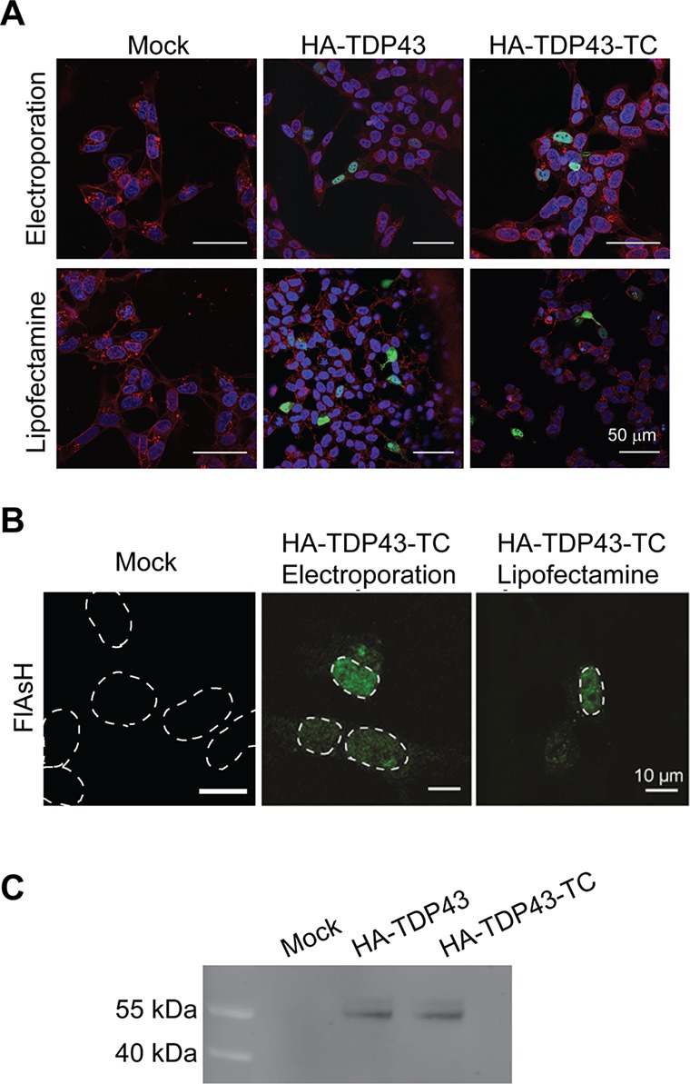
Figure 2.

Confocal images of SH-SY5Y cells transfected (by either electroporation or lipofection) to overexpress HA-TDP43 or HA-TDP43-TC, 24 h post-transfection. (A) Immunofluorescence images generated using an anti-HA antibody and an Alexa Fluor 488 secondary antibody (green) and Hoechst nuclear stain (blue). Membranes are stained with wheat germ agglutinin (WGA) Alexa Fluor 647 conjugate (red), and scale bars are 50 μm. (B) Fluorescence images after the addition of the FlAsH dye (24 h post-transfection). For the sake of clarity, the white dotted line denotes the nucleus. Images are representative of multiple independent experiments. (C) Immunoprecipitation followed by Western blot analysis of HA-TDP43 and HA-TDP43-TC isolated from SH-SY5Y cell lysates 24 h post-transfection. Mock transfections are cells transfected with buffer alone.
