Abstract
Objectives:
To develop and validate formulas for age and sex estimation based on the pulp cavity volume of teeth using cone beam CT.
Methods:
The sample was composed of 116 cone beam CT scans from Brazilian individuals of both sexes, ranging in age from 13 to 70 years. A total of 232 teeth (upper central incisors and canines) were evaluated. Two calibrated examiners determined pulp cavity volumes using the ITK-SNAP software. Pearson’s correlation test was used to assess the correlation between chronological age and pulp volume. Linear and logistic regression models were developed for age and sex estimation, respectively, and were validated in another sample of 72 teeth.
Results:
Pearson’s correlation coefficients between age and pulp volume were negative and significant (p < 0.0001) for both teeth (r = −0.8782 for central incisors and r = −0.8738 for canines). The age estimation formulas showed good determination coefficients (adjusted R² = 0.7614 to 0.8367). For sex estimation, when the age was known, the coefficients were also good (adjusted R² = 0.649 to 0.812). However, when the age was unknown, the coefficients of the sex estimation formulas were low (adjusted R² = 0.047 to 0.393). Validation showed high accuracy of age estimation in individuals older than 35 years, as well as high accuracy of sex estimation when the age was known.
Conclusions:
Our formulas provided excellent results and can be applied to the Brazilian population. The best results were observed for age estimation in females and for sex estimation when the age was known.
Keywords: forensic dentistry, cone-beam CT, secondary dentin, age determination by teeth, sex characteristics
Introduction
In forensic investigations, age and sex estimation is fundamental for the creation of the individual’s biological profile. In intact cadavers or cadavers reduced to pieces, age estimation helps in human identification by narrowing down potentially matching identities within the available sample.1 Similarly, sex estimation contributes to the identification of cases, mainly in skeletonization situations. Moreover, when it comes to living individuals, such estimations are relevant in different situations such as those within the context of civil law and in cases of refugees and asylum seekers when information concerning identification is missing.1 Similarly, in cases of criminal nature, age estimation is required for the imputation of criminal responsibility to determine the adult status.1
Teeth are widely used in forensic and anthropological investigations to determine the sex and age of human remains, as they are the best-preserved parts of the human body, regardless of the cause of death and state of preservation of the body.2,3 After completion of formation of the apical part of the root, eruption of the tooth in the oral cavity and beginning of the tooth’s functioning, the so-called physiological secondary dentin is deposited in the pulp cavity.4 Its deposition by odontoblasts is a continuous process that causes progressive narrowing of the pulp chamber in all teeth.4,5 As secondary dentin is deposited along the inner surfaces of the tooth, protecting it from environmental influences, pulp evaluation has the potential to exclude, at least in part, the effects of external factors.6
The deposition of secondary dentin can be assessed by destructive methods such as tooth sectioning for microscopic study, or by conservative methods such as imaging exams. Destructive methods are not acceptable for forensic purposes because they can cause the loss of evidence.1,7 Therefore, noninvasive imaging technologies such as cone beam CT (CBCT) are preferable for application in living individuals because of ethical issues.
Three-dimensional (3D) images demonstrate the actual morphological changes, and are therefore better suited for tooth-based age estimation than two-dimensional radiographic images such as periapical radiographs.3,4 In dental practice, CBCT offers good image quality and a lower radiation dose compared to multidetector CT, in addition to allowing multiplanar and accurate assessment of tooth volume.3,5 Within this context, the combination of CBCT images with neural network mapping has been suggested as an alternative nondestructive method for age estimation in order to reduce the inherent limitations of linear regression models. However, more data are needed to increase the applicability of this new method.8
The morphological changes that occur in the pulp cavity and the accumulation of dentin are promising age estimation indicators that are often used.9 Furthermore, one study performed sex estimation based on the total volume of the tooth, but we found no previous study that evaluated only pulp volume.3 Few studies in Brazil have investigated age estimation based on the pulp volume parameter. Since anthropological differences exist between ancestries, methods must be developed for a specific population because the application of foreign standards can result in a loss of accuracy.10
Therefore, this study aimed to develop and validate new formulas to estimate age and sex from the pulp volume of upper central incisors and canines using CBCT images of a Brazilian sample.
Methods and Materials
Sample
This project was approved by the local Research Ethics Committee (CAAE 79944817.4.0000.5418).
The BioEstat 5.0 software (Mamirauá Foundation, Belém, PA, Brazil) was used to calculate the sample power and size according to Porto et al,11 which showed a minimal difference between means of 8.5 for pulp volume, with a standard deviation of 5.0. Thus, assuming a 5% α value and 95% test power, a sample of 90 patients would be significant.
A total of 116 CBCT scans were selected from patients of both sexes ranging in age from 13 to 70 years, who had at least one upper central incisor and one upper canine. Thus, the final sample was composed of 232 teeth. Given the symmetry of the internal anatomy of the tooth demonstrated in a previous study,12 when the tooth on the right side did not meet the criteria, the tooth on the left side of the same patient was used. Thus, the sample was homogenous in terms of age and sex, with one patient for each sex and for each age in the range of 13–70 years. No race/phenotype information was available for the CBCT scans.
The inclusion criteria for selection of the sample were: CBCT scans containing the patient’s date of birth and sex, in addition to the date of completion of the examination, and the presence of at least one healthy permanent upper central incisor and one healthy permanent upper canine with fully formed roots and erupted in the oral cavity. CBCT scans including teeth with decay or cavities, root canal therapy, restorations, excessive tooth wear reaching the incisal/occlusal third of the tooth’s crown, orthodontic or prosthetic devices, pulp calcification, dental fracture, impaction, severe rotation or some other severe type of malocclusion, periodontitis, torn roots, developmental abnormalities, and periapical lesions were excluded. In addition, CBCT scans of low quality due to the significant formation of artifacts caused by the presence of materials with high atomic number were also excluded.
The scans were obtained between June 2012 and April 2018 with a Kodak K9500® scanner (Carestream Health, Rochester, RY) using a voxel size of 0.2 mm3 and 0.3 mm³ and the same energy parameters (90 kVp and 10 mA). The size of the field of view (FOV) was varied as indicated for each patient (15 × 9 cm and 20 × 18 cm).
Image analysis
The proprietary CS 3D Imaging® software (Carestream Health, Rochester, NY) was used for selecting the CBCT scans. The scans were then exported in the DICOM format to the ITK-SNAP 3.4.0 segmentation software (Cognitica, Philadelphia, PA) to measure the pulp volume of the selected teeth.
The evaluations were conducted by two previously calibrated dental surgeons with prior knowledge of CT and the use of the ITK-SNAP tools. Each examiner individually evaluated the scans using a laptop computer (15.6-inch LCD Full HD monitor) under low light conditions. For calibration, the two examiners analyzed a sample of 20 teeth (upper central incisors and canines) on 6 CBCT scans that did not compose the study sample. The teeth were evaluated on two different occasions at an interval of 15 days to obtain the pulp volumes. The intraexaminer agreement ranged from 0.994 to 1.0, while the interexaminer agreement ranged from 0.994 to 0.998. Therefore, the examiners were able to perform the image analysis.
The pulp volumes were determined with the software’s semi-automatic segmentation mode in three steps. First, the limits of any extension of the tooth to be examined were marked by the examiners in the multiplanar reconstructions, defining the region of interest (ROI) for segmentation (Figure 1a). In the second step, the threshold interval was selected by an interactive method according to previous studies.13,14 For this interactive selection, the operator determines the best threshold interval based on visual analysis of the anatomical delimitation between the hard structures of the tooth and the dental pulp for each CBCT scan. The default density range was adjusted (0 for the lower threshold and ranging from 1100 to 1300 for the upper threshold), so that the 3D model to be built would only have voxels with grey values within this interval (Figure 1b). In the third step, “seeds” were added to the entire pulp extension delimited in the multiplanar reconstructions so that the space corresponding to the dental pulp was filled based on the previously determined threshold interval (Figure 1c). Finally, the segmentation process was started gradually by selecting its velocity and end. After this process, the image of the 3D reconstruction of the pulp cavity was obtained (Figure 2). The volumes of the segmented structures were measured in cubic millimeter (mm³).
Figure 1.
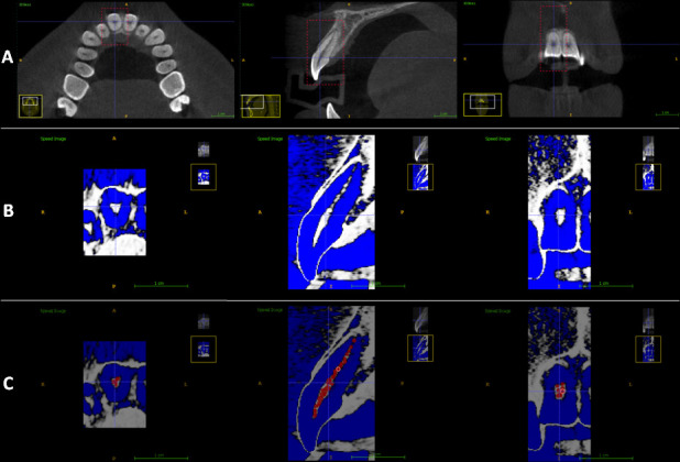
Steps of the segmentation process of a right upper central incisor using CBCT imaging. (a) Selection of the region of interest (ROI) in the axial, sagittal and coronal planes. (b) After adjustment of the default density range. (c) Addition of “seeds” throughout the pulp extension in the axial, sagittal and coronal planes.
Figure 2.
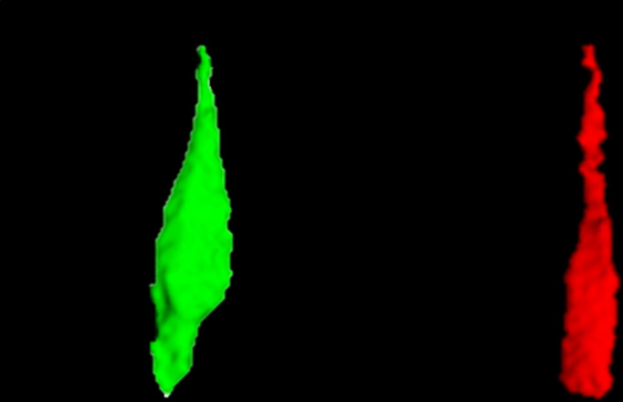
Image of the 3D reconstruction of the pulp of an upper right canine (in the left side) and of an upper right central incisor (in the right side).
Reproducibility
Thirty days after completion of the assessments, 20 teeth were randomly selected from the main sample and re-evaluated to obtain reproducibility.
Statistical analysis
The analyses were carried out using the BioEstat 5.3 (Instituto de Desenvolvimento Sustentável Mamirauá, Tefé, MA, Brazil) and the SPSS 23.0 (SPSS, Chicago, IL, USA) software. The intraclass correlation coefficient (ICC) was used as a measure of intra- and interexaminer agreement. Single and multiple linear regression models were developed to estimate age using pulp volume as the dependent variable when an upper canine, an upper central incisor or both types of teeth were present in the dental arch. Bivariate logistic regression models were developed to estimate sex using pulp volume as the dependent variable under the same conditions. Pearson’s correlation coefficient was used to assess the correlation between chronological age and pulp volume. For all analyses, the statistical significance level was set at p < 0.05.
Validation
For validation of the developed formulas, another sample was selected by applying the same previously established inclusion and exclusion criteria. This sample consisted of 36 CBCT scans, 18 males and 18 females, with three individuals per age group, totaling 72 teeth. The pulp volumes obtained for this sample were used in the formulas developed by analysis of the main sample, and age and sex were estimated. The age obtained with the formulas and the chronological age were compared by the paired Student t test and validation charts were drawn in Excel.
Results
The ICC for intraexaminer agreement was 0.9998 for examiner 1 and 0.9825 for examiner 2. The ICC for interexaminer agreement was 0.9997. These ICC values revealed excellent reproducibility of the volumetric measurements.15 Thus, the mean pulp volumes obtained by the examiners were used for linear regression and Pearson’s correlation analysis.
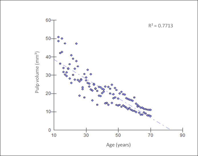
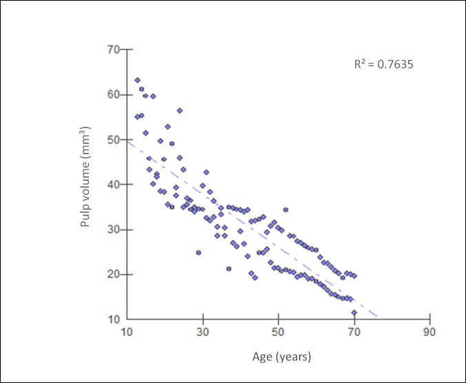
Pearson’s correlation coefficient between chronological age and pulp volume was negative and statistically significant (p < 0.0001) for both types of teeth (Table 1), with the observation of high correlations. The upper central incisor had a slightly higher coefficient (r = −0.8782; 95% CI = -0.91 to -0.83; R² = 0.7713) than the upper canine (r = −0.8738; 95% CI = -0.91 to -0.82; R² = 0.7635). Correlation graphs showing the correlation between chronological age and pulp cavity volume of upper central incisors and between chronological age and pulp cavity volume of upper canines are illustrated in Figures 3 and 4, respectively.
Table 1.
Correlation coefficients between age and pulp volume
| Tooth | Pearson correlation | IC 95% | R² | t value | p value |
| Upper central incisor | −0.8782 | −0.91 a −0.83 | 0.7713 | −19.609 | <0.0001 |
| Upper canine | −0.8738 | −0.91 a −0.82 | 0.7635 | −19.1829 | <0.0001 |
Figure 3.
Correlation graph showing the correlation between chronological age and the pulp volume of upper central incisors.
Figure 4.
Correlation graph showing the correlation between chronological age and the pulp volume of upper canines.
Table 2 shows the formulas used to estimate age when the pulp volume of one or both teeth are used and when sex is known or unknown. The best determination coefficients for the age estimation formulas were obtained when the pulp volume data of the upper canines or of both teeth were used and when sex was known (adjusted R² = 0.8228 to 0.8367). The coefficients were slightly lower when only the upper central incisors were used (adjusted R² = 0.7938 for females; adjusted R² = 0.8007 for males). However, when sex was unknown, slightly lower determination coefficients were obtained for central incisors, canines or both (adjusted R² = 0.7614 to 0.7779).
Table 2.
Equations to estimate age using pulp volume
| Tooth | Sex condition | Linear regression equation | R² adjusted | p values |
| Upper central incisor | Female | Age = 70.4276+(−1.449 x central incisor volume) | 0.7938 | <0.0001 |
| Male | Age = 78.8388+(−1.5712 x central incisor volume) | 0.8007 | <0.0001 | |
| Unknown sex | Age = 73.2547+(−1.4524 x central incisor volume) | 0.7693 | <0.0001 | |
| Upper canine | Female | Age = 80.0709+(−1.3924 x canine volume) | 0.8367 | <0.0001 |
| Male | Age = 90.2228+(−1.4232 x canine volume) | 0.8228 | <0.0001 | |
| Unknown sex | Age = 81.4635+(−1.2905 x canine volume) | 0.7614 | <0.0001 | |
| Both teeth | Female | Age = 79.4605+(−1.2857 x canine volume) + (−0.1175 x central incisor volume) |
0.8341 | canine 0.0003 |
| central incisor 0.7451 | ||||
| Male | Age = 87.3157+(−0.9881 x canine volume) + (−0.5045 x central incisor volume) |
0.8255 | canine 0.0041 | |
| central incisor 0.1774 | ||||
| Unknown sex | Age = 77.4387+(−0.5677 x canine volume) + (−0.8397 x central incisor volume) |
0.7779 | canine 0.0215 | |
| central incisor 0.0026 |
The sex estimation formulas are shown in Table 3. In general, the sex estimation formulas provided low coefficients when age was unknown (adjusted R² = 0.047 to 0.393). Conversely, when the age was known, the coefficients were similar to those found for age estimation (adjusted R² = 0.649 to 0.812). The logistic regression used 0.5 as cut-off point, i.e., when using the formulas to estimate sex, results > 0.5 indicate male, whereas results ≤ 0.5 indicate female.
Table 3.
Equations to estimate sex using pulp volume
| Tooth | Age condition | Logistic regression equation | R² adjusted | p values |
| Upper central incisor | Known age | Logit Sex = −33.258+(0.823 x central incisor volume) + (0.378 x age) | 0.649 | <0.0001 |
| Unknown age | Logit Sex = −0.840+(0.039 x central incisor volume) | 0.047 | 0.047 | |
| Upper canine | Known age | Logit Sex = −40.655+(0.818 x canine volume) + (0.395 x age) | 0.770 | <0.0001 |
| Unknown age | Logit Sex = −2.040+(0.069 x canine volume) | 0.148 | 0.001 | |
| Both teeth | Known age | Logit Sex = −41.614+(−0.747 x central incisor volume) + (1.411 x canine volume) + (0.364 x age) | 0.812 | central incisor <0.0001 |
| canine <0.0001 | ||||
| Unknown age | Logit Sex = −5.442+(−0.517 x central incisor volume) + (0.549 x canine volume) | 0.393 | central incisor <0.0001 | |
| canine <0.0001 |
Results of the validation
Table 4 shows the mean absolute error and p values when estimated and chronological ages were compared. The best age estimation results were obtained when sex was known. The lowest mean absolute error values were found for females when sex was known, while the highest mean absolute error values were observed for males when sex was unknown.
Table 4.
Value of p and mean absolute error for the age estimate, comparing the chronological age and the estimated age of the validation sample using the developed formulas
| Sex sample | Information available | Tooth type | p value | Mean Absolute Error |
| Male sample | With sex info | Upper central incisor | 0.4623 | 5.45297 |
| Upper canine | 0.6744 | 5.86164 | ||
| Both teeth | 0.5641 | 5.58014 | ||
| Without sex info | Upper central incisor | 0.3251 | 6.22535 | |
| Upper canine | 0.0678 | 7.05212 | ||
| Both teeth | 0.1479 | 6.39402 | ||
| Female sample | With sex info | Upper central incisor | 0.4241 | 4.21398 |
| Upper canine | 0.9287 | 4.34364 | ||
| Both teeth | 1.0 | 4.25219 | ||
| Without sex info | Upper central incisor | 0.0066 | 5.25634 | |
| Upper canine | 0.0075 | 5.96617 | ||
| Both teeth | 0.0028 | 5.36004 |
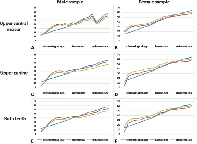
The validation plots shown in Figure 5 provide a better view of the comparison between real or chronological age and estimated age in situations where sex is known or not. As can be seen in the figure, age is generally overestimated in individuals up to the age of 35 years. After this age, excellent agreement exists between chronological age and age estimated with the formulas proposed, regardless of whether the sex is known or not.
Figure 5.
Comparison of chronological age (line 1) and estimated age when sex is known (line 2) and unknown (line 3). (a) Using only the upper central incisor of the male validation sample. (b) Using only the upper central incisor of the female validation sample. (c) Using only the upper canine of the male validation sample. (d) Using only the upper canine of the female validation sample. (e) Using the two types of teeth, upper central incisor and upper canine, of the male validation sample. (f) Using the two types of teeth, upper central incisor and upper canine, of the female validation sample.
The pulp volumes of the validation sample were also used in the sex estimation formulas. The estimated sex was compared to the patient’s actual sex and the percentage of accuracy is shown in Table 5. In general, high accuracy was found, especially when age was known. An exception was the use of canines in males without age information, which resulted in an accuracy of 28%.
Table 5.
Sex estimation of the validation sample using the developed formulas
| Sex sample | Information available | Tooth type | Accuracy |
| Male sample | With age info | Upper central incisor | 67% |
| Upper canine | 83% | ||
| Both teeth | 83% | ||
| Without age info | Upper central incisor | 72% | |
| Upper canine | 28% | ||
| Both teeth | 50% | ||
| Female sample | With age info | Upper central incisor | 94% |
| Upper canine | 94% | ||
| Both teeth | 94% | ||
| Without age info | Upper central incisor | 94% | |
| Upper canine | 83% | ||
| Both teeth | 83% |
Discussion
In our study, the correlation between chronological age and pulp volume of the two types of teeth was high, with a marginally higher correlation for the upper central incisors. High correlations for these types of teeth have also been reported in another study assessing the relationship between the pulp volume/tooth ratio of anterior upper and lower teeth and age.12 The authors observed a stronger correlation for upper central incisors and canines compared to other teeth and explained this result by the smaller internal anatomical variation of these teeth. A previous study that analyzed the pulp volume/tooth ratio of several types of teeth based on CBCT scans also found a higher correlation coefficient for the upper central incisors (R² = 0.532) than for the upper canines (R² = 0.153).16 However, it should be noted that our correlations refer to a Brazilian population and are more expressive than those reported in previous studies.17–20
In the current study, the age estimation formula using the upper canines provided a slightly higher determination coefficient for females than for males. The same was observed when both types of teeth were used, while the opposite occurred when the upper central incisors were used. This greater correlation for males compared to females was also reported in other studies.19,21 The former analyzed the upper central incisors and found high determination coefficients (R² = 0.851 for males and 0.776 for females), while the latter reported moderate determination coefficients for the upper canines (R² = 0.273 for males and 0.180 for females). Conversely, a recent study did not observe this difference between males and females. Furthermore, a highest coefficient of determination was obtained for the upper central incisor (R² = 0.70) when compared with the superior canine (R² = 0.53), although that no sex specific formulas were developed.22
The present study is the first to determine formulas for age estimation based on volumetric measurements of the dental pulp when the sex of the individual is unknown using either only one tooth (incisor or canine) or two types of teeth (incisor and canine). The determination coefficients of age estimation in these two conditions were low. The same was observed for sex estimation of the individual when the age is unknown using volumetric measurements of dental pulp of either one or both types of teeth. In contrast, the addition of a covariant (age or sex) to the formulas provided slightly higher determination coefficients. These novel results may serve as possible references for subsequent studies. Regarding the methodology, the sample size used in the present study is within the mean sample size used by other previous studies that aimed to estimate age and determine sex from pulp volume analysis using CBCT images.3,7,11,12,16,20
Validation confirmed that better age estimation results are achieved when the sex is known. The lowest mean absolute error values were obtained for females of the sample when sex was known and when only the central incisors were used, while the highest mean absolute error values were found for males when sex was unknown and when only the upper canines were used. In a previous study,17 a lower mean absolute error value (3.876) was found for the upper canines considering the total sample, i.e., both sexes.
Regarding the validation of the age estimation formulas, in general, an overestimation of age was observed for individuals younger than 35 years old when one or both types of teeth were used, regardless of knowing the sex. However, there was excellent harmony between the chronological and the estimated ages for the 35 years old or older individuals, when applying our formulas.
As observed for the validation of the age estimation formulas, the validation results of the sex estimation formulas were generally better when some other biological profile information was available, in this case, information about age. The sex estimation formulas showed high accuracy during validation, which was higher for the female sample using one or both types of teeth. For the male sample, when sex estimation was performed without the information of age, the use of canine volumetric measurements alone provided low accuracy. Thus, it is not recommended to use the pulp volume of upper canines without some other biological profile information for sex estimation.
The ITK-SNAP software used here has been validated by its developers and is a tool used to segment structures in neuroimaging and other applications.23 Another advantage is that it is a free software, which allows it to be widely used by forensic anthropology and medical professionals. This software enables 3D segmentation and has already been used in previous studies for age estimation by tooth analysis, with proven efficiency and reliability.4,23–25
Using 3D imaging methods, several authors have studied the correlation between secondary dentin formation and age based on the analysis of the tooth volume/pulp volume ratio. However, we used only pulp volume data, in agreement with the findings and recommendations of previous studies.4,24,26,27 Tooth volume can be reduced as a result of enamel attrition and its calculation is therefore less accurate, hindering the distinction between cortical alveoli and the space corresponding to the periodontal ligament and cementum.
Limitations of this study include the need for CBCT scans with two voxel and FOV sizes. In the present study, most of the CBCT scans used to perform the evaluations had smaller voxel (0.2 mm³) and FOV (15 × 9 cm) sizes available in the CBCT equipment used. However, it was necessary to include scans with different sizes of voxel (0.3 mm³) and FOV (20 × 18 cm) to obtain an equally distributed according to age and sex. The influence of these two technical parameters on volumetric measurements in different diagnostic tasks is controversial.21,28–30 However, Adisen et al, who investigated the effect of voxel resolution and FOV size on age estimation based on the volumetric measurements of teeth, found no significant differences between chronological age and the estimated age using different voxel (0.2 and 0.4 mm³) and FOV (5 × 5.5 cm and 23 × 25 cm) sizes.21
Finally, it is suggested that future studies could be conduct for age and sex estimation based on the pulp volume of other types of teeth and from other populations. Additionally, when possible, it is recommended that the formulas found in the present study may be associated with other methods with proven efficacy, such as through the pelvic bones for sex estimation or through the hand and wrist bones or dental development for age estimation.31–35
Conclusions
The formulas developed to estimate age and sex using the pulp volume of upper central incisors and upper canines provided good results and can be applied to the Brazilian population. In general, when the pulp volume of one or both teeth is used together with age or sex information, the other characteristic (sex or age) can be obtained with high accuracy using these formulas. For age estimation, better results are obtained for individuals older than 35 years and females. The validation confirmed our results.
Footnotes
Acknowledgment: The authors thank Professor Dr Fabio Ribeiro Guedes, responsible for the archives of the Dental Radiology Clinic of the Federal University of Rio de Janeiro’s School of Dentistry, for allowing access to and use of the cone-beam computed tomography data in this study. The study was supported in part by the Coordenação de Aperfeiçoamento de Pessoal de Nível Superior (CAPES), Brazil (Finance Code 001).
Disclosure: The authors declare no potential conflict of interest.
Contributor Information
Vanessa M Andrade, Email: vmoreira@pcivil.rj.gov.br.
Rocharles C Fontenele, Email: rocharlesf@gmail.com.
Andreia CB de Souza, Email: andreiacrisbreda@gmail.com.
Francisco C Groppo, Email: fcgroppo@unicamp.br.
Deborah Q Freitas, Email: deborahq@unicamp.br.
Eduardo D Junior, Email: darugejr@gmail.com.
REFERENCES
- 1.Karkhanis S, Mack P, Franklin D. Age estimation standards for a Western Australian population using the coronal pulp cavity index. Forensic Sci Int 2013; 231(1-3): 412.e1–412.e6. doi: 10.1016/j.forsciint.2013.04.004 [DOI] [PubMed] [Google Scholar]
- 2.Jeevan MB, Kale AD, Angadi PV, Hallikerimath S. Age estimation by pulp/tooth area ratio in canines: Cameriere's method assessed in an Indian sample using radiovisiography. Forensic Sci Int 2011; 204(1-3): 209.e1–209.e5. doi: 10.1016/j.forsciint.2010.08.017 [DOI] [PubMed] [Google Scholar]
- 3.Tardivo D, Sastre J, Ruquet M, Thollon L, Adalian P, Leonetti G, et al. Three-Dimensional modeling of the various volumes of canines to determine age and sex: a preliminary study. J Forensic Sci 2011; 56: 766–70. doi: 10.1111/j.1556-4029.2011.01720.x [DOI] [PubMed] [Google Scholar]
- 4.Ge Z-pu, Ma R-han, Li G, Zhang J-zong, Ma X-chen. Age estimation based on pulp chamber volume of first molars from cone-beam computed tomography images. Forensic Sci Int 2015; 253: 133.e1–133.e7. doi: 10.1016/j.forsciint.2015.05.004 [DOI] [PubMed] [Google Scholar]
- 5.Pinchi V, Pradella F, Buti J, Baldinotti C, Focardi M, Norelli G-A. A new age estimation procedure based on the 3D CBCT study of the pulp cavity and hard tissues of the teeth for forensic purposes: a pilot study. J Forensic Leg Med 2015; 36: 150–7. doi: 10.1016/j.jflm.2015.09.015 [DOI] [PubMed] [Google Scholar]
- 6.Jagannathan N, Neelakantan P, Thiruvengadam C, Ramani P, Premkumar P, Natesan A, et al. Age estimation in an Indian population using pulp/tooth volume ratio of mandibular canines obtained from cone beam computed tomography. J Forensic Odontostomatol 2011; 29: 1–6. [PMC free article] [PubMed] [Google Scholar]
- 7.Aboshi H, Takahashi T, Komuro T. Age estimation using microfocus X-ray computed tomography of lower premolars. Forensic Sci Int 2010; 200(1-3): 35–40. doi: 10.1016/j.forsciint.2010.03.024 [DOI] [PubMed] [Google Scholar]
- 8.Farhadian M, Salemi F, Saati S, Nafisi N. Dental age estimation using the pulp-to-tooth ratio in canines by neural networks. Imaging Sci Dent 2019; 49: 19–26. doi: 10.5624/isd.2019.49.1.19 [DOI] [PMC free article] [PubMed] [Google Scholar]
- 9.Du C, Zhu Y, Hong L. Age-Related changes in pulp cavity of incisors as a determinant for forensic age identification. J Forensic Sci 2011; 56 Suppl 1: S72–S76. doi: 10.1111/j.1556-4029.2010.01577.x [DOI] [PubMed] [Google Scholar]
- 10.Karkhanis S, Mack P, Franklin D, et al. Age estimation standards for a Western Australian population using the dental age estimation technique developed by Kvaal et al. Forensic Sci Int 2014; 235: 104.e1–104.e6. doi: 10.1016/j.forsciint.2013.12.008 [DOI] [PubMed] [Google Scholar]
- 11.Porto LVMG, Celestino da Silva Neto J, Anjos Pontual AD, Catunda RQ. Evaluation of volumetric changes of teeth in a Brazilian population by using cone beam computed tomography. J Forensic Leg Med 2015; 36: 4–9. doi: 10.1016/j.jflm.2015.07.007 [DOI] [PubMed] [Google Scholar]
- 12.Biuki N, Razi T, Faramarzi M. Relationship between pulp-tooth volume ratios and chronological age in different anterior teeth on CBCT. J Clin Exp Dent 2017; 9: e688–93. doi: 10.4317/jced.53654 [DOI] [PMC free article] [PubMed] [Google Scholar]
- 13.Weissheimer A, Menezes LMde, Sameshima GT, Enciso R, Pham J, Grauer D. Imaging software accuracy for 3-dimensional analysis of the upper airway. Am J Orthod Dentofacial Orthop 2012; 142: 801–13. doi: 10.1016/j.ajodo.2012.07.015 [DOI] [PubMed] [Google Scholar]
- 14.Farias Gomes A, de Oliveira Gamba T, Yamasaki MC, Groppo FC, Haiter Neto F, Possobon RdeF. Development and validation of a formula based on maxillary sinus measurements as a tool for sex estimation: a cone beam computed tomography study. Int J Legal Med 2019; 133: 1241–9. doi: 10.1007/s00414-018-1869-6 [DOI] [PubMed] [Google Scholar]
- 15.Landis JR, Koch GG. The measurement of observer agreement for categorical data. Biometrics 1977; 33: 159–74. doi: 10.2307/2529310 [DOI] [PubMed] [Google Scholar]
- 16.Gulsahi A, Kulah CK, Bakirarar B, Gulen O, Kamburoglu K. Age estimation based on pulp/tooth volume ratio measured on cone-beam CT images. Dentomaxillofac Radiol 2018; 47: 20170239. doi: 10.1259/dmfr.20170239 [DOI] [PMC free article] [PubMed] [Google Scholar]
- 17.Tardivo D, Sastre J, Catherine J-H, Leonetti G, Adalian P, Foti B. Gender determination of adult individuals by three-dimensional modeling of canines. J Forensic Sci 2015; 60: 1341–5. doi: 10.1111/1556-4029.12821 [DOI] [PubMed] [Google Scholar]
- 18.Hatice BD, Nihal A, Nursel A, Humeyra Ozge Y, Goksuluk D. Applicability of Cameriere's and Drusini's age estimation methods to a sample of Turkish adults. Dentomaxillofac Radiol 2017; 46: 20170026. doi: 10.1259/dmfr.20170026 [DOI] [PMC free article] [PubMed] [Google Scholar]
- 19.Asif MK, Nambiar P, Mani SA, Ibrahim NB, Khan IM, Sukumaran P. Dental age estimation employing CBCT scans enhanced with mimics software: comparison of two different approaches using pulp/tooth volumetric analysis. J Forensic Leg Med 2018; 54: 53–61. doi: 10.1016/j.jflm.2017.12.010 [DOI] [PubMed] [Google Scholar]
- 20.Marroquin TY, Karkhanis S, Kvaal SI, Kruger E, Tennant M. Overcoming population differences for dental age estimation in adults through pulp/tooth volume calculations: a pilot study. Aust J Forensic Sci 2018; 6: 1–8. doi: 10.1080/00450618.2018.1554089 [DOI] [Google Scholar]
- 21.Adisen MZ, Keles A, Yorubulut S, Nalcaci R. Age estimation by measuring maxillary canine pulp/tooth volume ratio on cone beam CT images with two different voxel sizes. Aust J Forensic Sci 2018; 25: 1–12. doi: 10.1080/00450618.2018.1474947 [DOI] [Google Scholar]
- 22.Asif MK, Nambiar P, Mani SA, Ibrahim NB, Khan IM, Lokman NB. Dental age estimation in Malaysian adults based on volumetric analysis of pulp/tooth ratio using CBCT data. Leg Med 2019; 36: 50–8. doi: 10.1016/j.legalmed.2018.10.005 [DOI] [PubMed] [Google Scholar]
- 23.Yushkevich PA, Piven J, Hazlett HC, Smith RG, Ho S, Gee JC, et al. User-guided 3D active contour segmentation of anatomical structures: significantly improved efficiency and reliability. Neuroimage 2006; 31: 1116–28. doi: 10.1016/j.neuroimage.2006.01.015 [DOI] [PubMed] [Google Scholar]
- 24.Ge Z-P, Yang P, Li G, Zhang J-Z, Ma X-C. Age estimation based on pulp cavity/chamber volume of 13 types of tooth from cone beam computed tomography images. Int J Legal Med 2016; 130: 1159–67. doi: 10.1007/s00414-016-1384-6 [DOI] [PubMed] [Google Scholar]
- 25.Villoria EM, Lenzi AR, Soares RV, Souki BQ, Sigurdsson A, Marques AP, et al. Post-Processing open-source software for the CBCT monitoring of periapical lesions healing following endodontic treatment: technical report of two cases. Dentomaxillofac Radiol 2017; 46: 20160293. doi: 10.1259/dmfr.20160293 [DOI] [PMC free article] [PubMed] [Google Scholar]
- 26.Someda H, Saka H, Matsunaga S, Ide Y, Nakahara K, Hirata S, et al. Age estimation based on three-dimensional measurement of mandibular central incisors in Japanese. Forensic Sci Int 2009; 185(1-3): 110–4. doi: 10.1016/j.forsciint.2009.01.001 [DOI] [PubMed] [Google Scholar]
- 27.Akay G, Gungor K, Gurcan S. The applicability of Kvaal methods and pulp/tooth volume ratio for age estimation of the Turkish adult population on cone beam computed tomography images. Aust J Forensic Sci 2019; 51: 251–65. doi: 10.1080/00450618.2017.1356872 [DOI] [Google Scholar]
- 28.Sönmez G, Koç C, Kamburoğlu K. Accuracy of linear and volumetric measurements of artificial ERR cavities by using CBCT images obtained at 4 different voxel sizes and measured by using 4 different software: an ex vivo research. Dentomaxillofac Radiol 2018; 47: 20170325. doi: 10.1259/dmfr.20170325 [DOI] [PMC free article] [PubMed] [Google Scholar]
- 29.Da Silveira PF, Fontana MP, Oliveira HW, Vizzotto MB, Montagner F, Silveira HL, et al. CBCT-based volume of simulated root resorption - influence of FOV and voxel size. Int Endod J 2015; 48: 959–65. doi: 10.1111/iej.12390 [DOI] [PubMed] [Google Scholar]
- 30.Maret D, Telmon N, Peters OA, Lepage B, Treil J, Inglèse JM, et al. Effect of voxel size on the accuracy of 3D reconstructions with cone beam CT. Dentomaxillofac Radiol 2012; 41: 649–55. doi: 10.1259/dmfr/81804525 [DOI] [PMC free article] [PubMed] [Google Scholar]
- 31.Bonczarowska JH, Bonicelli A, Papadomanolakis A, Kranioti EF. The posterior portion of the ilium as a sex indicator: a validation study. Forensic Sci Int 2019; 294: 216.e1–216.e6. doi: 10.1016/j.forsciint.2018.10.031 [DOI] [PubMed] [Google Scholar]
- 32.Zhan M-jun, Fan F, Qiu L-rong, Peng Z, Zhang K, Deng Z-hua. Estimation of stature and sex from sacrum and coccyx measurements by multidetector computed tomography in Chinese. Leg Med 2018; 34: 21–6. doi: 10.1016/j.legalmed.2018.07.003 [DOI] [PubMed] [Google Scholar]
- 33.Kumagai A, Willems G, Franco A, Thevissen P. Age estimation combining radiographic information of two dental and four skeletal predictors in children and subadults. Int J Legal Med 2018; 132: 1769–77. doi: 10.1007/s00414-018-1910-9 [DOI] [PubMed] [Google Scholar]
- 34.Panchbhai AS. Dental radiographic indicators, a key to age estimation. Dentomaxillofac Radiol 2011; 40: 199–212. doi: 10.1259/dmfr/19478385 [DOI] [PMC free article] [PubMed] [Google Scholar]
- 35.Birchler FA, Kiliaridis S, Combescure C, Vazquez L. Dental age assessment on panoramic radiographs in a Swiss population: a validation study of two prediction models. Dentomaxillofac Radiol 2016; 45: 20150137. doi: 10.1259/dmfr.20150137 [DOI] [PMC free article] [PubMed] [Google Scholar]


