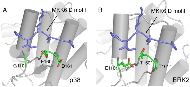
Figure 1.
A comparison of D motif binding sites on p38α and ERK2. (A) A crystal structure (PDB ID: 2y8o) of a docking peptide derived from MKK6 (purple) bound to p38α (gray). p38α residues adjacent to the binding site that differ from ERK2 are shown in green. (B) A superposition of the MKK6 D-motif (purple) on a crystal structure of ERK2 (gray, PDB ID: 2y9q). Residues in green differ from those on p38α. *Residue numbers from ERK2 are shifted up by one to match structurally equivalent positions on p38α.

