Key Points
Question
May acute optic neuritis help to assess neuroprotection and remyelination?
Findings
This cohort study found that trials testing neuroprotection using optical coherence tomography as an outcome should include 37 to 50 participants per arm within 15 days after onset to reveal a 50% reduction in 6-month peripapillary retinal nerve fiber layer or ganglion cell plus inner plexiform layer thinning; stratified randomization by sex and high-contrast visual acuity is highly recommended. Larger sample sizes may be needed for trials testing remyelination using 6-month latency changes in multifocal visual evoked potentials; the correlation of optical coherence tomographic measures with low-contrast letter acuity was stronger than with multifocal visual evoked potentials, although cohorts were not identical.
Meaning
Acute optic neuritis appears to be an appropriate condition to test neuroprotective and remyelinating therapies after acute inflammation.
This cohort study comprehensively assesses key biological and methodologic aspects of trials of acute optic neuritis for testing neuroprotection and remyelination in multiple sclerosis.
Abstract
Importance
Neuroprotective and remyelinating therapies are required for multiple sclerosis (MS), and acute optic neuritis (AON) is a potential condition to evaluate such treatments.
Objective
To comprehensively assess key biological and methodological aspects of AON trials for testing neuroprotection and remyelination in MS.
Design, Setting, and Participants
The AON-VisualPath prospective cohort study was conducted from February 2011 to November 2018 at the Hospital Clinic of University of Barcelona, Barcelona, Spain. Consecutive patients with AON were prospectively enrolled in the cohort and followed up for 18 months. Data analyses occurred from November 2018 to February 2019.
Exposures
Participants were followed up for 18 months using optical coherence tomography, visual acuity tests, and in a subset of 25 participants, multifocal visual evoked potentials.
Main Outcomes and Measures
Dynamic models of retinal changes and nerve conduction and their associations with visual end points; and eligibility criteria, stratification, and sample-size estimation for future trials.
Results
A total of 60 patients (50 women [83%]; median age, 34 years) with AON were included. The patients studied displayed early and intense inner retinal thinning, with a thinning rate of approximately 2.38 μm per week in the ganglion cell plus inner plexiform layer (GCIPL) during the first 4 weeks. Eyes with AON displayed a 6-month change in latency of about 20 milliseconds, while the expected change in the eyes of healthy participants by random variability was 0.13 (95% CI, −0.80 to 1.06) milliseconds. The strongest associations with visual end points were for the 6-month intereye difference in 2.5% low-contrast letter acuity, which was correlated with the peripapillary retinal nerve fiber layer thinning (adjusted R2, 0.57), GCIPL thinning (adjusted R2, 0.50), and changes in mfVEP latency (adjusted R2, 0.26). A 5-letter increment in high-contrast visual acuity at presentation (but not sex or age) was associated with 6-month retinal thinning (1.41 [95% CI, 0.60-2.23] μm less peripapillary retinal nerve fiber layer thinning thinning; P = .001; adjusted R2, 0.20; 0.86 [95% CI, 0.35-1.37] μm less GCIPL thinning; P = .001; adjusted R2, 0.19) but not any change in multifocal visual evoked potential latency. To demonstrate 50% efficacy in GCIPL thinning or change in multifocal visual evoked potential latency, a 6-month, 2-arm, parallel-group trial would need 37 or 50 participants per group to test a neuroprotective or remyelinating drug, respectively (power, 80%; α, .05).
Conclusions and Relevance
Acute optic neuritis is a suitable condition to test neuroprotective and remyelinating therapies after acute inflammation, providing sensitive markers to assess the effects on both processes and prospective visual recovery within a manageable timeframe and with a relatively small sample size.
Introduction
Multiple sclerosis (MS) is an inflammatory demyelinating and neurodegenerative disease of the central nervous system1 for which some disease-modifying drugs have been incorporated into clinical practice in recent years. This success has only been possible with anti-inflammatory drugs, and these have benefited from the use of new or enlarging T2 or gadolinium-enhancing lesions as suitable surrogate markers. Thus, to further expand the therapeutic repertoire for MS, it will be necessary to extend studies toward neuroprotection and remyelination.
Spectral-domain optical coherence tomography (SD-OCT) and visual evoked potentials (VEP) are sensitive, reproducible techniques capable of tracking prominent and short-term changes in neuroaxonal integrity provoked by acute optic neuritis (AON) as well as alterations to nerve conduction.2,3 Consequently, AON may be considered as an appropriate condition to test neuroprotective and remyelinating drugs in MS. Indeed, a few trials have already used AON,3,4,5,6,7,8 although with some controversial results.7,9,10 Methodologic limitations3,5 and a lack of standardized definitions for markers and end points limit the use of AON for drug development.
Hence, we aimed herein to comprehensively assess certain biological and methodological aspects of AON to validate it as a condition for assessing acute neuroprotection and remyelination. The specific aims were to (1) evaluate the dynamic changes in retinal thickness and nerve conduction; (2) assess the association of these changes with visual end points; and (3) obtain information to guide key methodologic aspects of future trials, such as eligibility criteria, the need for stratified randomization, and sample-size estimates.
Methods
Study Design and Participants
The first 60 consecutive patients with AON11 enrolled onto the Barcelona AON-VisualPath prospective cohort at the Hospital Clinic (University of Barcelona, Barcelona, Spain) were evaluated for eligibility from February 2011 to November 2018 (in the Supplement, the eMethods contains the protocol and eFigure 1, a study flowchart). An institutional review board (Comité Ético de Investigación Clínica del Hospital Clínic de Barcelona [Clinical Research Ethical Committee of Hospital Clinic of Barcelona]) approved the study, and all participants provided written informed consent. The article follows Strengthening the Reporting of Observational Studies in Epidemiology (STROBE)12 and Advised Protocol for Optical Coherence Tomography (OCT) Study Terminology and Elements (APOSTEL)13 recommendations.
Procedures
All participants underwent a monthly spectral-domain optical coherence tomography (SD-OCT) scan and a bimonthly test of visual function for up to 6 months. The protocol was updated in 2014 by removing the 3-month and 5-month SD-OCT assessments and including a weekly SD-OCT assessment during the first month and an additional 18-month assessment. Moreover, we incorporated multifocal VEP (mfVEP) evaluations at baseline, month 6, and month 18 (eMethods in the Supplement).
Statistical Analyses
Change in Retinal Thickness Using OCT
We described qualitative variables using absolute and relative numbers and quantitative variables using medians and interquartile ranges (IQRs). We plotted the absolute change in thickness of each macular retinal layer at each visit over time from the baseline value and from the time of the first symptom up to the 6-month visit. In terms of the peripapillary retinal nerve fiber layer (pRNFL) thickness, we estimated the intereye asymmetry with reference to the baseline value from the eye unaffected by AON. The candidate markers to track neuroprotection were the 6-month change in ganglion cell plus inner plexiform layer (GCIPL) and pRNFL thicknesses. Changes in the other layers were fit as complementary results. We modeled retinal changes using mixed-effect models using the Akaike Information Criterion14 and visual inspection of the raw data for model selection.
We performed 3 sensitivity analyses. First, we repeated the analyses using the relative changes ([Follow-up Value − Baseline Value] / Baseline Value) instead of the absolute changes to test the robustness of the results to possible floor effects attributable to prior retinal injury (eg, relapsing-remitting MS–AON or prior AON in the same eye). Second, we explored GCIPL dynamics using intereye asymmetry, a parameter recently reported to be more sensitive than the absolute differences in the affected eye GCIPL thickness using baseline value as a reference to capturing early damage.15 Third, we evaluated the assumption that retinal changes did not differ significantly between AON types and with the presence of disc edema (pRNFL). Finally, we illustrated the changes from baseline to 6 months and 6 months to 18 months using boxplots. Based on the assumption that the neuroaxonal injury extended retrogradely from the inner toward the outer layers, we explored the dynamics of the changes in inner nuclear layer, outer nuclear layer, and photoreceptors by tertiles of 1-month GCIPL thinning, the period when thinning is the greatest.2 We did not impute missing values and assumed missingness was at random.
Change in mfVEP
We examined the changes in mfVEP (amplitude and latency) in each of the 56 segments in both eyes over 18 months of follow-up. Changes in unaffected eyes relative to their baseline values were described recently,16 yet the underlying nature of these changes (injury or compensation) was difficult to interpret without normative values. Consequently, we used 2 complementary strategies to set the reference values. First, we calculated a z score for each segment using a sex-matched and age-matched population of healthy volunteers (n = 22), and we displayed the z scores over 18 months of follow-up. Second, we estimated the 6-month and 18-month changes relative to the baseline value for each participant and eye, except for the latency in the affected eye, for which we used the baseline value of the unaffected eye as a reference.16 We developed an in-house heatmap display (Python version 3.7 [Python Software Foundation]) for data visualization. We performed a short-term (1-month) test-retest study in the 22 healthy volunteers to define the magnitude of the changes expected in the mfVEP data that would be attributable to random variability.
Assessment of Visual Outcomes Based on Retinal Thickness and mfVEP Latency
We examined the association between the SD-OCT and mfVEP markers and the visual function tests, using linear (simple or spline) regression models after visual inspection of the data. Statistical analyses included stated robust regression if needed, based on the Cook distance. In addition to the 6-month score in the affected eye,3,7 we proposed a 6-month intereye asymmetry (affected eye score minus unaffected eye score) as an alternative visual end point.
We aimed to overcome 2 caveats associated with the definition of the former end point. First, unlike high-contrast visual acuity (HCVA)17 and Hardy-Rand-Rittler plates,18 there are no normative standards for low-contrast letter acuity (LCLA), the preferred clinical end point for AON. The available evidence suggests a wide range of normal LCLA values among healthy volunteers.19,20 In addition, participants with relapsing-remitting MS–AON may have prior visual impairment because of a diffuse MS burden.21,22 Consequently, the visual performance 6 months after AON onset in patients with MS may not reach the values found in healthy individuals, even in the case of complete visual recovery. As such, the intereye difference in LCLA of more than 7 letters and HCVA of more than 5 letters were considered significant in patients with MS.19 We did not find any intereye cutoff value for Hardy-Rand-Rittler plates. Patients with prior AON in the affected eye (n = 5) were not included in these analyses, and furthermore, patients with a prior AON in the fellow eye (n = 4) were not considered for intereye measures.
Sample Size, Randomization, and Eligibility Criteria for Phase II Trials
We calculated the sample sizes required for a 6-month, 2-arm, parallel-group trials to test drugs using an unpaired, 2-sample t test. Previous trials included small sample sizes, for which stratified randomization is recommended to achieve balanced groups. Consequently, we evaluated the associations of the changes in SD-OCT and mfVEP, with 4 prespecified baseline features: sex, age, HCVA in the affected eye, and disc edema. Men may have more severe retinal thinning after AON,23 and age and HCVA level at inclusion were also studied as modifiers of the therapeutic response in patients with AON.24 The presence of baseline disc edema may be relevant for pRNFL measures. We did not consider the time from onset to study inclusion, which is likely to be inversely associated with the severity of AON in observational studies (and is a confounder but not an effect modifier). However, we evaluated the influence of time from onset to inclusion on the estimated sample size.
All statistical analyses were done using R version 3.5.2 (R Foundation for Statistical Computing) and Stata version 14 (StataCorp) between November 2018 and February 2019. Any P value less than .05 was considered significant.
Results
Study Population
The study population included 60 patients (50 women [83%]; median [IQR] age, 33.96 [29.04-40.53] years; eTable 3 in the Supplement). The median (IQR) time since onset was 9 (6-15) days. Of the 60 patients, 43 (72%) experienced MS-associated AON, and 7 patients with relapsing-remitting MS–AON received disease-modifying drugs of mild to intermediate efficacy at baseline (interferon-beta-1b, n = 3; interferon-beta-44 1a, n = 1; glatiramer acetate, n = 2, and fingolimod, n = 1). During the 18-month follow-up period, the patients could start, change, or abandon disease-modifying drugs (eTable 1 in the Supplement). Two participants had a second AON episode in the follow-up period, and for these individuals, we only included the data prior to the second episode.
Changes in Retinal Thickness in Affected and Unaffected Eyes
We detected distinct phases of pRNFL thinning, the initial phase extending over the first approximate 45 days with positive intereye measures and a second one over the following 35 days or so with negative intereye measures. This was followed by a third phase of relatively mild thinning, which lasted until the end of the 200 days of the follow-up period (Figure 1A). Both the macular RNFL and GCIPL thinned during the first approximate 45 days after AON diagnosis (Figure 1B and C). This was anticipated by the model as 2.17 μm per week of GCIPL thinning during the initial 45 days (β = −0.31 [95% CI, −0.38 to −0.25] μm/day; P <.001), although the rate of GCIPL thinning then decreased to 0.22 μm per month (β = −0.007 [95% CI, −0.01 to −0.003] μm/day; P = .002). Moreover, similar dynamics were observed for intereye GCIPL thinning (eFigure 1 in the Supplement). This early thinning was confirmed in 29 individuals through weekly SD-OCT assessments within the first month of follow-up, in whom GCIPL thinning of 2.38 μm per week was seen (β, −0.3438 [95% CI, −0.42 to −0.26] μm/day; P < .001). We did not find any model that reflected the changes in the unaffected eyes.
Figure 1. Models of the Change in Retinal Layer Thicknesses During the 6-Month Follow-up Period.
The black points joined by dashed lines represent the individual trajectories of the changes in retinal thickness, the thicker curves represent the individual fit of the model, and the dark red line represents the population model. A, Peripapillary retinal nerve fiber layer (pRNFL); B, macular retinal nerve fiber layer (RNFL); C, ganglion cells plus inner plexiform layer (GCIPL); D, inner nuclear layer (INL); E, outer nuclear layer (ONL); and F, photoreceptors (PRL). The y-axis represents the absolute change (follow-up visit minus baseline) in the affected eye, except for the pRNFL, for which the intereye asymmetry refers to the baseline value in the unaffected eye. The x-axis represents the time in days from clinical onset. A and C, Linear spline mixed-effect models with 2 knots (A, 45 and 85 days) and 1 knot (C, 45 days); B and D-F, mixed-effect, third-order polynomials with all coefficients set as fixed and random effects. All models were fit using the lme4 package in R version 3.5.2 (R Foundation for Statistical Computing).
In the inner nuclear layer, we detected a transitory, mild early thickening during the first 3 months of follow-up (Figure 1D), whereas in this period, we observed outer nuclear layer and photoreceptor thickening that had only partially recovered by the end of the short-term (3-month) follow-up (Figure 1E and F). The inner nuclear layer and photoreceptor models only defined significant changes for participants with a thinning of the GCIPL greater than 10.72 μm (third tertile), and the models for the outer nuclear layer only reflected significant changes when GCIPL thinning was greater than 5.76 μm (second and third tertiles) (eFigure 3 in the Supplement). The dynamics of the relative changes were similar (eFigures 4 and 5 in the Supplement), and we found no differences among the types of AON (eFigure 6 in the Supplement) or disc edema (eFigure 7 in the Supplement). Finally, we observed mild thinning in all the macular layers from 6 to 18 months (eFigure 8 in the Supplement).
Changes in mfVEP Latency and Amplitude in Affected and Unaffected Eyes
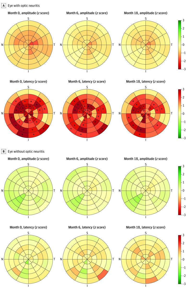
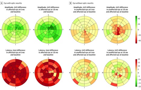
Compared with a sex-matched and age-matched population, affected eyes appeared to display a nonsignificant decrease in amplitude in most of the segments partially recovered during the follow-up period. In contrast, the mfVEP latency included 31 significant segments at baseline with a median z score of 2.60 (IQR, 2.29-3.10; P = .009), 28 significant segments at 6-month follow-up, with a median z score of 2.36 (IQR, 2.16-2.75; P = .02), and 20 significant segments at 18-month follow-up, with a median z score of 2.38 (IQR, 2.12-2.65; P = .02) (Figure 2). Relative to the baseline values, there was a nonsignificant increase in amplitudes, predominantly central at 6 months and diffuse at 18 months, in line with recovery of conduction block. Consistently, affected eyes showed a homogeneous increase in latency at 6 months that was only slightly recovered at 18 months (Figure 3).
Figure 2. Standarized (z Score) Amplitudes and Latencies in the 56 Segments of the Multifocal Visual Evoked Potentials in Affected and Unaffected Eyes.
A heatmap display developed in house to visualize the normalized (z score) latencies and amplitudes in the multifocal visual evoked potentials segments. Each segment represents the mean amplitude or latency of a patient’s eyes, with the data available from each visit normalized using the mean (SD) of the values obtained in a sex-matched and age-matched population of healthy volunteers (n = 22) to create a z score. A, Results for the affected eye; B, Results for the unaffected eye. A color scale is used to represent the magnitude and direction of the changes. The latencies and amplitude are adimensional, and the numbers represent the SD from the healthy population. Green represents improvement (normalized z scores in amplitude of more than zero and z scores of latency less than zero), while red represents worsening. In addition, the symbol + denotes all segments greater than 1.96 SD more than the mean; the symbol − was designated for all segments at least 1.96 SD less than the mean, but this was not applicable to any segment. I indicates inferior; N, nasal; S, superior; and T, temporal.
Figure 3. Change Over 6 Months and 18 Months in the Amplitude and Latency in the 56 Segments of the Multifocal Visual Evoked Potentials in Affected and Unaffected Eyes.
A heatmap developed by the authors to visualize the changes in latency and amplitude in the multifocal visual evoked potentials segments. Each segment represents the mean change (6-month and 18-month) relative to the baseline value of each participant and eye, except the latency in the affected eye, for which the baseline value of the unaffected eye was a reference. A, Results for the affected eye; B, Results for the unaffected eye. A color scale is used to represent the magnitude and direction of the changes. Amplitudes were measured in nanovolts and latencies as milliseconds. The numbers represent the 6-month and 18-month changes; green represents improvement (increase in amplitude and decrease in latency relative to the baseline), while red represents worsening. I indicates inferior; N, nasal; S, superior; and T, temporal.
Compared with a sex-matched and age-matched population, unaffected eyes appeared to display higher amplitudes, particularly at baseline, albeit not significantly higher, while no such pattern was evident in terms of the latencies (Figure 2). Figure 3 and eTable 2 in the Supplement show nonsignificant decreases in amplitudes in unaffected eyes compared with baseline, which is in line with the attenuation of the increase in amplitude in Figure 2. eFigure 9 in the Supplement shows short-term test-retest values of mfVEP in healthy volunteers.
Retinal Thickness, Nerve Conduction, and Visual Outcomes
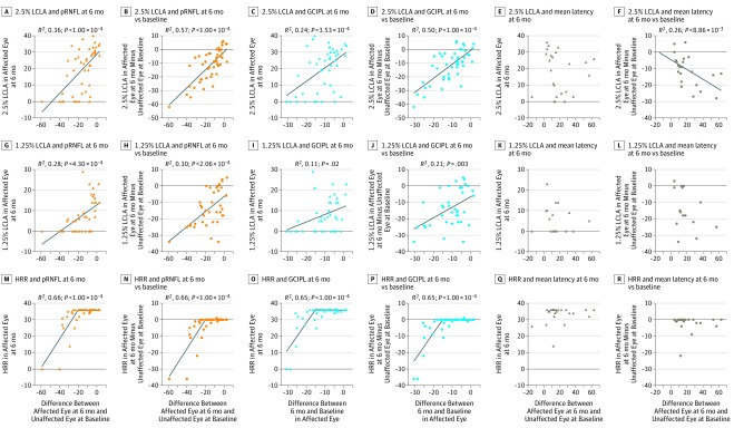
Most of the visual recovery was detected within 60 to 70 days (eFigure 10 in the Supplement), and because most participants (36 [80%]) had attained complete HCVA recovery after a 6-month follow-up, there appeared to be a ceiling effect. Consequently, we did not further explore HCVA as a clinical end point for AON. By contrast, only 19 participants (41.3%) and 13 participants (34.2%) achieved complete 2.5% LCLA and 1.25% LCLA recovery, respectively. Indeed, we found the highest correlations always involved 6-month intereye 2.5% LCLA with either GCIPL change (adjusted R2, 0.50) or pRNFL (adjusted R2, 0.57) (Figure 4). The 2.5% LCLA intereye difference augmented (worsened) by 0.66 letters (95% CI, 0.48-0.82; P < .001) per 1 μm of RNFL thinning and 1.01 letters (95% CI, 0.71-1.31; P < .001) per 1 μm of GCIPL thinning. Similarly, each 1 μm of RNFL and 1 μm of GCIPL thinning enhanced the 1.25% LCLA intereye difference by 0.46 letters (95% CI, 0.23-0.69 letters; P < .001; adjusted R2, 0.30) and 0.64 letters (95% CI, 0.24-1.04 letters; P < .003; adjusted R2, 0.30), respectively.
Figure 4. Assessment of 6-Month Visual Outcomes Based on the Change in Retinal Thickness and Latency During the Follow-up.
The different scatterplots represent the changes in the 3 surrogate markers and visual function tests at 6 months. The visual function tests include 2.5% low-contrast letter acuity (LCLA) (A-F), 1.25% low-contrast letter acuity (G-L), and color vision (M-R). Visual recovery is evaluated as the 6-month performance in affected eye (triangles) and the intereye differences, calculated as the affected eye value minus unaffected eye value (dots). The surrogate markers are the 6-month changes in peripapillary retinal nerve fiber layer thickness (orange), ganglion cells plus inner plexiform layer thickness (blue), and latency (brown). Each scatterplot includes the fit of a regression model, and where the model was significant, the adjusted R2 and P values of the model are displayed. The models are a linear regression models for LCLA, and a linear spline model with 1 knot each at 20 μm for peripapillary retinal nerve fiber layer (pRNFL) thinning and 15 μm for ganglion cells plus inner plexiform layer (GCIPL) thinning. HRR indicates Hardy-Rand-Rittler plates.
Finally, 33 patients (69%) achieved complete recovery of color vision. The Hardy-Rand-Rittler score decreased by 0.88 symbols (95% CI, 0.66-1.10 symbols; adjusted R2, 0.66; P < .001) for each 1 μm of pRNFL thinning after 20 μm and 1.63 symbols (95% CI, 1.21-2.06 symbols; adjusted R2, 0.65; P < .001) for each 1 μm of GCIPL thinning after 15 μm. The 6-month 2.5% LCLA intereye difference worsened by 0.31 letters (95% CI, 0.08-0.51 letters; P = .009; adjusted R2, 0.26) per 1-millisecond increase in latency over 6 months (Figure 4).
Sample Size, Randomization, and Eligibility Criteria for Phase II Trials
When we focused on sample-size calculations, a 6-month, 2-arm, parallel-group trial testing the benefits of a remyelinating drug on the change in mfVEP latency would need 50 to 163 participants per arm, depending on effect size (50% for 50 participants per arm and 30% for 163 participants per arm). This was assessed using a subset of 25 participants. A trial testing the efficacy of a neuroprotective drug that aimed to decrease GCIPL thinning using SD-OCT by same effect size would need 37 to 101 participants per arm (Table).
Table. Sample Size per Group for a 2-Arm, Parallel-Group Trial Testing Drugs and Retinal Thinning or the Multifocal Visual Evoked Potential Change in Latency as Surrogate Markers of Efficacya.
| Effect size, %b | Peripapillary Retinal Nerve Fiber Layer Thinning at 6 Mo | Ganglion Cell Plus Inner Plexiform Layer Thinning at 6 Mo | Change in Latency at 6 Mo | |||
|---|---|---|---|---|---|---|
| 80% Power | 90% Power | 80% Power | 90% Power | 80% Power | 90% Power | |
| 30 | 136 | 181 | 101 | 135 | 163 | 218 |
| 40 | 77 | 103 | 58 | 77 | 92 | 123 |
| 50 | 50 | 66 | 37 | 50 | 50 | 79 |
Settings included 1:1 randomization; α of .05; power of 80% or 90%; and means (SDs) for peripapillary retinal nerve fiber layer of −15.21277 (SD, 13.34645) μm, for ganglion cell plus inner plexiform layer of −10.96809 (8.295863) μm, and for latency of 18.10 (18:21) milliseconds. The statistical test used was an unpaired 2-sample t test. These sample size estimations did not account for any loss to follow-up or lack of compliance.
Effect size: percentage decrease in peripapillary retinal nerve fiber layer loss, ganglion cell plus inner plexiform layer loss, or latency increase comparing treatment with the placebo arm. The sample size was based on 47 participants for peripapillary retinal nerve fiber layer and ganglion cell plus inner plexiform layer and 25 participants for latency.
We also assessed the association between baseline features and the changes in retinal thinning and latency. Men displayed a nonsignificant difference in GCIPL thinning (β = −5.77 [95% CI, −11.79 to 0.24] μm; P = .06) and pRNFL thinning (β = −6.74 [95% CI, −16.61 to 3.12] μm; P = .18); women had nonsignificant differences in the same measures. Moreover, a 5-letter increment in HCVA in the affected eye at inclusion was associated with 1.41 μm less pRNFL thinning (95% CI, 0.60-2.23 μm; P = .001; adjusted R2, 0.20) and 0.86 μm less GCIPL thinning (95% CI, 0.35-1.37 μm; P = .001; adjusted R2, 0.19) during the 6-month follow-up. No association was found between age and disc edema and SD-OCT changes over 6 months. We did not observe any association between the baseline features and the changes in latency in the affected eyes.
With a sample of 101 participants per group, we would have 80% power to detect a difference of 3.29 μm (30% effect size) in GCIPL thinning between the null hypothesis that both group means were 10.97 μm and the alternative hypothesis that the mean of the intervention group was 7.67 μm, with an estimated SD of 8.29 and with a significance level of .05. Based on this model, a trial enrolling participants with a mean time after AON onset of 25 days would have found a mean GCIPL thinning of 5 μm during the follow-up period (Figure 1C). With all other elements (α, β, 30% effect size, and SD) being equal, the trial would have need 401 participants per arm.
Discussion
This study confirms the suitability of AON to test neuroprotective and remyelinating drugs after acute inflammation in a phase 2 trial. Trials testing neuroprotection may use either 6-month pRNFL or GCIPL thinning, which correlate strongly with the 2.5% LCLA, the best clinical end point. As such, a 6-month, parallel-group trial should enroll 37 to 50 participants per arm within the first 15 days after disease onset to reveal a 50% reduction in neuroaxonal injury. Moreover, this study also shows the importance of stratified randomization by sex and particularly HCVA when assessing neuroprotective effects. Trials testing remyelination may require larger sample size if they use the 6-month change in mfVEP latency, for which no associations were found with age, sex, HCVA, or disc edema at inclusion. The magnitude of association between 2.5% LCLA and mfVEP latency was half of the value found for retinal thinning.
Based on these results and previous studies,3,4,5,7 AON should be considered as an appropriate condition to test neuroprotective drugs, because it causes prominent neuroaxonal injury that can be readily and rapidly tracked using SD-OCT. To date, trials have focused on pRNFL thinning to test the outcomes of several drugs.6,7,8,25 Both pRNFL and GCIPL thinning may serve as suitable markers, although the first is simpler because segmentation is achieved robustly and automatically by all SD-OCT devices. In addition, correlations with visual end points were slightly stronger for pRNFL than GCIPL thinning. However, using this marker implies the exclusion of participants with prior AON in the fellow eye, which serves as a reference to estimate pRNFL thinning, thereby limiting the generalizability of the results. Moreover, pRNFL thinning has more variability than GCIPL because of disc edema, which negatively affects the sample size. The GCIPL showed no pseudoatrophy, and thus neurodegeneration could be tracked effectively from very early phases (with no need of intereye measures). Consequently, patients with vs those without prior AON in the fellow eye could be enrolled. This eases recruitment timelines and increases heterogeneity in AON trials, which is challenging but enhances generalizability of results. Nevertheless, the inclusion of patients with an affected fellow eye may impose an additional challenge for the structural-functional correlations in trials combining GCIPL thinning as a structural end point and 6-month intereye LCLA performance as a clinical end point. As another disadvantage, GCIPL segmentation is not fully automatic in all devices. Irrespective of the choice, we suggest stratifying randomization by sex and HCVA at onset and using an inclusion period shorter than 15 days because the sample size greatly increases with larger times. We believe that the benefit in terms of the smaller sample size required outweighs the difficulties of recruitment at such short times. Dynamic changes in HCVA suggested a relevant ceiling effect, while changes in 1.25% LCLA probably reflected floor effects, supporting the use of 2.5% LCLA as the primary clinical end point. Both GCIPL and pRNFL thinning displayed a strong linear correlation with 2.5% LCLA, particularly with intereye difference, and Hardy-Rand-Rittler levels, potentially representing a marker of disease severity.
As observed in previous studies,2,26 we found thickening of outer layers including the inner nuclear layer, outer nuclear layer, and particularly photoreceptors. These changes were proportional to early GCIPL thinning and only partially recovered during follow-up, suggesting that both inflammatory and neurodegenerative processes may have mediated these changes. Although appealing, their magnitude did not support their use as markers in AON trials.
Acute optic neuritis may be an appropriate condition to test remyelinating drugs, tracking the changes in latency with mfVEP or full-field VEP (ffVEP). To date, trials testing opicinumab3 and clemastine (NCT02521311) used the change in latency measured by ffVEP, which is technically simpler and faster than mfVEP. Nevertheless, mfVEP has certain advantages over ffVEP, including capturing a wider visual field and identifying changes in field segments, where capturing minor differences might be particularly relevant. In addition, this technique allows the homogeneity of the therapeutic response to be evaluated across the visual field. Here, affected eyes displayed a 6-month, 20-millisecond increase in latency relative to the baseline value of the fellow unaffected eyes, compared with the change of 0.13 (95% CI, −0.80 to 1.06) millisecond expected by random variability. However, the variability in the change of latency for mfVEP was larger than that found for retinal thinning on OCT, and this issue has a negative influence on sample-size estimations.
This study has several strengths. First, we enrolled consecutive patients admitted to our center to avoid a selection bias and enhance generalizability of the results. Second, SD-OCT and mfVEP were performed following standard protocols by technicians masked to the study details to minimize observational bias. Third, a population of healthy participants was also studied by mfVEP, for which normative values and reproducibility data are scarce. Fourth, we used statistical approaches robust to missing data (mixed-effect models) and outliers (robust regression), and we included sensitivity analyses to test key assumptions (floor effects and the type of AON).
Limitations
This study also has certain limitations, not least of which is that the data came from a single center. Hence, further multicentric studies will be needed to validate the conclusions. Moreover, we only included a single type of SD-OCT and mfVEP devices. The mfVEP data are also derived from a smaller sample (25 participants), which could have explained the weaker association between mfVEP and 2.5% LCLA. Alternatively, 2.5% LCLA may not be as appropriate a clinical end point for remyelination as it seems to be for neuroprotection. More importantly, this protocol did not include ffVEP assessments, so we could not assess the correlation between mfVEP and ffVEP measurements. The experience with mfVEP is still limited, and the results were driven from a small single-center data set. Altogether, mfVEP findings should be interpreted with caution and require validation. Moreover, we were unable to assess the influence of race/ethnicity, a feature that seems to influence the severity of retinal damage.17,27 Finally, these results should not be extrapolated to neither long-term neuroprotection nor demyelination, because the underlying pathogenetic mechanisms may be likely different.
Conclusions
Acute optic neuritis may help to test drugs that promote neuroprotection and remyelination under an acute inflammatory insult. The evidence obtained is more robust for neuroprotection than remyelination because the dynamic changes of retinal thinning are better characterized and the correlation with visual end points are stronger than for the change in mfVEP latency. Thus, further studies should focus on providing further evidence for remyelination.
eMethods. Methods
eTable 1. Baseline characteristics of the study population
eTable 2. Average amplitude and latencies (Z-scores), and the changes from baseline in ON-eyes and non-ON eyes
eTable 3. Demographic characteristics of participants in AON VisualPath cohort and healthy volunteers
eFigure 1. Patient eligibility and inclusion flowchart
eFigure 2. Model of the change in inter-eye asymmetry of the ganglion cells plus inner plexiform layer in the first 6 months
eFigure 3. Model of the change in inner nuclear layer, outer nuclear layer and photoreceptor layer classified by the ganglion cell plus inner plexiform layer thinning at the first assessment
eFigure 4. Overview of the relative changes in retinal layer thickness at a population level
eFigure 5. Models of the relative change in retinal layer thicknesses during the 6-month follow-up period
eFigure 6. Models of the change in retinal layer thickness over the 6-month follow-up classified by type of acute optic neuritis
eFigure 7. Models of the change in retinal layer thickness over the 6-month follow-up classified by presence of disk edema at baseline
eFigure 8. Box plot of the changes at 6-months and 6-18 months
eFigure 9. Short-term test-retest of multifocal visual evoked potentials in healthy volunteers
eFigure 10. Evolution of visual function in the acute phase of ON
References
- 1.Reich DS, Lucchinetti CF, Calabresi PA. Multiple Sclerosis. N Engl J Med. 2018;378(2):169-180. doi: 10.1056/NEJMra1401483 [DOI] [PMC free article] [PubMed] [Google Scholar]
- 2.Gabilondo I, Martínez-Lapiscina EH, Fraga-Pumar E, et al. . Dynamics of retinal injury after acute optic neuritis. Ann Neurol. 2015;77(3):517-528. doi: 10.1002/ana.24351 [DOI] [PubMed] [Google Scholar]
- 3.Cadavid D, Balcer L, Galetta S, et al. ; RENEW Study Investigators . Safety and efficacy of opicinumab in acute optic neuritis (RENEW): a randomised, placebo-controlled, phase 2 trial. Lancet Neurol. 2017;16(3):189-199. doi: 10.1016/S1474-4422(16)30377-5 [DOI] [PubMed] [Google Scholar]
- 4.Esfahani MR, Harandi ZA, Movasat M, et al. . Memantine for axonal loss of optic neuritis. Graefes Arch Clin Exp Ophthalmol. 2012;250(6):863-869. doi: 10.1007/s00417-011-1894-3 [DOI] [PubMed] [Google Scholar]
- 5.Tsakiri A, Kallenbach K, Fuglø D, Wanscher B, Larsson H, Frederiksen J. Simvastatin improves final visual outcome in acute optic neuritis: a randomized study. Mult Scler. 2012;18(1):72-81. doi: 10.1177/1352458511415452 [DOI] [PubMed] [Google Scholar]
- 6.Salari M, Janghorbani M, Etemadifar M, Dehghani A, Razmjoo H, Naderian G. Effects of vitamin D on retinal nerve fiber layer in vitamin D deficient patients with optic neuritis: preliminary findings of a randomized, placebo-controlled trial. J Res Med Sci. 2015;20(4):372-378. [PMC free article] [PubMed] [Google Scholar]
- 7.Raftopoulos R, Hickman SJ, Toosy A, et al. . Phenytoin for neuroprotection in patients with acute optic neuritis: a randomised, placebo-controlled, phase 2 trial. Lancet Neurol. 2016;15(3):259-269. doi: 10.1016/S1474-4422(16)00004-1 [DOI] [PubMed] [Google Scholar]
- 8.McKee JB, Cottriall CL, Elston J, et al. . Amiloride does not protect retinal nerve fibre layer thickness in optic neuritis in a phase 2 randomised controlled trial. Mult Scler. 2019;25(2):246-255. doi: 10.1177/1352458517742979 [DOI] [PubMed] [Google Scholar]
- 9.Saidha S, Calabresi PA. Phenytoin in acute optic neuritis: neuroprotective or not? Lancet Neurol. 2016;15(3):233-235. doi: 10.1016/S1474-4422(16)00024-7 [DOI] [PubMed] [Google Scholar]
- 10.Martinez-Lapiscina EH, Andorra M, Sanchez-Dalmau B, Villoslada P. Phenytoin for neuroprotection. Lancet Neurol. 2016;15(9):901-902. doi: 10.1016/S1474-4422(16)30093-X [DOI] [PubMed] [Google Scholar]
- 11.Petzold A, Wattjes MP, Costello F, et al. . The investigation of acute optic neuritis: a review and proposed protocol. Nat Rev Neurol. 2014;10(8):447-458. doi: 10.1038/nrneurol.2014.108 [DOI] [PubMed] [Google Scholar]
- 12.von Elm E, Altman DG, Egger M, Pocock SJ, Gøtzsche PC, Vandenbroucke JP; STROBE Initiative . The Strengthening the Reporting of Observational Studies in Epidemiology (STROBE) statement: guidelines for reporting observational studies. Lancet. 2007;370(9596):1453-1457. doi: 10.1016/S0140-6736(07)61602-X [DOI] [PubMed] [Google Scholar]
- 13.Cruz-Herranz A, Balk LJ, Oberwahrenbrock T, et al. ; IMSVISUAL consortium . The APOSTEL recommendations for reporting quantitative optical coherence tomography studies. Neurology. 2016;86(24):2303-2309. doi: 10.1212/WNL.0000000000002774 [DOI] [PMC free article] [PubMed] [Google Scholar]
- 14.Akaike H. A new look at the statistical model identification. IEEE Trans Automat Contr. 1974;19(6):716-723. doi: 10.1109/TAC.1974.1100705 [DOI] [Google Scholar]
- 15.Brandt AU, Specovius S, Oberwahrenbrock T, Zimmermann HG, Paul F, Costello F. Frequent retinal ganglion cell damage after acute optic neuritis. Mult Scler Relat Disord. 2018;22:141-147. doi: 10.1016/j.msard.2018.04.006 [DOI] [PubMed] [Google Scholar]
- 16.Klistorner A, Chai Y, Leocani L, et al. ; RENEW MF-VEP Investigators . Assessment of opicinumab in acute optic neuritis using multifocal visual evoked potential. CNS Drugs. 2018;32(12):1159-1171. doi: 10.1007/s40263-018-0575-8 [DOI] [PMC free article] [PubMed] [Google Scholar]
- 17.Beck RW, Cleary PA, Anderson MM Jr, et al. ; The Optic Neuritis Study Group . A randomized, controlled trial of corticosteroids in the treatment of acute optic neuritis. N Engl J Med. 1992;326(9):581-588. doi: 10.1056/NEJM199202273260901 [DOI] [PubMed] [Google Scholar]
- 18.Cole BL, Lian KY, Lakkis C. The new Richmond HRR pseudoisochromatic test for colour vision is better than the Ishihara test. Clin Exp Optom. 2006;89(2):73-80. doi: 10.1111/j.1444-0938.2006.00015.x [DOI] [PubMed] [Google Scholar]
- 19.Balcer LJ, Baier ML, Pelak VS, et al. . New low-contrast vision charts: reliability and test characteristics in patients with multiple sclerosis. Mult Scler. 2000;6(3):163-171. [DOI] [PubMed] [Google Scholar]
- 20.Pineles SL, Birch EE, Talman LS, et al. . One eye or two: a comparison of binocular and monocular low-contrast acuity testing in multiple sclerosis. Am J Ophthalmol. 2011;152(1):133-140. doi: 10.1016/j.ajo.2011.01.023 [DOI] [PMC free article] [PubMed] [Google Scholar]
- 21.Martinez-Lapiscina EH, Arnow S, Wilson JA, et al. ; IMSVISUAL consortium . Retinal thickness measured with optical coherence tomography and risk of disability worsening in multiple sclerosis: a cohort study. Lancet Neurol. 2016;15(6):574-584. doi: 10.1016/S1474-4422(16)00068-5 [DOI] [PubMed] [Google Scholar]
- 22.Petzold A, Balcer LJ, Calabresi PA, et al. ; ERN-EYE IMSVISUAL . Retinal layer segmentation in multiple sclerosis: a systematic review and meta-analysis. Lancet Neurol. 2017;16(10):797-812. doi: 10.1016/S1474-4422(17)30278-8 [DOI] [PubMed] [Google Scholar]
- 23.Costello F, Hodge W, Pan YI, et al. . Sex-specific differences in retinal nerve fiber layer thinning after acute optic neuritis. Neurology. 2012;79(18):1866-1872. doi: 10.1212/WNL.0b013e318271f755 [DOI] [PMC free article] [PubMed] [Google Scholar]
- 24.Cadavid D, Balcer L, Galetta S, et al. ; RENEW Study Investigators . Predictors of response to opicinumab in acute optic neuritis. Ann Clin Transl Neurol. 2018;5(10):1154-1162. doi: 10.1002/acn3.620 [DOI] [PMC free article] [PubMed] [Google Scholar]
- 25.Sühs KW, Hein K, Sättler MB, et al. . A randomized, double-blind, phase 2 study of erythropoietin in optic neuritis. Ann Neurol. 2012;72(2):199-210. doi: 10.1002/ana.23573 [DOI] [PubMed] [Google Scholar]
- 26.Al-Louzi OA, Bhargava P, Newsome SD, et al. . Outer retinal changes following acute optic neuritis. Mult Scler. 2016;22(3):362-372. doi: 10.1177/1352458515590646 [DOI] [PMC free article] [PubMed] [Google Scholar]
- 27.Kimbrough DJ, Sotirchos ES, Wilson JA, et al. . Retinal damage and vision loss in African American multiple sclerosis patients. Ann Neurol. 2015;77(2):228-236. doi: 10.1002/ana.24308 [DOI] [PMC free article] [PubMed] [Google Scholar]
Associated Data
This section collects any data citations, data availability statements, or supplementary materials included in this article.
Supplementary Materials
eMethods. Methods
eTable 1. Baseline characteristics of the study population
eTable 2. Average amplitude and latencies (Z-scores), and the changes from baseline in ON-eyes and non-ON eyes
eTable 3. Demographic characteristics of participants in AON VisualPath cohort and healthy volunteers
eFigure 1. Patient eligibility and inclusion flowchart
eFigure 2. Model of the change in inter-eye asymmetry of the ganglion cells plus inner plexiform layer in the first 6 months
eFigure 3. Model of the change in inner nuclear layer, outer nuclear layer and photoreceptor layer classified by the ganglion cell plus inner plexiform layer thinning at the first assessment
eFigure 4. Overview of the relative changes in retinal layer thickness at a population level
eFigure 5. Models of the relative change in retinal layer thicknesses during the 6-month follow-up period
eFigure 6. Models of the change in retinal layer thickness over the 6-month follow-up classified by type of acute optic neuritis
eFigure 7. Models of the change in retinal layer thickness over the 6-month follow-up classified by presence of disk edema at baseline
eFigure 8. Box plot of the changes at 6-months and 6-18 months
eFigure 9. Short-term test-retest of multifocal visual evoked potentials in healthy volunteers
eFigure 10. Evolution of visual function in the acute phase of ON


