Figure 1.
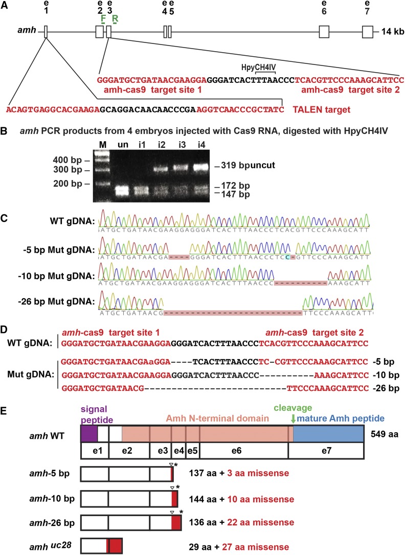
CRISPR/Cas9-induced amh mutants. (A) 14 kb of the amh locus showing two CRISPR target sites (red letters) in exon 3. PCR primers, forward (F) and reverse (R) (green). (B) Assay for injected CRISPR efficacy. PCR analysis of four G0 injected embryos at 1 dpf using genotyping primers F and R shows a 319-bp fragment in wild types that digested with HpyCH4IV to produce fragments of 172 and 147 bp; this site disappeared from amh genes in a large portion of cells in CRISPR-injected embryos. (C) Sequence traces from genomic DNA from a wild-type fish and from three stable mutant lines carrying −5, −10, and −26 bp deletions. (D) Sequences of genomic DNAs from a wild-type fish and three stable mutant lines (Mut). (E) Predicted structure of Amh protein showing the location of the mutation (triangle), the predicted out-of-frame portion (red), and the premature stop codon (*). Protein coding domains: signal peptide, purple; Amh amino-terminal domain, salmon; cleavage site, green arrow; mature Amh peptide, blue. i1-i4, CRISPR-injected 24 hpf embryos; M, length marker; un, uninjected 24 hpf embryos; WT, wild type.

