Abstract
Opioid receptor agonists are known to alter the activity of membrane ionic conductances and receptor-activated channels in CNS neurons and, via these mechanisms, to modulate neuronal excitability and synaptic transmission. In neuronal-like cell lines opioids also have been reported to induce intracellular Ca2+ signals and to alter Ca2+signals evoked by membrane depolarization; these effects on intracellular Ca2+ may provide an additional mechanism through which opioids modulate neuronal activity. However, opioid effects on resting or stimulated intracellular Ca2+ levels have not been demonstrated in native CNS neurons. Thus, we investigated opioid effects on intracellular Ca2+ in cultured rat hippocampal neurons by using fura-2-based microscopic Ca2+ imaging. The opioid receptor agonistd-Ala2-N-Me-Phe4,Gly-ol5-enkephalin (DAMGO; 1 μm) dramatically increased the amplitude of spontaneous intracellular Ca2+ oscillations in the hippocampal neurons, with synchronization of the Ca2+ oscillations across neurons in a given field. The effects of DAMGO were blocked by the opioid receptor antagonist naloxone (1 μm) and were dependent on functional NMDA receptors and L-type Ca2+ channels. In parallel whole-cell recordings, DAMGO enhanced spontaneous, synaptically driven NMDA receptor-mediated burst events, depolarizing responses to exogenous NMDA and current-evoked Ca2+ spikes. These results show that the activation of opioid receptors can augment several components of neuronal Ca2+ signaling pathways significantly and, as a consequence, enhance intracellular Ca2+ signals. These results provide evidence of a novel neuronal mechanism of opioid action on CNS neuronal networks that may contribute to both short- and long-term effects of opioids.
Keywords: glutamate receptors, synaptic transmission, calcium channels, NMDA receptors, intracellular calcium, opioid receptors
Pharmacological studies demonstrate that opiates and opioid peptides regulate neuronal activity in a variety of ways, dictated in part by the opioid receptor subtypes that are involved. The most widespread effect of opioids in the mammalian CNS is the reduction of excitatory and inhibitory synaptic transmission via the presynaptic inhibition of transmitter release (North and Lovinger, 1993). In addition, opioids can act postsynaptically to inhibit neuronal activity by opening K+channels (North et al., 1987; Madison and Nicoll, 1988; Wimpey and Chavkin, 1991; Moore et al., 1994) and thereby hyperpolarizing neurons or by inhibiting voltage-sensitive Ca2+channels (Gross and MacDonald, 1987; Seward et al., 1991; Schroeder and McCleskey, 1993; Moises et al., 1994) and thereby reducing depolarizing drive. Opioids also can excite neurons directly by closing K+ channels (Shen and Crain, 1990; Moore et al., 1994; Baraban et al., 1995) or by augmenting NMDA receptor-mediated responses (Chen and Huang, 1991; Martin et al., 1997) or indirectly by disinhibition (Zieglgansberger et al., 1979; Nicoll et al., 1980; Cohen et al., 1992).
Many of the effects of opioids on neuronal excitability occur via the direct actions of opioid receptor-coupled G-proteins or via the regulation of cAMP levels (Childers, 1993; North, 1993). In addition, recent studies indicate that opioids can influence the levels of other second messengers, including intracellular Ca2+ (Tomura et al., 1992; Jin et al., 1994; Kaneko et al., 1994; Tang et al., 1994, 1995; Fields et al., 1995; Wandless et al., 1996; Connor et al., 1997) and inositol 1,4,5-trisphosphate (IP3) (Johnson et al., 1994;Smart et al., 1994); these second messengers may provide additional pathways for opioid control of neuronal activity. Opioid effects on intracellular Ca2+ are of particular interest because of the wide-ranging role of intracellular Ca2+ in a variety of cellular processes such as protein phosphorylation, membrane excitability, synaptic transmission, synaptic plasticity, genomic expression, cell growth and differentiation, and neurotoxicity (Siesjo, 1994; Petersen and Kasai, 1996; Ghosh and Greenberg, 1998).
Although results from studies of opioid regulation of intracellular Ca2+ in neuronal-like cell lines could have profound implications for CNS neuronal function and viability, little is known about opioid effects on the pathways that regulate intracellular Ca2+ levels in native CNS neurons. Therefore, we have examined the effect of the opioid receptor agonistd-Ala2-N-Me-Phe4,Gly-ol5-enkephalin (DAMGO; 1 μm) on baseline Ca2+ levels and on physiological events known to increase intracellular Ca2+ in primary cultures of rat hippocampal neurons. Neurons in the hippocampus are known to contain opioids, to express opioid receptors, and to show altered function when they are exposed to exogenously applied opioids (Simmons and Chavkin, 1996).
We find that the activation of opioid receptors by DAMGO potentiates and synchronizes intracellular Ca2+oscillations generated by the network synaptic activity in the hippocampal cultures and that these effects of DAMGO involve the NMDA subtype of glutamate receptors and L-type voltage-sensitive Ca2+ channels. DAMGO produced only small changes in baseline intracellular Ca2+levels when synaptic transmission was blocked pharmacologically. The intracellular Ca2+ oscillations generated by opioids could serve as important regulators of a variety of neuronal functions.
MATERIALS AND METHODS
Cell culture. Modified organotypic cultures were prepared from 20 d embryonic rat hippocampi according to methods reported previously (Gruol, 1983; Urrutia and Gruol, 1992). Briefly, hippocampi were isolated from the brain, minced, gently triturated, and plated on Matrigel-coated (Collaborative Biochemicals, Bedford, MA) coverglasses in tissue culture dishes containing Minimal Essential Medium (MEM) with Earle's salts and l-glutamine (Life Technologies, Gaithersburg, MD), d-glucose (5 gm/l), 10% fetal calf serum, and 10% horse serum. The cultures were maintained at 37°C in a 5% CO2 humidified atmosphere for up to 1 month. Medium was changed twice weekly by using MEM with 10% horse serum. Brief treatment with 5-fluorodeoxyuridine (20 μg/ml; 3 d) added at the first medium change retarded the growth of non-neuronal cells.
Immunohistochemistry. Cultures were immunostained with antibodies to microtubular-associated protein-2 (MAP-2; Boehringer Mannheim, Indianapolis, IN), glial fibrillary acidic protein (GFAP; Boehringer Mannheim), γ-aminobutyric acid (GABA; Incstar, Stillwater, MN), or glutamate (Chemicon, Temecula, CA). The immunohistochemistry protocol followed methods published previously (Gruol and Crimi, 1988). Briefly, the cells were fixed for 30 min at room temperature in buffered fixative, were permeabilized with 0.25% Triton X-100 (Sigma, St. Louis, MO) in PBS, pH 7.4, and were incubated overnight in primary antibody at 4°C. Immunoreactivity was detected the next day via an immunoperoxidase reaction by using the materials and procedures provided in the Vectastain Elite kit (Vector Laboratories, Burlingame, CA).
Intracellular Ca2+ measurement.Intracellular Ca2+ levels were determined for individual hippocampal neurons in selected microscopic fields by using standard fura-2 digital imaging methods described previously (Qiu et al., 1995). Hippocampal neurons were loaded with 3 μm fura-2 AM in physiological saline containing 0.02% pluronic F-127 (Molecular Probes, Eugene, OR) for 30 min, followed by incubation in physiological saline to allow for de-esterification of the fura-2 AM. For experiments the coverglass containing the neurons was mounted in a chamber attached to the stage of an inverted fluorescent microscope used for fura-2 imaging. The chamber contained physiological saline with reduced Mg2+ to enable activation of the NMDA receptors at normal resting membrane potentials and with glycine (5 μm), a coagonist at the NMDA receptor. The composition of the saline was (in mm): 140 NaCl, 3.5 KCl, 0.4 KH2PO4, 1.25 Na2HPO4, 2.2 CaCl2, 0.030 MgCl2, 10 glucose, and 10 HEPES-NaOH, pH 7.3. In some studies ion channel blockers or transmitter receptor agonists were included in the recording saline. Dye loading and experiments were performed at room temperature (21–23°C).
Live video images of microscopic fields illuminated at 340 and 380 nm wavelengths were recorded with a SIT-66 video camera (DAGE-MTI, Michigan City, IN) and digitized by computer. Data points were collected at 3–5 sec intervals, depending on the experiment. For each data point eight frames were averaged per wavelength. Ratio images of individual neurons in each field were formed by a pixel-by-pixel division of the averaged images collected at 340 and 380 nm (340/380 nm). Real time digitized display, image acquisition, and the calculation of intracellular Ca2+ levels were made with MCID imaging software (Imaging Research, St. Catharine's, Ontario). Intracellular Ca2+levels were estimated from the following formula:
| Equation 1 |
where R is the ratio value,Rmin is the ratio for a Ca2+ free solution,Rmax is the ratio for a saturated Ca2+ solution,Kd is 135 (the dissociation constant for fura-2), Fo is the intensity of a Ca2+ free solution at 380 nm, andFs is the intensity of a saturated Ca2+ solution at 380 nm. The low level of background fluorescence eliminated the need for background subtraction. Calibration was done by using fura salt (100 μm) in solutions of known Ca2+ concentration (Molecular Probes kit C-3009). In vitro calibration produced inconsistent results and thus was not used. Typical Rmax,Rmin, andFo/Fsvalues were 0.61, 2.85, and 2.5, respectively.
Measurements were made of resting Ca2+levels, the amplitude of spontaneous Ca2+oscillations, and the amplitude of Ca2+signals produced by the application of NMDA. For the spontaneous Ca2+ oscillations the amplitude of individual oscillations (peak to trough) was measured with Axograph software (Axon Instruments, Foster City, CA). Approximately eight oscillations were measured per cell. In some studies the data from control and drug conditions were obtained from the same field of neurons. However, in most cases different fields were used for each condition to avoid the possibility of UV damage to the neurons. For intracellular Ca2+ signals elicited by exogenously applied NMDA, measurements were made of the peak amplitude of the Ca2+ signal, defined as the difference between resting Ca2+ levels and the maximum increase in Ca2+ after stimulation with NMDA.
Intracellular Ca2+ measurements (amplitude of the Ca2+ oscillation or peak amplitude of the Ca2+ signal to NMDA) from individual neurons were normalized to the mean value for the respective measurement under control conditions in neurons in the same culture dish. Data from several cultures were pooled and analyzed for statistical significance (p < 0.05) by one-way ANOVA, followed by the Fisher post hoc test for multiple comparisons. In general, results were obtained from at least three culture sets for each treatment group. For each culture set one to three culture dishes were analyzed for each treatment group, with three to five microscopic fields (5–15 neurons per field) examined per culture dish. Compiled data are expressed as mean ± SEM, withn denoting the number of neurons that were studied.
Electrophysiological studies. To identify the effects of opioid receptor agonists on electrophysiological measurements, we made whole-cell recordings in the somatic region of pyramidal-like neurons by using the nystatin/perforated-patch method. Before recording, the culture medium was replaced with reduced Mg2+ saline (with 5 μmglycine) to parallel the recording conditions used in the Ca2+ imaging experiments (see above). In some studies ion channel blockers or transmitter receptor agonists were included in the recording saline. The tips of the patch pipettes (4–6mΩ resistances) were filled with recording solution containing (in mm): 6 NaCl, 154 K+-gluconate, 2 MgCl2, 10 glucose, 1 BAPTA, 0.5 CaCl2, and 10 HEPES-KOH, pH 7.3. The patch pipettes were backfilled with the same solution but containing nystatin (200 μg/ml; stock, 50 mg/ml DMSO; Sigma). Recordings were made with the Axopatch-1C amplifier (Axon Instruments) and monitored on a polygraph, computer, and oscilloscope. For higher resolution of fast events, selected data were recorded on FM tape (Racal, Irvine, CA) for playback at reduced tape speed onto a polygraph recorder. Recordings were made at room temperature (21–23°C).
Measurements were made of resting membrane potential, input resistance, current-evoked spiking, spontaneous burst activity, and the amplitude and duration of the membrane responses to NMDA or AMPA. Typically, one neuron was studied per culture dish, first under baseline conditions and then in the presence of an opioid receptor agonist. Data from several cultures were pooled and analyzed for statistically significant differences (p < 0.05) by using the paired Student's t test.
Drug application. Opioid receptor agonists were added to the recording saline by bath addition or bath exchange. The studies focused on the opioid receptor agonist DAMGO (Sigma), which was tested at a standard dose of 1 μm in most experiments. The neurons were exposed to the opioid receptor agonists for 5–20 min before measurements were made.
Several receptor antagonists and ion channel blockers were tested by bath addition or bath exchange; these included naloxone, tetrodotoxin (TTX), picrotoxin (all from Sigma), (S)-α-methyl-4-carboxyphenylglycine (MCPG), 6,7-dinitroquinoxaline-2,3-dione (DNQX), 6-nitro-7-sulfamoylbenzo[t]quinoxaline-2,3-dione (NBQX),d(−)-2-amino-5-phosphono-pentanoic acid (d-AP5; all from Tocris Cookson, Ballwin, MO), nimodipine (Research Biochemicals, Natick, MA), ω-agatoxin IVA (Pfizer, Groton, CT), and ω-conotoxin GVIA (Peptides International, Louisville, KY).
In some experiments the exogenous application of NMDA (10–200 μm) or AMPA (1–2 μm; S isomer used; both from Tocris Cookson) was used. For these studies NMDA or AMPA was dissolved in bath saline and applied by a brief (1 sec) microperfusion pulse from a pipette (1–3 μm tip diameter) placed under visual control near the target neurons. A dye (fast green) was included in the agonist solution to monitor neuronal exposure to the agonist.
RESULTS
Hippocampal neurons exhibit intracellular Ca2+oscillations that are enhanced by the opioid receptor agonist DAMGO
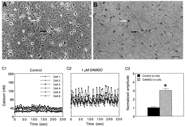
Hippocampal neurons were studied in cultures ranging in age from 6 to 37 d in vitro (DIV). The neurons were identified by morphological criteria developed from immunohistochemical studies by using an antibody to MAP-2, a cytoskeletal protein found in neurons. Immunostaining with an antibody to the transmitter glutamate or GABA showed that both glutamate-containing (data not shown) and GABA-containing neurons (Fig.1A,B) were present in the cultures. The neurons formed extensive processes and synaptic connections in culture and exhibited physiological properties reflective of hippocampal neurons in vivo, including network synaptic activity (see below).
Fig. 1.
Effect of DAMGO on baseline intracellular Ca2+ oscillations in cultured hippocampal neurons. Shown are phase contrast (A) and bright-field (B) micrographs of hippocampal neurons in cultures immunostained with an antibody to the neurotransmitter GABA. The cultures contain a variety of neuronal types based on morphological characteristics and immunostaining for GABA, glutamate, and peptides (e.g., somatostatin; D. Gruol, unpublished data). The neurons grow on a background of astrocytes, identified by immunostaining with an antibody to GFAP. The white arrow points to a neuron immunostained with an antibody for GABA; the dark arrowpoints to a neuron that did not immunostain with the antibody. Calibration bar, 60 μm. C1, C2, Representative recordings of intracellular Ca2+ oscillations in six cultured hippocampal neurons in a microscopic field (different field than in A) under baseline control conditions (C1) and after the addition of DAMGO to the bath saline (C2). Each neuron is represented by a different symbol. Small intracellular Ca2+ oscillations were observed under baseline conditions, and these oscillations were enhanced dramatically in amplitude by the addition of DAMGO to the bath saline. DAMGO also synchronized the oscillations among the neurons in the field. C3, Mean values ± SEM for the amplitude of the intracellular Ca2+ oscillations (peak to trough measurement) in a population of control and DAMGO-treated neurons in the same culture dish as the recordings shown in C1 andC2. *Significant difference from control (p < 0.05; ANOVA). In these and all other experiments the Mg2+ concentration of the bath saline was 30 μm, and the bath saline contained glycine (5 μm).
Intracellular Ca2+ levels were measured in the somatic region of the cultured hippocampal neurons under control conditions and after the addition of the opioid receptor agonist DAMGO to the bath saline. Under control conditions the spontaneous intracellular Ca2+ oscillations of varying amplitude were observed in the majority of neurons that were studied. The Ca2+ oscillations often were synchronized among several neurons in a microscopic field, suggesting that network synaptic activity played a role in the generation of the oscillations. Bath application of TTX, a treatment that blocks synaptic transmission in the hippocampal cultures, blocked the intracellular Ca2+ oscillations (data not shown), consistent with a dependence of the oscillations on network synaptic activity.
When applied to the bath saline at a standard test dose of 1 μm, DAMGO significantly increased the amplitude of the baseline Ca2+ oscillations (Fig.1C; >10 culture sets tested). In addition, DAMGO further synchronized the Ca2+ oscillations among the neurons in a microscopic field (Fig. 1C). However, DAMGO did not induce intracellular Ca2+oscillations in cultures pretreated with TTX.
The enhancement of the intracellular Ca2+oscillations by DAMGO was reversed by the addition of the opioid receptor antagonist naloxone (1 μm) to the bath saline (five culture sets tested; Fig. 2). Moreover, the addition of naloxone to the bath before DAMGO blocked the enhancement of the spontaneous Ca2+oscillations by DAMGO (Fig. 2B). Naloxone alone had no effect on baseline Ca2+ oscillations (Fig. 2B). These results show that opioids can modulate intracellular Ca2+ oscillations in hippocampal neurons and that opioid receptors mediate these effects.
Fig. 2.
The effect of DAMGO was blocked by the opioid receptor antagonist naloxone. A, Representative recordings of Ca2+ oscillations in cultured hippocampal neurons under baseline control conditions, in the presence of DAMGO, and after the addition of naloxone to cultures treated with DAMGO. The recordings are from three different microscopic fields; five neurons are shown for each condition. Naloxone reversed the enhancing effects of DAMGO on the Ca2+ oscillations.B, Mean ± SEM normalized amplitude of Ca2+ oscillations for control, DAMGO alone, naloxone alone, and DAMGO plus naloxone (Nal/DAMGO) conditions. In other experiments, naloxone before DAMGO blocked the effects of DAMGO (data not shown). Oscillations were measured as in Figure 1. Results represent the data from three experiments. *Significant difference from control (p < 0.05; ANOVA).
Involvement of glutamate receptors in the regulation of intracellular Ca2+ oscillations by DAMGO
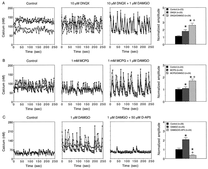
Baseline Ca2+ oscillations and the effect of DAMGO on the oscillations were blocked by TTX, consistent with an involvement of network synaptic activity in these functions. Network synaptic activity in the hippocampal cultures involves excitatory synaptic transmission mediated by glutamate receptors. Therefore, it was of interest to determine whether the enhancement of intracellular Ca2+ oscillations by DAMGO was dependent on one or more of the glutamate receptor subtypes known to be expressed by hippocampal neurons. Bath application of DNQX (10 μm; five culture sets tested) or NBQX (10 μm; one culture set tested), antagonists at the AMPA subtype of glutamate receptor (AMPAR), increased the amplitude of baseline intracellular Ca2+ oscillations. These results suggest that AMPARs are not required for the generation of the intracellular Ca2+ oscillations but can modulate the oscillations. Presumably, AMPAR antagonists block activation of inhibitory GABAergic interneurons, resulting in disinhibition of the network synaptic activity. In the presence of an AMPAR antagonist, DAMGO still elicited prominent alterations in baseline intracellular Ca2+ oscillations (Fig. 3A).
Fig. 3.
Effects of glutamate receptor antagonists on the enhancement of intracellular Ca2+ oscillations by DAMGO. A, B, Representative recordings of intracellular Ca2+ oscillations in cultured hippocampal neurons under baseline control conditions, after the addition of the AMPAR antagonist DNQX (A) or the mGluR antagonist MCPG (B) to the bath saline, and after the subsequent addition of DAMGO (antagonist present). Mean values ± SEM for the amplitude of the intracellular Ca2+ oscillations (peak to trough measurement) in a population of neurons studied under the same conditions as in the representative recordings are shown at the right. Blocking AMPARs enhanced the intracellular Ca2+oscillations but did not block the effects of DAMGO. Blocking mGluRs had only minor effects on baseline intracellular Ca2+ oscillations and did not block the effects of DAMGO. C, Studies with the NMDAR antagonistd-AP5. Shown are representative recordings of intracellular Ca2+ oscillations in cultured hippocampal neurons under baseline control conditions, after the addition of DAMGO to the bath saline, and after the subsequent addition of d-AP5. Blocking NMDARs reversed the effects of DAMGO on intracellular Ca2+ oscillations. d-AP5 also reduced baseline oscillations. In other experiments the addition ofd-AP5 before DAMGO reduced baseline oscillations and blocked enhancement of the intracellular Ca2+oscillations by DAMGO (data not shown). Mean values ± SEM for the amplitude of the intracellular Ca2+ oscillations (peak to trough measurement) in a population of neurons studied under the same conditions as the representative recordings are shown at theright. For all three antagonists the representative recordings are from different microscopic fields in the same culture dish; five neurons are shown for each condition. In the graphs of mean values the amplitude of the oscillations in the various treatment groups was normalized to the mean amplitude of oscillations under the respective baseline control conditions. *Significant difference from control values (p < 0.05; ANOVA). +, Significant difference from DNQX (A), MCPG (B), or DAMGO (C).
The metabotropic glutamate receptor (mGluR) antagonist MCPG (1 mm; four culture sets tested) produced small changes in the amplitude or degree of synchrony of baseline intracellular Ca2+ oscillations but did not block the effects of DAMGO on intracellular Ca2+oscillations (Fig. 3B). In contrast, the NMDAR antagonist d-AP5 (50 μm; four culture sets tested) significantly reduced baseline intracellular Ca2+ oscillations and blocked the enhancement of the Ca2+ oscillations by DAMGO (data not shown). The addition of d-AP5 after DAMGO reversed the enhancement of the Ca2+ oscillations by DAMGO (Fig.3C). Finally, in a related series of studies, neither the GABAA receptor antagonist picrotoxin nor the GABAB receptor antagonist CGP55845A blocked the action of DAMGO to enhance intracellular Ca2+ oscillations (data not shown). Taken together, these results show that NMDAR-mediated responses elicited by network synaptic activity play a prominent role in the generation of baseline intracellular Ca2+ oscillations and in the augmentation of these oscillations by DAMGO, whereas synaptic events mediated by AMPARs, mGluRs, GABAA, or GABAB receptors are not essential for either of these functions.
Involvement of Ca2+ channels in the regulation of intracellular Ca2+ oscillations by DAMGO
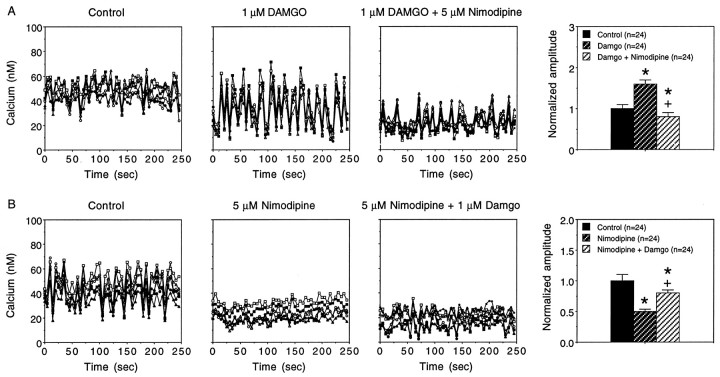
Activation of NMDARs depolarizes hippocampal neurons and, if the depolarization is large enough, can elicit action potentials. Voltage-sensitive Ca2+ channels contribute to the action potentials. Thus, network synaptic activity in the hippocampal cultures could activate voltage-sensitive Ca2+ channels, resulting in Ca2+ influx that contributes to the intracellular Ca2+ oscillations. To determine whether voltage-sensitive Ca2+channels play a role in the intracellular Ca2+ oscillations, we tested the effects of L- and P-type Ca2+ channel blockers on the baseline Ca2+ oscillations and the ability of DAMGO to enhance the oscillations. The P-type Ca2+ channel blocker ω-agatoxin IVA (200 nm; two culture sets tested) almost completely blocked baseline Ca2+ oscillations. Moreover, DAMGO did not induce intracellular Ca2+oscillations in cultures pretreated with ω-agatoxin IVA (data not shown). These results are similar to results obtained with TTX treatment and may reflect a blockade of synaptic transmission via ω-agatoxin IVA-mediated inhibition of presynaptic Ca2+ channels. Nimodipine (5 μm; three culture sets tested), an L-type Ca2+ channel blocker, had variable effects on baseline intracellular Ca2+oscillations. However, nimodipine significantly reduced the enhancement of the intracellular Ca2+ oscillations by DAMGO. Representative data are shown in Figure4. These results indicate that L-type Ca2+ channels contribute to baseline Ca2+ oscillations and play a prominent role in the enhancement of intracellular Ca2+ oscillations by DAMGO.
Fig. 4.
Effect of nimodipine on the modulation of intracellular Ca2+ oscillations by DAMGO.A, Representative recordings of intracellular Ca2+ oscillations under baseline control conditions, in the presence DAMGO, and after the addition of the L-type channel Ca2+ blocker nimodipine to the bath saline. Recordings are from different microscopic fields in the same culture dish; five neurons are shown for each condition. Nimodipine blocked the enhanced intracellular Ca2+ oscillations produced by DAMGO. Mean values ± SEM for the amplitude of the intracellular Ca2+ oscillations (peak to trough measurement) in a population of neurons under the same conditions as the representative recordings (same culture dish as representative recordings) are shown to the right. B, Representative recordings of intracellular Ca2+ oscillations under baseline control conditions, after the addition of the L-type channel Ca2+ blocker nimodipine to the bath saline, and after the subsequent addition of DAMGO to the bath. Recordings are from different microscopic fields in the same culture dish; five neurons are shown for each condition. Nimodipine blocked baseline intracellular Ca2+ oscillations and reduced the enhancement of the oscillations by DAMGO. Mean values ± SEM for the amplitude of the intracellular Ca2+ oscillations (peak to trough measurement) in a population of neurons under the same conditions as the representative recordings (same culture dish as representative recordings) are shown to the right. In the graphs of mean values the amplitudes of the intracellular Ca2+oscillations in the various treatment groups were normalized to the mean amplitude under baseline control conditions. *Significant difference from control values (p > 0.05; ANOVA). +, Significant difference from DAMGO for DAMGO/Nimodipine; Nimodipine for Nimodipine/DAMGO.
Effect of DAMGO on membrane properties and spontaneous network electrical activity
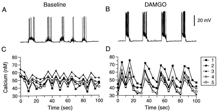
In current-clamp recordings the cultured hippocampal neurons exhibit prominent spontaneous burst events composed of synaptic potentials and action potentials. This activity was blocked by TTX (data not shown). The intracellular Ca2+oscillations also were blocked by TTX, suggesting that the burst events contribute to generation of the intracellular Ca2+ oscillations. Technical limitations prevented recording of the intracellular Ca2+ oscillations and burst events in the same neurons. However, comparison of intracellular Ca2+ oscillations and burst activity confirmed similarities in time course and frequency (Fig.5; see below). Therefore, we used electrophysiological recordings of the burst activity under a variety of conditions to help elucidate the mechanisms responsible for the effects of DAMGO on the intracellular Ca2+oscillations.
Fig. 5.
DAMGO alters spontaneous, synaptically driven burst activity in hippocampal neurons. A,B, Electrophysiological recordings of spontaneous burst activity in a cultured pyramidal-like hippocampal neuron under baseline control conditions and after the addition of DAMGO (1 μm) to the bath saline. The neuron exhibited repetitive burst activity under control conditions. The amplitude of the membrane depolarization during the burst and the number of spikes that were evoked were enhanced in the presence of DAMGO. C, D, For comparison purposes, representative recordings of intracellular Ca2+ oscillations are shown under the same conditions as in electrophysiological studies. The Ca2+ recordings are from in five neurons in a microscopic field. The time scale is the same for the intracellular Ca2+ and electrophysiological recordings. Note the similarity in the pattern of activity of spike bursts and intracellular Ca2+ oscillations in the presence of DAMGO.
All electrophysiological recordings were performed as outlined for the Ca2+ imaging experiments, and the same doses of agonists and antagonists were used. Mean resting membrane potential (RMP) values under the various conditions tested in the electrophysiological recordings are shown in Table1. In general, there was no significant effect of the treatments on RMP. However, the addition of DAMGO to the bath saline significantly enhanced the spontaneous burst events, as evidenced by an increase in amplitude of the burst depolarizations and/or the number of action potentials comprising the burst event (Fig.5, Table 1). The intrinsic excitability of the neurons, as determined by the number of current-evoked spikes, was not altered by DAMGO (Table1). In addition, DAMGO did not significantly alter input resistance (determined from the slope of current–voltage curves) measured at membrane potentials depolarized from rest (Table 1), indicating that alterations in passive membrane properties were unlikely to account for the enhancement of the burst events by DAMGO. DAMGO did decrease input resistance at membrane potentials hyperpolarized to rest (Table 1), perhaps because of the opioid activation of K+ conductances, as has been shown to occur in acutely isolated hippocampal neurons (Wimpey and Chavkin, 1991).
Table 1.
Effect of DAMGO on electrophysiological measurements
| Treatment | Electrophysiological measure | |||||
|---|---|---|---|---|---|---|
| Vm (mV) | Rin(hyperpol) MΩ | Rin (depol) MΩ | Burst amplitude (mV) | Burst frequency (bursts/min) | Spike count1-a | |
| Control | −67 ± 2 (7)b | ND | ND | 15 ± 2 (7) | 4 ± 1 (7) | 8 ± 0 (2) |
| DAMGO | −65 ± 1* (7) | ND | ND | 18 ± 3* (7) | 4 ± 1 (7) | 8 ± 1 (2) |
| NBQX | −64 ± 2 (14) | 658 ± 102 (14) | 305 ± 58 (9) | 16 ± 2 (14) | 3 ± 0 (15) | 8 ± 1 (10) |
| NBQX/DAMGO | −64 ± 2 (14) | 535 ± 80* (14) | 293 ± 51 (9) | 19 ± 2∧ (14) | 3 ± 1 (15) | 8 ± 1 (10) |
| NBQX/Picro | −68 ± 1 (15) | 768 ± 67 (14) | 414 ± 32 (14) | 23 ± 2 (6) | 2.0 ± 1 (6) | ND |
| NBQX/Picro/DAMGO | −67 ± 1 (15) | 699 ± 63* (14) | 392 ± 22 (14) | 32 ± 5* (6) | 2.0 ± 1 (6) | ND |
| NBQX/Picro/TTX | −67 ± 1 (7) | 731 ± 106 (7) | 441 ± 66 (7) | NA | NA | NA |
| NBQX/Picro/TTX/DAMGO | −66 ± 1 (7) | 620 ± 91* (7) | 396 ± 41 (7) | NA | NA | NA |
Spikes elicited by a 50 pA depolarizing current pulse. bNumbers in parentheses are the number of cells tested. *Significantly different from the respective baseline control (i.e., without DAMGO; p < 0.05; paired t test). ∧Trend toward a significant difference from baseline control (0.05 ≤ p < 0.10; paired t test). Picro, Picrotoxin; ND, not determined; NA, not applicable.
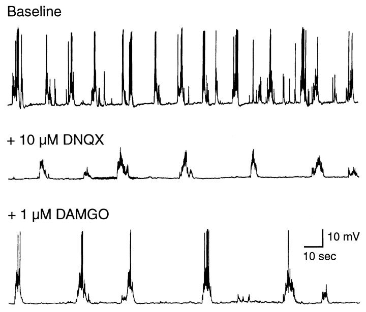
In the Ca2+ imaging studies, NMDARs were found to play a major role in the generation of baseline and DAMGO-induced intracellular Ca2+oscillations, whereas AMPARs were not essential (see Fig. 3). In electrophysiological recordings the AMPAR antagonists NBQX (5 μm) or DNQX (10 μm) produced some alterations in the general form of the spontaneous burst events. However, subsequent exposure to DAMGO still enhanced the spontaneous burst events by increasing the amplitude of the burst depolarization and/or increasing the number of action potentials comprising the burst event (Fig. 6, Table 1). In the presence of an AMPAR antagonist the addition of the NMDAR antagonistd-AP5 to the bath saline abolished the burst potentials observed under baseline conditions or in the presence of DAMGO (data not shown), indicating that NMDARs are essential for the generation of the burst events.
Fig. 6.
AMPARs are not required for the actions of DAMGO. Shown are electrophysiological recordings of spontaneous burst activity in a cultured hippocampal neuron under baseline control conditions after the addition of the AMPAR antagonist DNQX to the bath saline, followed by the subsequent addition of 1 μm DAMGO. DNQX produced some alterations in baseline activity but did not block the ability of DAMGO to enhance the amplitude and spike number of spontaneous burst events.
In addition to excitatory transmission mediated by glutamate receptors, inhibitory input mediated by GABAA receptors (GABAARs) plays an important role in synaptic transmission in the hippocampus. Immunostaining with an antibody to GABA showed that GABA-containing neurons are present in the hippocampal cultures (see Fig. 1A,B), and these neurons could contribute an inhibitory influence to the burst events. Opiates are known to depress inhibitory GABA systems via both μ- and δ-opioid receptors in hippocampal slice preparations (Nicoll et al., 1980;Lupica and Dunwiddie, 1991; Cohen et al., 1992; Capogna et al., 1993;Lupica, 1995). As a consequence, inhibitory synaptic input from the GABA-containing interneurons to the pyramidal neurons is reduced and there is an overall increase in pyramidal neuron excitability [i.e., a disinhibitory mechanism; (Zieglgansberger et al., 1979)]. To determine whether such a disinhibitory mechanism mediated the enhancement of the burst events by DAMGO, we investigated the ability of the GABAAR antagonist picrotoxin to block the enhancement of the burst events by DAMGO.
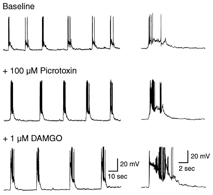
In cultures pretreated with NBQX (5 μm) to block AMPARs and simplify the synaptic network activity, the addition of picrotoxin (100 μm) to the bath saline increased the amplitude of the spontaneous burst events (Fig. 7). However, the subsequent addition of DAMGO further enhanced the amplitude of the burst events (Fig. 7, Table 1). These results indicate that DAMGO can enhance spontaneous NMDAR-mediated burst events in the cultured hippocampal neurons and that this enhancement is not attributable to a disinhibitory mechanism.
Fig. 7.
GABAARs are not required for the actions of DAMGO. Shown are electrophysiological recordings of spontaneous burst activity in a cultured hippocampal neuron under baseline control conditions (5 μm NBQX is present to block AMPARs) and after the addition of the GABAAR antagonist picrotoxin to the bath saline, followed by the subsequent addition of 1 μm DAMGO (NBQX and picrotoxin are still present). In the presence of NBQX and picrotoxin, DAMGO still enhanced the amplitude and spike number of spontaneous burst events.
DAMGO enhances the membrane response and intracellular Ca2+ signals to exogenously applied NMDA
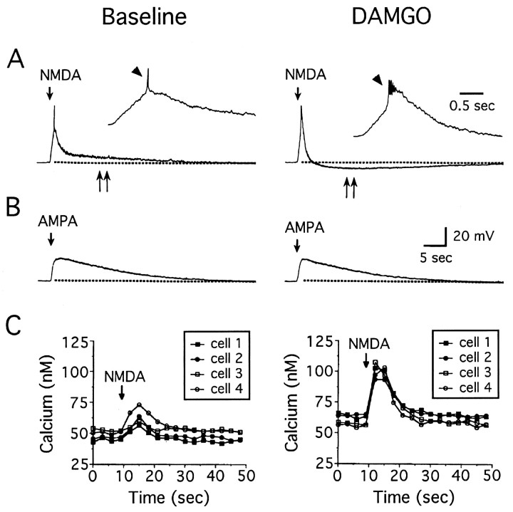
The above results show that DAMGO can enhance spontaneous, synaptically driven burst events and intracellular Ca2+ oscillations in cultured hippocampal neurons and that these effects involve NMDARs. Opiates are known to alter hippocampal synaptic transmission via both pre- and postsynaptic opioid receptors (Simmons and Chavkin, 1996). To determine whether postsynaptic opioid receptors are involved in the effects of DAMGO on the cultured hippocampal neurons, we tested the effect of DAMGO on membrane responses produced by brief microperfusion application of NMDA (10–200 μm; 0.25–1 sec pulse) to the hippocampal neurons. In these studies the bath saline contained TTX (0.5 μm), NBQX (5 μm), and picrotoxin (100 μm) to block Na+ channels, AMPARs, and GABAARs, respectively. Micropressure application of NMDA elicited a prominent membrane depolarization under these conditions (Fig.8A). The addition of DAMGO to the bath saline significantly increased (p < 0.05; paired t test) the amplitude and significantly decreased (p < 0.05; paired t test) the duration of the depolarization to NMDA in 13 of 15 neurons tested (Fig. 8A). Mean values for the peak amplitude of the membrane depolarization to NMDA for the 13 DAMGO-sensitive neurons were 40 ± 2 mV under control conditions and 45 ± 2 mV in the presence of DAMGO. Mean values for the duration of the membrane depolarization to NMDA in the same population of neurons were 55 ± 6 sec under control conditions and 35 ± 6 sec in the presence of DAMGO.
Fig. 8.
DAMGO selectively enhances the membrane depolarization elicited by the exogenous application of NMDA.A, Representative recordings of membrane depolarizations elicited in two hippocampal neurons by brief (1 sec) micropressure application of NMDA or AMPA (at the arrows) under control conditions and after the addition of DAMGO to the bath saline. DAMGO (1 μm) increased the amplitude and decreased the duration of the membrane depolarization to NMDA. DAMGO also induced an increase in Ca2+ spiking (arrowhead) and a prominent AHP (double arrows) after the depolarizing phase of the response to NMDA. The response to AMPA was unaltered by DAMGO. C, In parallel Ca2+ imaging experiments DAMGO enhanced the intracellular Ca2+ signal to NMDA. Graphs show four cells in a control microscopic field and four cells in another microscopic field in the same culture dish after the addition of DAMGO to the recording saline.
In 5 of the 13 DAMGO-sensitive neurons, DAMGO altered the recovery phase of the depolarization to DAMGO by revealing (Fig.8A) or enhancing an afterhyperpolarization (AHP) that followed the depolarizing phase of the membrane response to NMDA. Measurement of the AHP in these neurons showed that the amplitude and the duration of AHP were increased significantly by DAMGO. The mean amplitude of the AHP was 0.4 ± 0.4 mV under control conditions as compared with 3.8 ± 1.2 mV after the addition of DAMGO to the bath saline. The mean duration of the AHP was 29 ± 29 sec under control conditions as compared with 102 ± 28 sec after the addition of DAMGO to the bath saline.
In parallel Ca2+ imaging studies DAMGO significantly enhanced the intracellular Ca2+ signal elicited by the exogenous application of NMDA to the hippocampal neurons (Fig. 8C), consistent with the larger membrane response observed in the electrophysiological studies (Fig. 8A). Mean values for the peak amplitude of the intracellular Ca2+ signal to NMDA (resting levels subtracted) were 32 ± 1 nm(n = 184) under control conditions and 40 ± 1 nm (n = 235) in the presence of DAMGO (p < 0.05; unpaired t test), an ∼28% increase. DAMGO also produced a small but significant increase in resting Ca2+ levels in these studies. The mean resting Ca2+ level was 45 ± 1 nm (n = 184) under control conditions and 52 ± 1 nm(n = 235) in the presence of DAMGO (p < 0.05; unpaired t test), an ∼13% increase.
DAMGO does not alter the membrane response to exogenously applied AMPA
In addition to NMDARs, AMPARs are involved in glutamate-mediated synaptic transmission in the hippocampal cultures. Thus, it was of interest to determine whether the effects of DAMGO were selective for membrane depolarizations mediated by NMDARs or whether membrane depolarizations mediated by AMPARs could be affected as well. For these studies the bath saline contained TTX (0.5 μm),d-AP5 (100 μm), and picrotoxin (100 μm) to block Na+ channels, NMDARs, and GABAARs, respectively. Brief microperfusion of AMPA (1–2 μm; 1 sec pulse; applied as for NMDA) elicited a membrane depolarization in all seven cultured hippocampal neurons that were tested (Fig. 8B). The addition of DAMGO to the bath saline had no effect on the membrane depolarization to AMPA (Fig. 8B); mean values for the peak amplitude and duration of the response to AMPA were 24 ± 4 mV and 68 ± 12 sec, respectively, under both conditions (p > 0.05; paired t test).
DAMGO enhances Ca2+ spiking
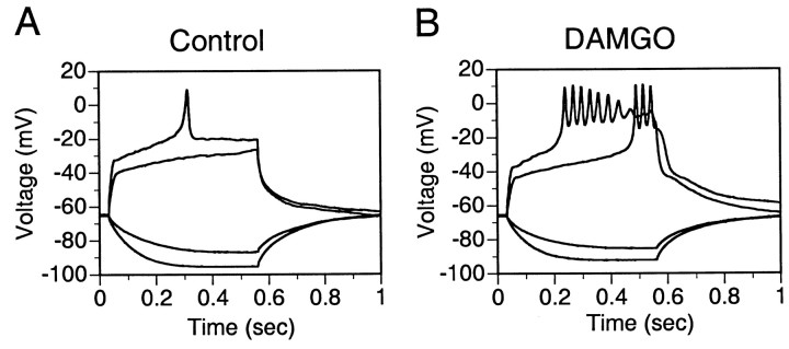
Large membrane depolarizations in response to NMDA application elicited Ca2+-dependent spikes in the cultured hippocampal neurons (TTX, NBQX, and picrotoxin present in the recording medium; Fig. 8A). The Ca2+ spiking was more pronounced after the addition of DAMGO to the recording medium (Fig. 8A), possibly because of the enhancement of the depolarization to NMDA by DAMGO. Alternatively, DAMGO could act to enhance Ca2+ spiking by affecting either Ca2+ or K+currents directly. To test this possibility, we examined the effect of DAMGO on Ca2+ spiking elicited by depolarizing current pulses. In 6 of 11 neurons that showed Ca2+ spiking under baseline conditions, DAMGO increased the number or amplitude of the Ca2+ spikes (Fig.9). In the remaining five neurons the Ca2+ spikes were unaltered or slightly depressed by DAMGO. Nimodipine (1–5 μm) reduced the Ca2+ spikes in six of six neurons that were tested (data not shown), suggesting that L-type Ca2+ channels contributed to the Ca2+ spiking. Nimodipine did not alter the enhancement of the membrane depolarization to NMDA by DAMGO, indicating that the actions of DAMGO on network synaptic activity involve multiple mechanisms. The mean values for the peak amplitude of the membrane depolarization to NMDA in five neurons tested were 34 ± 3 mV under control conditions, 40 ± 4 mV in the presence of DAMGO, and 43 ± 2 after the addition of nimodipine to the recording saline (DAMGO still present).
Fig. 9.
DAMGO enhances Ca2+ spiking.A, B, Voltage responses elicited by intracellular application of two depolarizing (+150 and +250 pA) and two hyperpolarizing (−30 and −50 pA) current pulses in a hippocampal neuron under control conditions and after the addition of DAMGO to the bath saline. TTX, NBQX, and picrotoxin were present in the bath saline. DAMGO increased the Ca2+ spiking elicited by the depolarizing current pulses.
DISCUSSION
In this study we identified a novel opioid mechanism in cultured hippocampal neurons that may play a role in opioid regulation of neuronal activity in synaptic pathways in situ. The opioid receptor agonist DAMGO significantly increased the amplitude of TTX-sensitive intracellular Ca2+oscillations in the cultured hippocampal neurons, with synchronization of the Ca2+ oscillations across neurons in a given field. These actions of DAMGO were blocked by the opioid receptor antagonist naloxone, indicating that opioid receptors, presumably μ-opioid receptors, mediated the opioid effects. Spontaneous TTX-sensitive burst depolarizations also were shown to be enhanced by DAMGO in the cultured hippocampal neurons and to be likely initiators of the Ca2+ oscillations. Studies with selective glutamate receptor antagonists revealed that functional NMDARs were required for the enhancement of intracellular Ca2+ oscillations and burst events by DAMGO, whereas AMPARs and mGluRs were not essential. Further studies showed that intracellular Ca2+ signals and depolarizing responses evoked by exogenously applied NMDA were augmented by DAMGO, supporting a link between opioid receptors and NMDARs in the hippocampal neurons. The NMDARs presumably provide a major pathway for the Ca2+ influx that contributes to the intracellular Ca2+oscillations. In addition, L-type Ca2+channels were identified as important contributors to the enhancement of the Ca2+ oscillations by DAMGO, and their involvement appears to result from an enhancement of Ca2+-dependent spiking by DAMGO. Taken together, these results show that the activation of opioid receptors can influence several components of neuronal Ca2+ signaling pathways in cultured hippocampal neurons and, as a consequence, enhance intracellular Ca2+ levels.
Several lines of evidence indicate that the spontaneous intracellular Ca2+ oscillations and burst depolarizations observed under baseline conditions (low Mg2+) and in the presence of DAMGO were generated by network synaptic activity. The intracellular Ca2+ oscillations and burst depolarizations showed a similar pattern of activity, sensitivity to TTX (which blocks synaptic transmission), and sensitivity to transmitter receptor antagonists. These observations are consistent with studies of cultured cortical neurons (Rose et al., 1990; Murphy et al., 1992; Robinson et al., 1993) and cultured hippocampal neurons (Shen et al., 1996), in which spontaneous periodic intracellular Ca2+ oscillations were shown to correlate with spontaneous synaptic potentials and to be blocked by TTX. In studies of cultured cortical neurons Rose et al. (1990) found that the spontaneous intracellular Ca2+oscillations could be blocked by the NMDAR antagonistd-AP5, as in the current study. In contrast, Shen et al. (1996) reported that spontaneous intracellular Ca2+ oscillations in cultured hippocampal neurons were blocked by the AMPA/kainate receptor antagonist CNQX, whereas d-AP5 caused only a partial depression. These combined results show that intracellular Ca2+ oscillations in CNS neurons can be mediated by NMDA and/or non-NMDA subtypes of glutamatergic receptors. Similar conclusions were reached in studies of cultured cortical neurons by Murphy et al. (1992) and Robinson et al. (1993). Several factors are likely to play a role in determining the relative contribution of NMDARs and non-NMDARs to the intracellular Ca2+ oscillations and associated synaptic events in the cultured neurons, such as the neuronal composition of the cultures, the circuitry established, and the experimental conditions (e.g., Mg2+ level in the bath saline).
DAMGO increased the amplitude of the intracellular Ca2+ oscillations in the cultured hippocampal neurons. DAMGO also enhanced spontaneous burst events, consistent with a role of the burst events in the generation of the intracellular Ca2+ oscillations. The enhancement of burst events by DAMGO could arise from an increase in excitatory synaptic transmission or a decrease in inhibitory synaptic transmission (i.e., a disinhibitory mechanism). Blockade of GABAAR-mediated responses with picrotoxin enhanced the amplitude of the spontaneous burst events in the hippocampal neurons, showing that GABA-containing neurons provided an inhibitory component to the network synaptic activity. However, treatment with picrotoxin did not prevent the augmentation of the burst events by DAMGO. Thus, an opioid disinhibitory mechanism does not appear to explain our results.
Although GABAARs were not required for the enhancement of the spontaneous Ca2+oscillations and burst events by DAMGO in the cultured hippocampal neurons, our results show that glutamate and NMDARs play a critical role. The near-total blockade of opioid-induced intracellular Ca2+ oscillations by the NMDAR antagonistd-AP5, an action that mimicked the effects of TTX, suggests that NMDARs presynaptic (i.e., “upstream”) to the imaged neurons could be involved in the enhanced intracellular Ca2+ oscillations and burst events. Alternatively, the opioid enhancement of the Ca2+ oscillations could arise from potentiation of postsynaptic currents mediated by NMDARs, as described by Chen and Huang (1991) and Martin et al. (1997). This possibility is supported by our finding of DAMGO potentiation of membrane depolarizations elicited by exogenous application of NMDA (but not AMPA) to the hippocampal neurons. The potentiation of NMDA responses by opioid receptor agonists in spinal neurons appears to be mediated by the activation of protein kinase C (PKC; Chen and Huang, 1991). A similar PKC-dependent effect could be involved in the present findings.
In the current study the P-type Ca2+channel antagonist ω-agatoxin IVA blocked the spontaneous and opioid-induced intracellular Ca2+oscillations in the cultured hippocampal neurons. P-type Ca2+ channels are implicated in transmitter release (Luebke et al., 1993), supporting a role for synaptic transmission in the generation of the Ca2+ oscillations. Moreover, the synchronization of the Ca2+ oscillations across neurons in a microscopic field strongly supports the participation of presynaptic elements, probably via the network or feedback interconnectivity functions of the cultures. Opioid regulation of N- and P/Q-types of Ca2+ channels has been demonstrated in native neurons (Gross and MacDonald, 1987;Schroeder and McCleskey, 1993; Moises et al., 1994) and has been thought to contribute to opioid depression of synaptic transmission (North and Lovinger, 1993). Although an inhibition of synaptic responses by DAMGO was not observed in the current study, opioid actions at N- or P/Q-type Ca2+ channels could contribute in a covert way to the effects of DAMGO on network synaptic activity.
In addition to the regulation of N and P/Q Ca2+ channels, opioid receptors have been shown to regulate L-type Ca2+ channels. Thus, opioids increase intracellular Ca2+by inducing Ca2+ influx via dihydropyridine-sensitive (i.e., L-type) Ca2+ channels in neuronal-like cell lines that express opioid receptors (Jin et al., 1992; Tang et al., 1994). In the current study the L-type Ca2+ channel blocker nimodipine reduced the enhancement of intracellular Ca2+ oscillations by DAMGO with variable effects on baseline Ca2+ oscillations. This result suggests that postsynaptic L-type Ca2+ channels are involved in the DAMGO enhancement of the intracellular Ca2+oscillations, because L-type Ca2+ channels are reported to be localized predominantly postsynaptically in hippocampal neurons (Westenbroek et al., 1990). A role for postsynaptic Ca2+ channels is supported further by our finding that DAMGO enhanced the Ca2+-dependent spikes evoked by intracellular depolarizing current injections, and these spikes were blocked by the L-type Ca2+ channel blocker nimodipine.
Recent studies in neuron-like cell lines and transfected cells have shown that opioid receptors are coupled to various signaling pathways involved in Ca2+ homeostasis, including inhibition (Jin et al., 1992, 1993; Fields et al., 1995) or activation (Jin et al., 1992; Tang et al., 1994) of Ca2+ channels, mobilization of Ca2+ from intracellular stores (Tomura et al., 1992; Jin et al., 1994; Fields et al., 1995; Connor et al., 1997), and activation of phospholipase C (Okajima et al., 1993; Johnson et al., 1994), the enzyme responsible for generation of the Ca2+ mobilizing messenger inositol 1,4,5-trisphosphate (IP3). In the current studies the potential involvement of intracellular Ca2+ stores in the enhancement of the intracellular Ca2+ oscillations by DAMGO was not evaluated. However, in the presence of GABAAR and glutamate receptor antagonists, DAMGO elicited a small increase in baseline Ca2+levels, an effect that could involve the release of Ca2+ from intracellular stores. Actions of DAMGO on other Ca2+-sensitive pathways or components such as membrane pumps or transport systems cannot be eliminated at this time. Membrane potential changes are unlikely to mediate the effects on baseline Ca2+levels, because DAMGO did not alter resting membrane potential when synaptic transmission was blocked by GABAAR and glutamate receptor antagonists.
The dual action of opioids in augmenting responses mediated by NMDARs and L-type Ca2+ channels provides a novel mechanism through which opioids can control the excitability of CNS neurons. NMDARs and L-type Ca2+ channels often are activated simultaneously by synaptic signals and could interact synergistically to influence excitatory drive and regulate biochemical pathways controlled by intracellular Ca2+ concentrations. Moreover, Ca2+ oscillations are known to be a particularly effective regulatory mechanism for the control of gene expression (Dolmetsch et al., 1998; Li et al., 1998). By virtue of the large number of cellular processes regulated by Ca2+, increased intracellular Ca2+ or enhanced Ca2+ oscillatory activity could play a major role in the opioid regulation of CNS neuronal function. Such second messenger regulation could account for some of the long-term effects of opioids such as opioid effects on synaptic plasticity and genomic regulation (Nestler and Aghajanian, 1997).
Footnotes
This work was supported by National Institutes of Health Grants AA06420 (D.L.G.), DA03665 (G.R.S.), and Fullbright Fellowship 18619 (R.P.). We thank Jody Caguioa for technical assistance and performing some of the experiments, Floriska Chizer for secretarial assistance, Pfizer for the gift of ω-agatoxin IVA, and Novartis for the gift of CGP55845A.
Correspondence should be addressed to Dr. Donna Gruol, Department of Neuropharmacology, CVN 11, The Scripps Research Institute, 10550 North Torrey Pines Road, La Jolla, CA 92037.
Dr. Przewlocki's present address: Department of Molecular Neuropharmacology, Institute of Pharmacology, SMETNA 12, 31-343 Krakow, Poland.
REFERENCES
- 1.Baraban SC, Lothman EW, Lee A, Guyenet PG. Kappa opioid receptor-mediated suppression of voltage-activated potassium current in a catecholamine neuronal cell line. J Pharmacol Exp Ther. 1995;273:927–933. [PubMed] [Google Scholar]
- 2.Capogna M, Gahwiler BH, Thompson SM. Mechanism of μ-opioid receptor-mediated presynaptic inhibition in the rat hippocampus in vitro. J Physiol (Lond) 1993;470:539–558. doi: 10.1113/jphysiol.1993.sp019874. [DOI] [PMC free article] [PubMed] [Google Scholar]
- 3.Chen L, Huang LY. Sustained potentiation of NMDA receptor-mediated glutamate responses through activation of protein kinase C by a μ-opioid. Neuron. 1991;7:319–326. doi: 10.1016/0896-6273(91)90270-a. [DOI] [PubMed] [Google Scholar]
- 4.Childers SR. Opioid receptor-coupled second messenger systems. Handbook Exp Pharmacol. 1993;104:189–206. [Google Scholar]
- 5.Cohen G, Doze V, Madison D. Opioid inhibition of GABA release from presynaptic terminals of rat hippocampal interneurons. Neuron. 1992;9:325–335. doi: 10.1016/0896-6273(92)90171-9. [DOI] [PubMed] [Google Scholar]
- 6.Connor MA, Keir MJ, Henderson G. δ-Opioid receptor mobilization of intracellular calcium in SH-SY5Y cells: lack of evidence for δ-receptor subtypes. Neuropharmacology. 1997;36:125–133. doi: 10.1016/s0028-3908(96)00144-x. [DOI] [PubMed] [Google Scholar]
- 7.Dolmetsch RE, Xu K, Lewis RS. Calcium oscillations increase the efficiency and specificity of gene expression. Nature. 1998;392:933–936. doi: 10.1038/31960. [DOI] [PubMed] [Google Scholar]
- 8.Fields A, Gafni M, Oron Y, Sarne Y. Multiple effects of opiates on intracellular calcium level and on calcium uptake in three neuronal cell lines. Brain Res. 1995;687:94–102. doi: 10.1016/0006-8993(95)00475-6. [DOI] [PubMed] [Google Scholar]
- 9.Ghosh A, Greenberg ME. Calcium signaling in neurons: molecular mechanisms and cellular consequences. Science. 1998;268:239–247. doi: 10.1126/science.7716515. [DOI] [PubMed] [Google Scholar]
- 10.Gross RA, MacDonald RL. Dynorphin A selectively reduces a large transient (N-type) calcium current of mouse dorsal root ganglion neurons in cell culture. Proc Natl Acad Sci USA. 1987;84:5469–5473. doi: 10.1073/pnas.84.15.5469. [DOI] [PMC free article] [PubMed] [Google Scholar]
- 11.Gruol DL. Cultured cerebellar neurons: endogenous and exogenous components of Purkinje cell activity and membrane response to putative transmitters. Brain Res. 1983;263:223–241. doi: 10.1016/0006-8993(83)90315-3. [DOI] [PubMed] [Google Scholar]
- 12.Gruol DL, Crimi CP. Morphological and physiological properties of rat cerebellar neurons in mature and developing cultures. Dev Brain Res. 1988;41:135–145. doi: 10.1016/0165-3806(88)90177-0. [DOI] [PubMed] [Google Scholar]
- 13.Jin W, Lee NM, Loh HH, Thayer SA. Dual excitatory and inhibitory effects of opioids on intracellular calcium in neuroblastoma × glioma hybrid NG108-15 cells. Mol Pharmacol. 1992;42:1083–1089. [PubMed] [Google Scholar]
- 14.Jin W, Lee NM, Loh HH, Thayer SA. Opioid-induced inhibition of voltage-gated calcium channels parallels expression of ω-conotoxin-sensitive channel subtype during differentiation of NG108-15 cells. Brain Res. 1993;607:17–22. doi: 10.1016/0006-8993(93)91484-a. [DOI] [PubMed] [Google Scholar]
- 15.Jin W, Lee NM, Loh HH, Thayer SA. Opioids mobilize calcium from inositol 1,4,5-trisphosphate-sensitive stores in NG108-15 cells. J Neurosci. 1994;14:1920–1929. doi: 10.1523/JNEUROSCI.14-04-01920.1994. [DOI] [PMC free article] [PubMed] [Google Scholar]
- 16.Johnson PS, Wang JB, Wang WF, Uhl GR. Expressed μ-opiate receptor couples to adenylate cyclase and phosphatidyl inositol turnover. NeuroReport. 1994;5:507–509. doi: 10.1097/00001756-199401120-00035. [DOI] [PubMed] [Google Scholar]
- 17.Kaneko S, Nakamura S, Adachi K, Akaike A, Satoh M. Mobilization of intracellular Ca2+ and stimulation of cyclic AMP production by κ-opioid receptors expressed in Xenopus oocytes. Mol Brain Res. 1994;27:258–264. doi: 10.1016/0169-328x(94)90008-6. [DOI] [PubMed] [Google Scholar]
- 18.Li W-H, Llopis J, Whitney M, Zlokarnik G, Tsien RY. Cell permeant caged InsP3 ester shows that Ca2+ spike frequency can optimize gene expression. Nature. 1998;392:936–941. doi: 10.1038/31965. [DOI] [PubMed] [Google Scholar]
- 19.Luebke J, Dunlap K, Turner T. Multiple calcium channel types control glutamatergic synaptic transmission in the hippocampus. Neuron. 1993;11:895–902. doi: 10.1016/0896-6273(93)90119-c. [DOI] [PubMed] [Google Scholar]
- 20.Lupica C. δ and μ enkephalins inhibit spontaneous GABA-mediated IPSCs via a cyclic AMP-independent mechanism in the rat hippocampus. J Neurosci. 1995;15:737–749. doi: 10.1523/JNEUROSCI.15-01-00737.1995. [DOI] [PMC free article] [PubMed] [Google Scholar]
- 21.Lupica CR, Dunwiddie TV. Differential effects of mu and delta receptor-selective opioid agonists on feedforward and feedback GABAergic inhibition in hippocampal brain slices. Synapse. 1991;8:237–248. doi: 10.1002/syn.890080402. [DOI] [PubMed] [Google Scholar]
- 22.Madison DV, Nicoll RA. Enkephalin hyperpolarizes interneurons in the rat hippocampus. J Physiol (Lond) 1988;398:123–130. doi: 10.1113/jphysiol.1988.sp017033. [DOI] [PMC free article] [PubMed] [Google Scholar]
- 23.Martin G, Nie Z, Siggins GR. μ-Opioid receptors modulate NMDA receptor-mediated responses in nucleus accumbens neurons. J Neurosci. 1997;17:11–22. doi: 10.1523/JNEUROSCI.17-01-00011.1997. [DOI] [PMC free article] [PubMed] [Google Scholar]
- 24.Moises HC, Rusin KI, MacDonald RL. Mu- and kappa-opioid receptors selectively reduce the same transient components of high-threshold calcium current in rat dorsal root ganglion sensory neurons. J Neurosci. 1994;14:5903–5916. doi: 10.1523/JNEUROSCI.14-10-05903.1994. [DOI] [PMC free article] [PubMed] [Google Scholar]
- 25.Moore SD, Madamba SG, Schweitzer P, Siggins GR. Voltage-dependent effects of opioid peptides on hippocampal CA3 pyramidal neurons in vitro. J Neurosci. 1994;14:809–820. doi: 10.1523/JNEUROSCI.14-02-00809.1994. [DOI] [PMC free article] [PubMed] [Google Scholar]
- 26.Murphy TH, Blatter LA, Wier WG, Baraban JM. Spontaneous synchronous synaptic calcium transients in cultured cortical neurons. J Neurosci. 1992;12:4834–4845. doi: 10.1523/JNEUROSCI.12-12-04834.1992. [DOI] [PMC free article] [PubMed] [Google Scholar]
- 27.Nestler EJ, Aghajanian GK. Molecular and cellular basis of addiction. Science. 1997;278:58–63. doi: 10.1126/science.278.5335.58. [DOI] [PubMed] [Google Scholar]
- 28.Nicoll RA, Alger BE, Jahr CE. Enkephalin blocks inhibitory pathways in the vertebrate CNS. Nature. 1980;287:22–25. doi: 10.1038/287022a0. [DOI] [PubMed] [Google Scholar]
- 29.North RA. Opioid actions on membrane ion channels. Handbook Exp Pharmacol. 1993;104:773–796. [Google Scholar]
- 30.North R, Lovinger D. Presynaptic actions of opioids. In: Dunwiddie T, editor. Presynaptic receptors in the mammalian brain. Birkenhauser; Boston: 1993. pp. 71–76. [Google Scholar]
- 31.North RA, Williams JT, Surprenant A, Christie MJ. Mu and delta receptors belong to a family of receptors that are coupled to potassium channels. Proc Natl Acad Sci USA. 1987;84:5487–5491. doi: 10.1073/pnas.84.15.5487. [DOI] [PMC free article] [PubMed] [Google Scholar]
- 32.Okajima F, Tomura H, Kondo Y. Enkephalin activates the phospholipase C/Ca2+ system through cross-talk between opioid receptors and P2-purinergic or bradykinin receptors in NG 108-15 cells. A permissive role for pertussis toxin-sensitive G-proteins. Biochem J. 1993;290:241–247. doi: 10.1042/bj2900241. [DOI] [PMC free article] [PubMed] [Google Scholar]
- 33.Petersen OH, Kasai H, editors. Seminars in the neurosciences: neuronal calcium signaling, pp 259–334. Academic; New York: 1996. [Google Scholar]
- 34.Qiu Z, Parsons KL, Gruol DL. Interleukin-6 selectively enhances the intracellular calcium response to NMDA in developing CNS neurons. J Neurosci. 1995;15:6688–6699. doi: 10.1523/JNEUROSCI.15-10-06688.1995. [DOI] [PMC free article] [PubMed] [Google Scholar]
- 35.Robinson HPC, Kawahara M, Jimbo Y, Torimitsu K, Kuroda Y, Kawana A. Periodic synchronized bursting and intracellular calcium transients elicited by low magnesium in cultured cortical neurons. J Neurophysiol. 1993;70:1606–1616. doi: 10.1152/jn.1993.70.4.1606. [DOI] [PubMed] [Google Scholar]
- 36.Rose K, Christine CW, Choi DW. Magnesium removal induced paroxysmal neuronal firing and NMDA receptor-mediated neuronal degeneration in cortical cultures. Neurosci Lett. 1990;115:313–317. doi: 10.1016/0304-3940(90)90474-n. [DOI] [PubMed] [Google Scholar]
- 37.Schroeder J, McCleskey E. Inhibition of Ca2+ currents by a μ-opioid in a defined subset of rat sensory neurons. J Neurosci. 1993;13:867–873. doi: 10.1523/JNEUROSCI.13-02-00867.1993. [DOI] [PMC free article] [PubMed] [Google Scholar]
- 38.Seward E, Hammond C, Henderson G. μ-Opioid receptor-mediated inhibition of the N-type calcium channel current. Proc R Soc Lond [Biol] 1991;244:129–135. doi: 10.1098/rspb.1991.0061. [DOI] [PubMed] [Google Scholar]
- 39.Shen K-F, Crain SM. Dynorphin prolongs the action potential of mouse sensory ganglion neurons by decreasing a potassium conductance whereas another specific kappa opioid does so by increasing a calcium conductance. Neuropharmacology. 1990;29:343–349. [PubMed] [Google Scholar]
- 40.Shen M, Piser TM, Seybold VS, Thayer SA. Cannabinoid receptor agonists inhibit glutamatergic synaptic transmission in rat hippocampal cultures. J Neurosci. 1996;16:4322–4334. doi: 10.1523/JNEUROSCI.16-14-04322.1996. [DOI] [PMC free article] [PubMed] [Google Scholar]
- 41.Siesjo BK. Calcium-mediated processes in neuronal degeneration. Ann NY Acad Sci. 1994;747:140–161. doi: 10.1111/j.1749-6632.1994.tb44406.x. [DOI] [PubMed] [Google Scholar]
- 42.Simmons ML, Chavkin C. Endogenous opioid regulation of hippocampal function. Int Rev Neurobiol. 1996;39:145–196. doi: 10.1016/s0074-7742(08)60666-2. [DOI] [PubMed] [Google Scholar]
- 43.Smart D, Smith G, Lambert DG. μ-Opioid receptor stimulation of inositol (1,4,5) trisphosphate formation via a pertussis toxin-sensitive G-protein. J Neurochem. 1994;62:1009–1014. doi: 10.1046/j.1471-4159.1994.62031009.x. [DOI] [PubMed] [Google Scholar]
- 44.Tang T, Kiang JG, Cox BM. Opioids acting through delta receptors elicit a transient increase in the intracellular free calcium concentration in dorsal root ganglion–neuroblastoma hybrid ND8-47 cells. J Pharmacol Exp Ther. 1994;270:40–46. [PubMed] [Google Scholar]
- 45.Tang T, Kiang JG, Cote T, Cox BM. Opioid-induced increase in [Ca2+]i in ND8-47 neuroblastoma × dorsal root ganglion hybrid cells is mediated through G-protein-coupled delta-opioid receptors and sensitized by chronic exposure to opioid. J Neurochem. 1995;65:1612–1621. doi: 10.1046/j.1471-4159.1995.65041612.x. [DOI] [PubMed] [Google Scholar]
- 46.Tomura H, Okajima F, Kondo Y. Enkephalin induces Ca2+ mobilization in single cells of bradykinin-sensitized differentiated neuroblastoma hybridoma (NG108-15) cells. Neurosci Lett. 1992;148:93–96. doi: 10.1016/0304-3940(92)90812-l. [DOI] [PubMed] [Google Scholar]
- 47.Urrutia A, Gruol DL. Acute alcohol alters the excitability of cerebellar Purkinje neurons and hippocampal neurons in culture. Brain Res. 1992;569:26–37. doi: 10.1016/0006-8993(92)90365-g. [DOI] [PubMed] [Google Scholar]
- 48.Wandless AL, Smart D, Lambert DG. Fentanyl increases intracellular Ca2+ concentrations in SH-SY5Y cells. Br J Anaesth. 1996;76:461–463. doi: 10.1093/bja/76.3.461. [DOI] [PubMed] [Google Scholar]
- 49.Westenbroek RE, Ahlijanian MK, Catterall WA. Clustering of L-type Ca2+ channels at the base of major dendrites in hippocampal pyramidal neurons. Nature. 1990;347:281–284. doi: 10.1038/347281a0. [DOI] [PubMed] [Google Scholar]
- 50.Wimpey TL, Chavkin C. Opioids activate both an inward rectifier and a novel voltage-gated potassium conductance in the hippocampal formation. Neuron. 1991;6:281–289. doi: 10.1016/0896-6273(91)90363-5. [DOI] [PubMed] [Google Scholar]
- 51.Zieglgansberger W, French ED, Siggins GR, Bloom FE. Opioid peptides may excite hippocampal pyramidal neurons by inhibiting adjacent inhibitory interneurons. Science. 1979;205:415–417. doi: 10.1126/science.451610. [DOI] [PubMed] [Google Scholar]


