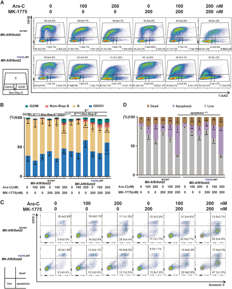
Fig. 6.
Checkpoint inhibition alters the cell cycle and promotes apoptosis in cells treated with chemotherapeutic agents. Mll-Af9 or Mll-Af9/Setd2F2478L/WT AML cells treated with Ara-C, MK-1775, or a combination, as indicated, for 24 h. a Cell cycle phase distributions were determined via BrdU incorporation for 40 min. b The percentage of cells at various stages of the cell cycle (G2/M, S, G0/G1, and non-Rep-S) in (a) was calculated and illustrated. Non-Rep-S represents the non-replicating S phase. c Apoptosis of the cells in quadrant 3 was evaluated by Annexin V/7-AAD staining. d Graphical representation of the percentage of apoptotic cells in (c). The stacked bar graphs indicate the mean percentage of viable (“live”), early apoptotic (“apoptosis”), and late apoptotic (“dead”) cells. Three biological replicates of each genotype are performed in triplicate and the data are presented as the mean ± SD values. *P < 0.05; **P < 0.01; ***P < 0.001; ****P < 0.0001

