Abstract
This case demonstrates extraosseous 99m-technetium methylene diphosphonate (99m-Tc-MDP) accumulation from a gastrointestinal stromal tumor (GIST). A 75-year-old woman underwent a temporal bone CT for conductive hearing loss that showed sclerosis in the right occipital condyle. Follow-up 99m-Tc-MDP bone scan for osseous metastases instead showed a mass-like extraosseous accumulation of 99m-Tc MDP in the anterior left upper quadrant. Differential included gastric cancer, lymphoma, metastatic melanoma, systemic hypercalcemia or heterotopic mesenteric ossification. Contrast CT showed a well-circumscribed mass arising from the stomach and subsequent pathology confirmed GIST. These tumors rarely can contain osteoclast-like giant cells and should be considered for extra-osseous 99m-Tc-MDP accumulation.
Keywords: GIST, extraosseous, MDP
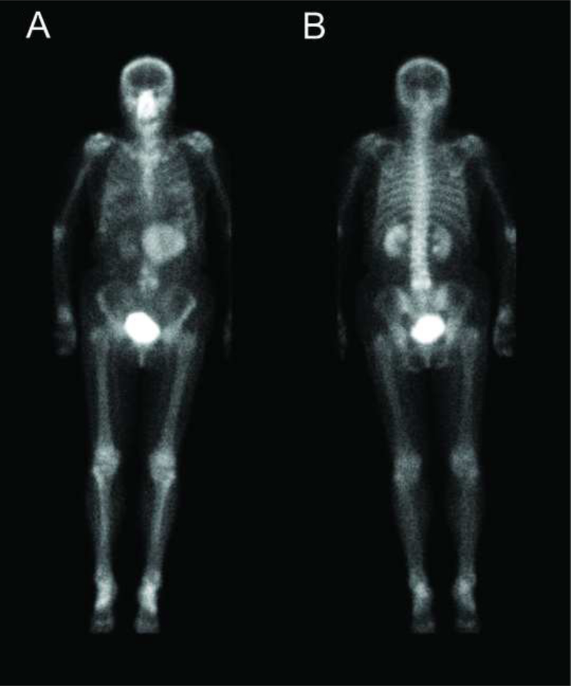
Figure 1 –
Temporal bone CT in a 75-year-old patient for mixed sensorineural and conductive hearing loss demonstrated a nonspecific sclerotic focus in the right occipital condyle. Anterior and posterior planar images from a subsequent whole body 99m-Tc-MDP bone scan were negative for suspicious osseous accumulation of radiotracer, but instead demonstrated a large discrete mass-like accumulation of extra-osseous 99m-Tc-MDP in the anterior left upper quadrant. Differential for this finding in an elderly patient included gastric lymphoma or cancer (1,2), metastatic melanoma (3,4) and heterotopic mesenteric ossification, such as from prior gastric bypass surgery (5,6). Gastric 99m-Tc-MDP uptake also has been observed in sarcoidosis (7) and diseases that cause hypercalcemia (8), including multiple myeloma (9), hyperparathyroidism (10) and vitamin D intoxication (11). Isolated splenic infarction and solitary metastasis from a mucinous ovarian or colon cancer seemed unlikely (1). A contrast-enhanced CT was obtained for further characterization.
Figure 2 –
Axial, sagittal and coronal contrast CT images of the abdomen and pelvis showed a large, well-circumscribed, heterogeneous necrotic mass arising from the greater curvature of the stomach most consistent with a gastrointestinal stromal tumor (GIST) (12). Endoscopic ultrasound-guided biopsy demonstrated spindle cells with strong CD117 and DOG-1 immunoperoxidase staining confirming the diagnosis. Previous literature has described unusual presentations of GIST on PET/CT (13), but this case represents the first example of a GIST initially detected by 99m-Tc-MDP bone scan. Malignant stromal tumors, such as GIST, can rarely contain osteoclast-like giant cells (14) that may explain the observed 99m-Tc-MDP accumulation in this case. This patient is currently undergoing imatinib mesylate therapy prior to surgical resection.
Acknowledgments
Support: TMS supported by NIH/NIBIB T32 EB001631-05
Footnotes
Publisher's Disclaimer: This is a PDF file of an unedited manuscript that has been accepted for publication. As a service to our customers we are providing this early version of the manuscript. The manuscript will undergo copyediting, typesetting, and review of the resulting proof before it is published in its final citable form. Please note that during the production process errors may be discovered which could affect the content, and all legal disclaimers that apply to the journal pertain.
References
- 1.Mettler FA and Guiberteau MJ: Skeletal System. In: Essentials of Nuclear Medicine Imaging. Philadelphia, Saunders: Elsevier, 2006, pp 243–292. [Google Scholar]
- 2.Watanabe N, Haida M, Mikuni I, et al. Bone imaging in advanced gastric cancer. Clin Nucl Med. 1998;23:384- [DOI] [PubMed] [Google Scholar]
- 3.Wheat D, McCarthy P Metastatic pulmonary, gastric, and renal calcification demonstrated on bone scintigraphy in a patient with malignant melanoma and renal failure. Clin Nucl Med. 1998;23:824–827. [DOI] [PubMed] [Google Scholar]
- 4.Garty I, Risescu J, Rosen G, et al. Unusual extraosseous tumoral accumulation of 99mTc-MDP. Eur J Nucl Med. 1985;10:362–365. [DOI] [PubMed] [Google Scholar]
- 5.Wilson JD, Montague CJ, Salcuni P, et al. Heterotopic mesenteric ossification (‘intraabdominal myositis ossificans’): report of five cases. Am J Surg Pathol. 1999;23:1464–1470. [DOI] [PubMed] [Google Scholar]
- 6.Yushuva A, Nagda P, Suzuki K, et al. Heterotopic mesenteric ossification following gastric bypass surgery: case series and review of literature. Obes Surg. 2010;20:1312–1315. [DOI] [PubMed] [Google Scholar]
- 7.Gezici A, van Duijnhoven EM, Bakker SJ, et al. Lung and gastric uptake in bone scintigraphy of sarcoidosis. J Nucl Med. 1996;37:1530–1532. [PubMed] [Google Scholar]
- 8.Delcourt E, Baudoux M, Neve P Tc-99m-MDP bone scanning detection of gastric calcification. Clin Nucl Med. 1980;5:546–547. [DOI] [PubMed] [Google Scholar]
- 9.Reitz MD, Vasinrapee P, Mishkin FS. Myocardial, pulmonary, and gastric uptake of technetium-99m MDP in a patient with multiple myeloma and hypercalcemia. Clin Nucl Med. 1986;11:730- [DOI] [PubMed] [Google Scholar]
- 10.Hwang GJ, Lee JD, Park CY, et al. Reversible extraskeletal uptake of bone scanning in primary hyperparathyroidism. J Nucl Med. 1996;37:469–471. [PubMed] [Google Scholar]
- 11.Corstens F, Kerremans A, Claessens R Resolution of massive technetium-99m methylene diphosphonate uptake in the stomach in vitamin D intoxication. J Nucl Med. 1986;27:219–222. [PubMed] [Google Scholar]
- 12.Burkill GJ, Badran M, Al-Muderis O, et al. Malignant gastrointestinal stromal tumor: distribution, imaging features, and pattern of metastatic spread. Radiology. 2003;226:527–532. [DOI] [PubMed] [Google Scholar]
- 13.Wong C, Chu T, Khong P Unusual features of gastrointestinal stromal tumor on PET/CT and CT. Clinical Nuclear Medicine. 2011;36:e1–e7. [DOI] [PubMed] [Google Scholar]
- 14.Insabato L, Di VD, Ciancia G, et al. Malignant gastrointestinal leiomyosarcoma and gastrointestinal stromal tumor with prominent osteoclast-like giant cells. Arch Pathol Lab Med. 2004;128:440–443. [DOI] [PubMed] [Google Scholar]


