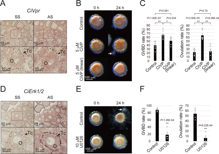
Figure 3. CiVP and CiErk1/2 promote both GVBD and ovulation.
(A) In situ hybridization using sense strand (SS) or antisense strand (AS) DIG-labeled probes and an NBT/BCIP system for visualizing CiVpr indicated its localization in late stage II oocytes (O). Enlarged images of red-boxed areas in the upper panels are shown below. (B) Representative images of follicles before and after treatment with CiVP for 24 hr. GVBD and ovulation of late stage II follicles were enhanced in the presence of 5 μM CiVP (t-value: −4.19; **, p=1.84E-03 in GVBD and t-value: −6.97; **, p=3.85E-05 in ovulation), whereas not in the presence of 5 μM inactive linear CiVP (t-value: −1.94; p=0.081 in GVBD and t-value: −0.27; p=0.79 in ovulation). (C) GVBD and ovulation rates were calculated from six independent experiments (n = 6; approximately 20 follicles per experiment) and found to be significantly upregulated in CiVP-treated follicles, indicating that CiVP promotes both oocyte maturation and ovulation. Data were analyzed by Student’s t test and are shown as mean ± SEM with data points. (D) In situ hybridization for CiErk1/2 indicated its localization in oocytes (O) and test cells (Tc). (E) Spontaneous GVBD and ovulation of early stage III follicles were inhibited in the presence of 10 μM U0126, an MEK inhibitor. (F) GVBD and ovulation rates from four independent experiments (n = 4) indicated that CiErk1/2 regulates both oocyte maturation (t-value: 8.72; **, p=1.26E-04) and ovulation (t-value: 6.52; **, p=6.22E-04). Data were analyzed and are shown as in (C). White arrows (B, E) indicate outer follicular cells remaining alongside the follicle. Scale bars in (A, D) and (B, E) represent 50 and 100 μm, respectively.


