. 2019 Oct 1;8:e49062. doi: 10.7554/eLife.49062

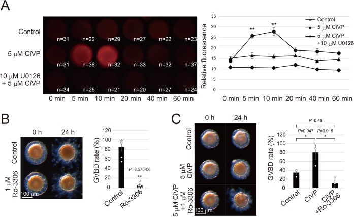
Figure 4. CiVP promotes GVBD via CiErk1/2-CiMPF pathway.

(A) Indicated numbers of numbers of de-folliculated oocytes pretreated with or without 10 μM U0126 were stimulated with 5 μM CiVP for 0, 5, 10, 20, 40, and 60 min. Immunofluorescence using anti-phosphorylated ERK1/2 antibody indicated specific activation of CiErk1/2 at 5 to 10 min after CiVP stimulation (F = 14.91; p=2.12E-12), which was not observed in control (F = 1.05; p=0.39) or U0126-pretreated oocytes (F = 2.13; p=0.065). Representative images are shown (left). Signal intensities were quantified using Fiji software (right), and significant activation was verified by one-way factorial ANOVA followed by Tukey’s post hoc test (**, P<0.01). (B) Treatment with 1 μM Ro-3306, a selective Cdc2 inhibitor, significantly inhibited GVBD in late stage III follicles (t-value: 9.12; **, p=3.67E-06). (C) CiVP-induced GVBD of late stage II follicles (t-value: −2.83; *, p=0.047) was disrupted by Ro-3306 (t-value: 4.12; *, p=0.015). Representative images of follicles and GVBD rates are shown. Data from six (B, n = 6) and three (C, n = 3) independent experiments were analyzed by Student’s t test and are shown as mean ± SEM with data points.

Figure 4—source data 1. Source data for Figure 4B,C, Figure 4—figure supplement 3A and C.
Percentages of GVBD follicles after incubating with CiVP and/or a Cdc2 inhibitor, Ro-3306 (Figure 4B and C). Relative expression values of the CiCcnb and CiCdk1 genes to CiUbac1 (RNA-seq data, Figure 4—figure supplement 3A) and relative values of in vitro CiCdk1 activity are also shown (Figure 4—figure supplement 3C).
DOI: 10.7554/eLife.49062.019

Figure 4.

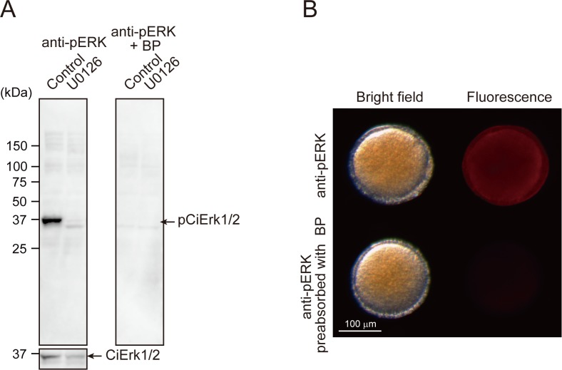
Figure 4—figure supplement 1. Specificity of the anti-pERK1/2 antibody.

Figure 4—figure supplement 1.

(A) A total of 30 μg of soluble protein from stage IV follicles pre-treated with or without 10 μM U0126 for 1 hr were subjected to Western blotting. A single band of endogenous pCiErk1/2 was detected by anti-pERK1/2 antibody, whereas no bands were detected in the follicles pre-treated with U0126 (left). Similarly, the band specific to pCiErk1/2 was abolished by pre-incubation of anti-pERK1/2 antibody with blocking peptide (BP), confirming the specificity of the anti-pERK1/2 antibody and the inhibitory effects of U0126 on pCiErk1/2. Anti-ERK1/2 antibody was used as an internal control. Similar results were obtained by immunostaining using de-folliculated oocytes (B).
Figure 4—figure supplement 2. DEG-profile in MEK-inhibited follicles.

Figure 4—figure supplement 2.

RNA-seq was performed using 150 ng of total RNA from the U0126-treated, early stage III follicles. Three independent sets of follicles were used (n = 3). Data were shown as Figure 2—figure supplement 1. Scatter plot shows various genes that were upregulated (>2 fold) or downregulated (<0.5 fold) upon MEK inhibition.
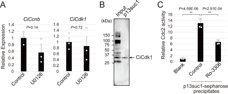
Figure 4—figure supplement 3. Ciona MPF is responsible for CiVP-directed oocyte maturation.

Figure 4—figure supplement 3.

(A) RNA-seq indicated that the expression levels of the MPF component, Ciona cyclin B (CiCcnb) and CiCdc2 (CiCdk1) were not altered in MEK-inhibited follicles, suggesting the post-translation regulation of MPF. Data were analyzed by Student’s t test compared with control (t-value: 1.84; p=0.14 for CiCcnb and t-value: 0.39; p=0.72 for CiCdk1) and are shown as mean ± SEM (n = 3). (B) Western blotting using anti-PSTAIR antibody for precipitates of p13suc1-sepharose confirmed the specific enrichment of CiCdk1. (C) Significant enzymatic activity of CiCdk1 was measured (t-value: −15.48; **, p=4.59E-06). In vitro reaction with 1 μM Ro-3306, a Cdc2 inhibitor, significantly inhibited CiCdk1 activity (t-value: 7.50; **, p=2.91E-04). Data were analyzed by Student’s t test (n = 4).