Abstract
PURPOSE:
To describe a case series of periocular lobular capillary hemangiomas in adults, outlining characteristic clinical and histopathological patterns.
MATERIALS AND METHODS:
This was a retrospective case series of 16 patients with review of clinical and histopathological features.
RESULTS:
Eleven male and five female patients were diagnosed with periocular lobular capillary hemangioma at a median age of 38 years (mean, 41 years; range, 21–71 years). The median tumor basal diameter was 6 mm (mean, 7 mm; range, 3–14 mm) and all were well circumscribed. They arose over the course of weeks to months and developed most commonly in the eyelid region (n = 10), followed by the conjunctiva (n = 6). Excisional biopsy of the lesion was done in all cases. On histopathology, the tumors were composed of repeating units of capillary-sized lobules lined by plump endothelial cells. Lesion recurrence was noted in one case.
CONCLUSION:
Lobular capillary hemangiomas are common benign vascular tumors of periocular region in adults. Clinicohistopathological features and clinical presentation of these lesions are distinctive. Excisional biopsy was curative with recurrence noted rarely.
Keywords: Capillary hemangioma, conjunctiva, eye, eyelid, lobular capillary hemangioma, pyogenic granuloma
Introduction
Lobular capillary hemangioma is a common benign vascular tumor.[1] Pyogenic granuloma has been subsumed into the category of lobular capillary hemangioma, with distinctive clinical and histomorphological features. In this report, we review 16 acquired periocular lobular capillary hemangiomas in adults with histopathological correlation.
Materials And Methods
This was a retrospective study conducted at the Operation Eyesight Universal Institute for Eye Cancer, L V Prasad Eye Institute, Hyderabad, Telangana, India. Institutional review board approval was obtained for the study. The report adhered to the ethical principles outlined in the Declaration of Helsinki as amended in 2013. All adult cases (>16 years) with histopathology confirmed diagnosis of lobular capillary hemangioma from May 2013 to May 2018 were included in the study. Those with inadequate data or lack of histopathology correlation were excluded from the study.
The data extracted from the medical records included age at presentation, gender, location of tumor, presence of local stimulants, and clinical diagnosis. Final histopathology diagnosis of each case was recorded from the histopathology records. The correlation between clinical and histopathology diagnoses was also reviewed.
Results
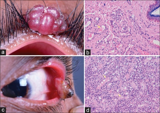
Of 16 cases included in the study, 11 were male and 5 were female with a mean age of 41 years (median, 38 years; range, 21–71 years). No patient had a history of pain, trauma, prior surgery, or systemic vascular disease. The mean duration of symptoms was 2 months. All eyelid lesions originated from the upper eyelid. Of the 6 conjunctival lesions [Figure 1], nasal quadrant (n = 4, 67%) was commonly involved, followed by temporal quadrant (n = 2, 33%). The mean tumor basal diameter was 7 mm (median, 6 mm; range, 3–14 mm). Of the 16 lesions, 10 (62%) developed in the eyelid [Figure 1] and 6 (38%) in the conjunctiva. Pedunculated lesions were seen in 88% (n = 14) of the patients, whereas the lesions were sessile in 12% (n = 2) of the patients. All the patients underwent excisional biopsy of the lesions. Recurrence was noted in 1 (6%) patient after 6 months who had undergone excision of three lesions.
Figure 1.

Eyelid and conjunctival lobular capillary hemangioma, (a) lobular capillary hemangioma arising from the upper eyelid of an adult male, who underwent excisional biopsy, (b) histopathology of excised specimen showing small caliber proliferating blood vessels (yellow arrow) filled with blood and adjacent stroma showing inflammatory cells, (c) lobular capillary hemangioma arising from the conjunctiva of an adult female, who underwent excisional biopsy, (d) histopathology of excised specimen showing small caliber proliferating blood vessels (yellow arrow) filled with blood separated by fibrocollagenous tissue
All patients had similar appearance of the lesion histopathologically. The lesions were composed microscopically of proliferating capillary-sized vascular channels. At high magnification, plump endothelial cells enclosing compressed lumens were seen [Figure 1]. There was no evidence of inflammatory cells or epithelioid giant cells.
Discussion
Lobular capillary hemangioma was first described by Poncet and Dor in 1897,[1] but has since rarely been reported in the periocular region. In 1904, Hartzell et al. coined the term “pyogenic granuloma” to describe the tumor because they believed that it represented a nonspecific granulation tissue response to any pyogenic variant.[2] In 1980, Mills proposed the term “lobular capillary hemangioma” to represent an essential, underlying lesion of a pyogenic granuloma.[3] The classification and terminology of pyogenic granuloma has been the source of considerable confusion. In the recent classification of vascular tumors by ISSVA in 2018, the term pyogenic granuloma (aka lobular capillary hemangioma) was included under benign vascular tumors.[4]
It is believed that periocular lobular capillary hemangioma is a reactive neoplasm caused by surplus restoration reaction to a local stimulus. It arises in response to various stimuli such as irritants or minor trauma or surgery.[5] However, in this case series, no such history of inciting agent was present in any of the patients.
Periocular lobular capillary hemangioma can arise in the eyelid, conjunctiva, or cornea.[6] Previous studies showed conjunctiva as the most common location.[1] In a series of 1643 conjunctival tumors, pyogenic granulomas were seen in 11 (<1%) patients.[7] In our case series, eyelid (62%) was commonly involved, followed by conjunctiva (38%). Majority of the lesions were pedunculated.
Microscopically, they are composed of acute and chronic inflammatory cells interspersed between the fibroblasts, fibrocytes, and lobules of proliferating capillaries. Epithelioid giant cells are not present, which are significant for granulomatous inflammation.[8] In the present case series, none of the patients had inflammatory cells or multinucleated giant cells on histopathology. All were composed of proliferating capillary-sized channels lined by characteristic plump endothelial cells. Clinicopathological correlation was seen in 88% in the present case series.
Treatment includes surgical excision and removal of the known inciting agent or irritant. Conjunctival lesions of small size respond to topical corticosteroids, but many cases ultimately require surgical excision. In 1997, Tay et al. reported a series of pediatric patients with small lobular capillary hemangiomas successfully treated with pulsed dye laser.[9] The authors found that shave excisional biopsy using monopolar cautery is usually effective. Recurrence was rarely seen. In the rare case of recurrence and continued growth of conjunctival lesions, low-dose plaque radiotherapy is effective.[10] In the present study, recurrence was noted in only one patient which resolved completely with repeat surgical excision.
Conclusion
Lobular capillary hemangiomas are common benign vascular tumors of periocular region. The authors believe that “pyogenic granuloma” is a misnomer because it does not form pus or contain epithelioid giant cells that are universally seen in granulomatous inflammation. The authors recommend the use of more accurate term “lobular capillary hemangioma” in describing this lesion rather than the conventional term “pyogenic granuloma.”
Financial support and sponsorship
Support was provided by Operation Eyesight Universal Institute for Eye Cancer and Hyderabad Eye Research Foundation, Hyderabad.
Conflicts of interest
There are no conflicts of interest.
References
- 1.Poncet A, Dor L. Botryomycosis humaine. Rev. de chir. 1897;18:996. [Google Scholar]
- 2.Hartzell MB. Granuloma pyogenicum (botryomycosis of French authors) J Cutan Dis. 1904;22:520–3. [Google Scholar]
- 3.Mills SE, Cooper PH, Fechner RE. Lobular capillary hemangioma: The underlying lesion of pyogenic granuloma. A study of 73 cases from the oral and nasal mucous membranes. Am J Surg Pathol. 1980;4:470–9. [PubMed] [Google Scholar]
- 4.ISSVA Classification for Vascular Anomalies, 20th ISSVA Workshop, Melbourne. 2014. Apr, [Last accessed on 2019 Feb 15]. Available from: http://www.issva.org/UserFiles/file/ISSVA-Classification-2018.pdf .
- 5.Soll SM, Lisman RD, Charles NC, Palu RN. Pyogenic granuloma after transconjunctival blepharoplasty: A case report. Ophthalmic Plast Reconstr Surg. 1993;9:298–301. doi: 10.1097/00002341-199312000-00013. [DOI] [PubMed] [Google Scholar]
- 6.Cameron JA, Mahmood MA. Pyogenic granulomas of the cornea. Ophthalmology. 1995;102:1681–7. doi: 10.1016/s0161-6420(95)30809-3. [DOI] [PubMed] [Google Scholar]
- 7.Shields CL, Demirci H, Karatza E, Shields JA. Clinical survey of 1643 melanocytic and nonmelanocytic conjunctival tumors. Ophthalmology. 2004;111:1747–54. doi: 10.1016/j.ophtha.2004.02.013. [DOI] [PubMed] [Google Scholar]
- 8.Stagner AM, Jakobiec FA. A critical analysis of eleven periocular lobular capillary hemangiomas in adults. Am J Ophthalmol. 2016;165:164–73. doi: 10.1016/j.ajo.2016.03.010. [DOI] [PubMed] [Google Scholar]
- 9.Tay YK, Weston WL, Morelli JG. Treatment of pyogenic granuloma in children with the flashlamp-pumped pulsed dye laser. Pediatrics. 1997;99:368–70. doi: 10.1542/peds.99.3.368. [DOI] [PubMed] [Google Scholar]
- 10.Gündüz K, Shields CL, Shields JA, Zhao DY. Plaque radiation therapy for recurrent conjunctival pyogenic granuloma. Arch Ophthalmol. 1998;116:538–9. doi: 10.1001/archopht.116.4.538. [DOI] [PubMed] [Google Scholar]


