A 72-year-old woman presented 6 months after the onset of falls and ataxia, which had progressed to wheelchair dependence, followed by the onset of bizarre behavior and rapid cognitive deterioration, for which she was hospitalized. Neurologic examination revealed marked disorientation, impairments in recent memory and attention, paranoia, disorganization of thought, dysarthria, symmetric waxy limb rigidity, ataxia of all limbs, and profound gait ataxia. Her medical history was notable only for an implanted cardiac pacemaker, precluding brain MR imaging.
Creutzfeldt-Jakob disease (CJD) was suspected clinically. A thorough search for alternative causes of rapidly progressive dementia was negative.1 CSF was noninflammatory but showed elevation of neuron-specific enolase and 14–3–3 protein. Electroencephalography (EEG) showed generalized slowing.
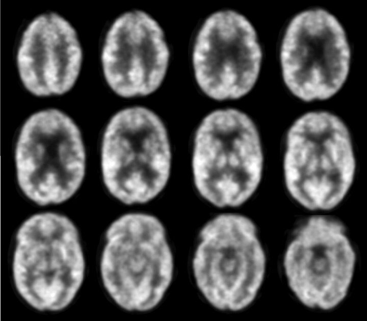
A brain [18F] fluorodeoxyglucose–positron-emission tomography (PET) scan revealed multiple scattered areas of focal cortical hypometabolism in the frontal, parietal, temporal, and occipital lobes. Additionally, there was marked hypometabolism in the bilateral caudate nuclei and heterogeneously in the cerebellum, midbrain, and upper pons (Fig 1). This pattern of basal ganglia and patchy cortical hypometabolism without white matter involvement is analogous to the anatomic distribution of MR imaging findings in CJD1,2 and thus is strongly supportive of the clinical diagnosis.
Fig 1.
PET scans show basal ganglia and patchy cortical hypometabolism, supportive of the clinical diagnosis of CJD.
During 2 weeks of hospitalization, the patient developed hallucinations, worsened limb ataxia, swallowing apraxia, and ultimately akinetic mutism. There was no response to a brief trial of high-dose corticosteroids. She was discharged to hospice care with a diagnosis of probable CJD, and she died 1 month later.
The terminal prognosis of CJD necessitates being reasonably certain before rendering a premortem diagnosis. Our patient's clinical presentation was typical for CJD, in the absence of alternative explanations, making the pretest probability for CJD high. The sensitivities and specificities (SE, Sp) for the usual ancillary tests are as follows: Periodic sharp wave complexes on EEG are specific (Sp ∼ 90%), but EEG is insensitive (SE ∼ 50%); the nonspecific “generalized slowing” seen in our case is typical.1 CSF 14–3–3 testing in recent studies shows modest statistical utility (SE ∼ 48%, Sp ∼ 65%), though other CSF markers such as neuron-specific enolase may be more reliable.1,3 Specific MR imaging fluid-attenuated inversion recovery and diffusion-weighted imaging patterns have emerged as reliable key diagnostic indicators (SE ∼ 92%; Sp, 94%), in which key findings include cortical hyperintensity restricted to the cortex (“cortical ribbon sign”), striatum, or both.1,2
The diagnostic utility of PET imaging is not well-established for CJD. However, we suspect that cases like ours, in which MR imaging is not feasible, are fairly common. The PET findings in our case are in agreement with those in a handful of other reported CJD cases in which PET imaging was performed.4 These imaging findings are distinct from findings in other common neurodegenerative diseases such as Alzheimer disease and Lewy body dementia3 but are anatomically analogous to the distribution of abnormalities seen in CJD on MR imaging. This case presents a clinical situation in which PET imaging provided strong supporting evidence for a diagnosis of probable CJD in the absence of MR imaging and suggests a future use for PET in the evaluation of rapidly progressive dementia.
References
- 1. Geschwind MD, Shu H, Haman A, et al. Rapidly progressive dementia. Ann Neurol 2008;64:97–108 [DOI] [PMC free article] [PubMed] [Google Scholar]
- 2. Shiga Y, Miyazawa K, Sato S, et al. Diffusion-weighted MRI abnormalities as an early diagnostic marker for Creutzfeldt-Jakob disease. Neurology 2004;63:443–49 [DOI] [PubMed] [Google Scholar]
- 3. Geschwind MD, Martindale J, Miller D, et al. Challenging the clinical utility of the 14–3-3 protein for the diagnosis of sporadic Creutzfeldt-Jakob disease. Arch Neurol 2003;60:813–16 [DOI] [PubMed] [Google Scholar]
- 4. Henkel K, Zerr I, Hertel A, et al. Positron emission tomography with [(18)F]FDG in the diagnosis of Creutzfeldt-Jakob disease (CJD). J Neurol 2002;249:699–705 [DOI] [PubMed] [Google Scholar]


