

Abstract
Background and Objectives:
The radial echoendoscope is still widely used for the investigation of abnormalities in the gastrointestinal wall and for stone detection in the extrahepatic biliary tree. Due to aging patient population, EUS is frequently performed in fragile and elderly individuals. We aimed to compare the maneuverability and image quality of a new thin radial echoendoscope to the current one.
Patients and Methods:
This evaluation was conducted at a referral academic EUS center. The new radial echoendoscope has a thinner shaft and distal tube and a shorter bending section compared to the previous one. Patients referred for diagnostic EUS with a radial echoendoscope were enrolled. Indications included pancreaticobiliary disease, esophagogastric abnormalities, and neoplasms and rectal cancer staging.
Results:
We enrolled 177 patients (122 pancreaticobiliary, 48 esophagogastric, and 7 rectal cases). Overall, the new echoendoscope performed better than the previous model in terms of maneuverability during esophageal intubation and transition from the duodenal bulb to the second portion. On the other hand, pylorus traversing was comparable to the current radial echoendoscope. No loss in image quality was appreciated at predefined stations (esophagus, stomach, and duodenum) compared to the current model. On the other hand, image penetration depth in tissue harmonic mode was significantly improved with the new echoendoscope.
Conclusions:
A new thinner radial echoendoscope showed improved maneuverability compared to the existing version. Image quality was also improved thanks to increased penetration depth in the tissue harmonic mode. We speculate that this new echoendoscope may allow for safer and faster EUS examination, which may prove useful in an aging patient population.
Keywords: Bending section, EUS, image quality, procedure safety, radial echoendoscope
INTRODUCTION
EUS has been used for decades for the diagnosis and staging of gastrointestinal and pancreaticobiliary neoplasms, along with benign conditions such as common bile duct stones.[1]
The radial echoendoscope was first introduced to the market in the early 80s by Olympus (Olympus Corp., Tokyo, Japan). At that time, EUS was mainly used for investigation of the gastric wall, portal hypertension, pancreatic parenchyma, and biliary tree.[2,3,4,5,6,7]
In the early 90s, the innovative linear echoendoscope by Pentax (Pentax Corp., Tokyo, Japan) allowed the performance of the first EUS-guided fine-needle aspiration.[8,9] In subsequent years, linear echoendoscopes gained increasing popularity over radial ones due to the wide demand for tissue acquisition and to the advent of several EUS-guided interventions.[10,11,12,13,14,15] More recently, a third type of instrument was invented, namely the forward view echoendoscope, allowing further therapeutic interventions in areas of the gastrointestinal tract that are not easily accessible with the oblique-viewing linear echoendoscope.[16,17]
Nevertheless, in all these years, the radial echoendoscope has not become obsolete as it renders complete and straightforward imaging of abnormalities in the gastrointestinal wall and the common bile duct. Many experts claim that the radial echoendoscope cannot be lacking in EUS centers as it allows complementing the linear echoendoscope for a thorough anatomical investigation.[18,19,20,21,22,23,24,25,26] When common bile duct exploration is the main indication to detect biliary stones, the radial echoendoscope grants a swift and accurate examination.[27,28]
Echoendoscopes are the most difficult endoscopes to maneuver as they are thicker and more rigid than their optical-only counterparts. With aging population, certain areas of the gastrointestinal tract may become more difficult to traverse with echoendoscopes due to increased tissue fragility. For this reason, slimmer and less rigid instruments are advocated.
We aimed to evaluate the thin Olympus radial echoendoscope GF-Y0016-UE in a standard clinical setting according to several indications. In particular, maneuverability and EUS images were analyzed and compared to the standard Olympus radial echoendoscope GF-UE160-AL5 that is currently available.
PATIENTS AND METHODS
Our institution is an academic referral EUS center in Northern Italy, at the University of Bologna in Imola. We perform an average of 600 EUS examinations per year, including all major types of diagnostic and therapeutic procedures.
We evaluated the Olympus radial GF-Y0016-UE (Olympus Corp., Tokyo, Japan) during the second half of 2017. When compared to the GF-UE160-AL5, the insertion tube and the distal end diameter of the new echoendoscope are thinner and the bending section is shorter [Table 1 and Figure 1].
Table 1.
Comparison between standard and new radial echoendoscope
| GF-Y0016-UE | GF-UE160-AL5 | |
|---|---|---|
| Optical system | ||
| Field of view | 100° | 100° |
| Direction of view | 50° forward oblique | 55° forward oblique |
| Outer diameter (mm) | ||
| Distal end | Φ13.4 | Φ13.8 |
| Insertion tube | Φ10.9 | Φ11.8 |
| Channel inner diameter (mm) | Φ2.2 | Φ2.2 |
| Working length (mm) | 1250 | 1250 |
| Angulation range | U: 130°D/R/L: 90° | U: 130°D/R/L: 90° |
| Ultrasound scanning range | 360° | 360° |
| Balloon function | Yes | Yes |
| Ultrasound cable | Detachable | Nondetachable |
Figure 1.

Head-to-head comparison of the new GF-Y0016-UE (right) with the GF-UE160-AL5 (left). Notice the shorter bending section of the new echoendoscope
Every patient referred for diagnostic EUS with a radial echoendoscope was enrolled in this retrospective analysis of a prospectively maintained database. EUS examinations were performed on patients lying on their left lateral decubitus and with conscious sedation using fentanyl and midazolam. All patients signed informed consent to grant permission to evaluate their data for anonymous research purposes. We did not consult with the institutional review board in view of the retrospective nature of the study.
The echoendoscopes' maneuverability and image quality were analyzed and compared to a similar cohort of patients who had undergone EUS with the standard radial echoendoscope during the first half of 2017.
All the procedures were performed by one of three experienced operators (PF, AL, and MS). Before starting enrollment, all endoscopists met to find an agreement related to the study standards of care and definitions.
Echoendoscope maneuverability was evaluated at three levels: intubation of cervical esophagus, traversing of the pylorus, and transition from the first to the second duodenal portion. In case of rectal examinations, ease of passage of the rectosigmoid junction was also evaluated. All the maneuvers were analyzed according to a qualitative scale using the standard radial echoendoscope as a reference (superior to/equal to/inferior to).
Image quality was assessed at four standard stations (second part of the duodenum, duodenal bulb, stomach, and esophagus) and recorded as excellent/good/fair and compared to the standard echoendoscope as well. Excellent was defined as a clean EUS view of the target organ with an adequate resolution; good was defined as a minimal loss in image resolution, but still adequate for the diagnosis; fair was defined as a significant loss in image quality, barely adequate for the diagnosis.
RESULTS
We enrolled 177 patients during the study period. According to the indications, 122 patients underwent EUS for pancreaticobiliary examination, 48 patients were referred for esophagogastric abnormalities, and seven patients for rectal cancer staging.
Detailed results are shown in Table 2. In brief, overall imaging quality of the new echoendoscope was rated excellent in the majority of cases. In particular, image penetration depth in tissue harmonic enhancement of the GF-Y0016-UE was greater than that obtained with the standard echoendoscope thanks to its blending with B-mode imaging in the deep scanning areas.
Table 2.
Patients population and imaging results
| Indication | # Cases; M/F; median age | EUS diagnosis | Overall imaging quality | Image artifacts |
|---|---|---|---|---|
| Pancreaticobiliary | 122; 60/62; 67 (range 22-89) | 40 CBD stones | 98% excellent 2% good | 2% minor air artifacts |
| 54 Normal findings | 100% excellent | none | ||
| 28 Neoplasms | 95% excellent 5% good | 5% minor air artifacts | ||
| Esophagogastric | 48; 23/25; 67.5 (range 44-88) | 5 Esophageal cancer | 100% excellent | none |
| 15 Normal findings | 100% excellent | none | ||
| 16 SMTs | 100% excellent | none | ||
| Rectal | 7; 3/4; 66 (range 55-78) | 7 Rectal cancer | 95% excellent 5% good | 5% minor air artifacts |
M/F: Male/Female; CBD: Common bile duct; SMTs: Submucosal tumors
While the maneuverability of the GF-Y0016-UE was deemed equivalent to the standard echoendoscope for traversing the pylorus, esophageal intubation and passage from the first to the second duodenal portion were rated easier with the new echoendoscope than with the standard one [Table 3]. Similarly, passage from the rectum to the sigmoid colon was also considered easier with the new model compared to the previous one.
Table 3.
Maneuvrability of the GF-Y0016-UE
| Comparison with GF-UE160-AL5 | Esophageal intubation (%) | Pylorus traversing (%) | Duodenal transition (%) |
|---|---|---|---|
| Superior to | 140/177 (79) | 20/177 (11) | 132/177 (75) |
| Equal to | 37/177 (21) | 157/177 (89) | 45/177 (25) |
| Inferior to | 0/177 | 0/177 | 0/177 |
All the patients tolerated well the procedures under conscious sedation with a mean dose of 100 mcg of fentanyl and 2.5 mg of midazolam. No adverse events occurred during the study period.
DISCUSSION
We evaluated a new radial echoendoscope that is thinner and has a shorter bending section compared to the previous model. We showed that the maneuverability of the new echoendoscope was improved at certain critical steps of EUS examination, such as esophageal intubation and passage from the duodenal bulb to the second portion.
On the other hand, image quality was not inferior to the preexisting model and was considered superior in the tissue harmonic enhancement mode thanks to the increased penetration depth.
The radial echoendoscope has represented the mainstay of EUS examinations for at least two decades after its introduction to the market. All the earliest publications and meetings dealt with radial examination for the staging of esophagogastric cancer and lymphoma, portal hypertension assessment, and differential diagnosis of submucosal tumors [Figures 2 and 3]. Subsequently, EUS exploration of the common bile duct for stones detection emerged as the main indication for radial imaging.
Figure 2.

Among the earliest publications in EUS were reports about the gastrointestinal wall layer structure
Figure 3.
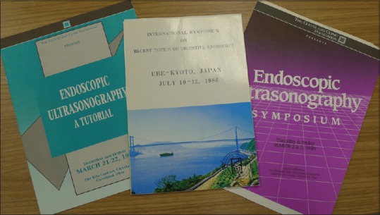
Proceedings of some of the earliest international meetings in EUS
Radial EUS is still considered a powerful diagnostic tool at major referral centers as it allows for quick and accurate exploration of the extrahepatic biliary tree and is a useful complement to linear EUS in difficult pancreaticobiliary cases. Moreover, it is optimal for the staging of esophageal, gastric, and rectal cancer.
However, the radial echoendoscope is stiff and rigid and is regarded as the most difficult endoscope to maneuver through the gastrointestinal tract. For this reason, a thinner echoendoscope would be desirable in an aging patient population who is more fragile and potentially more exposed to adverse events. In theory, the improved maneuverability of the new echoendoscope could allow for safer examinations and better patient tolerance. Therefore, the need for patient sedation could be diminished thereby limiting the risk of drug-related adverse events.
The improved penetration depth of tissue harmonic was of particular interest. The smaller penetration depth of the radial echoendoscope has been considered its main limitation when compared to the linear one. Tissue harmonic enhancement allows for improved EUS imaging as it emphasizes the boundaries between structures and lesions with different acoustic impedance. The better acoustical properties of the GF-Y0016-UE under evaluation could represent a useful tool to improve the diagnostic and staging accuracy [Figure 4].
Figure 4.

A typical image obtained with the new radial echoendoscope. Pancreatic head cancer (PC) is shown infiltrating the common bile duct (CBD) an the portal vein (PV). The gallbladder (GB) is dilated and contains sludge
As far as rectal cancer staging was concerned, we found it easier to reach the sigmoid colon with the GF-Y0016-UE than with the GF-UE160-AL5. Visualization through the sigmoid is essential for iliac lymph nodes detection to perform a complete locoregional staging. We also noticed that overall endoscopic exploration and the visualization of the papilla of Vater had improved thanks to the greater maneuverability of the echoendoscope, thereby potentially reducing the need for resorting to esophagogastroduodenoscopy before EUS.
Our study has several limitations. First, due to its retrospective design, it is open to inherent methodological flaws and inaccurate data collection. However, we arranged a structured database to gather all relevant study information and images. In addition, the analysis of the echoendoscope's maneuverability and image quality was subjective and prone to evaluation bias, although it was performed by experienced endosonographers with longstanding experience of EUS examination using the radial echoendoscope.
CONCLUSION
We tested the GF-Y0016-UE radial echoendoscope in a group of patients with pancreaticobiliary, esophagogastric, and rectal indications. Compared to the previous radial echoendoscope, the new model has a thinner shaft and distal tube and a shorter bending section. The new echoendoscope showed improved maneuverability particularly during some critical examination steps such as esophageal intubation and transition from the duodenal bulb to the second portion. Moreover, image quality was improved thanks to increased penetration depth in the tissue harmonic mode. We speculate that this new echoendoscope may allow for safer and faster EUS examination that is advantageous in an aging patient population, which is more fragile and exposed to adverse events.
Financial support and sponsorship
Nil.
Conflicts of interest
Olympus Corp. loaned us the echoendoscope that was used for this study.
REFERENCES
- 1.Palazzo L. How to perform EUS in the pancreaticobiliary area. Minerva Med. 2014;105:371–89. [PubMed] [Google Scholar]
- 2.Dimagno EP, Regan PT, Clain JE, et al. Human endoscopic ultrasonography. Gastroenterology. 1982;83:824–9. [PubMed] [Google Scholar]
- 3.Caletti G, Bolondi L, Labò G. Ultrasonic endoscopy – The gastrointestinal wall. Scand J Gastroenterol Suppl. 1984;102:5–8. [PubMed] [Google Scholar]
- 4.Caletti G, Bolondi L, Labò G. Anatomical aspects in ultrasonic endoscopy for the stomach. Scand J Gastroenterol Suppl. 1984;94:34–42. [PubMed] [Google Scholar]
- 5.Caletti GC, Lorena Z, Bolondi L, et al. Impact of endoscopic ultrasonography on diagnosis and treatment of primary gastric lymphoma. Surgery. 1988;103:315–20. [PubMed] [Google Scholar]
- 6.Caletti G, Brocchi E, Zani L, et al. The important role of EUS in the assessment of patients with portal hypertension. Gastrointest Endosc. 1988;34:154–5. doi: 10.1016/s0016-5107(88)71294-8. [DOI] [PubMed] [Google Scholar]
- 7.Tio LT, Blank LE, Wijers OB, et al. Staging and prognosis using endosonography in patients with inoperable esophageal carcinoma treated with combined intraluminal and external irradiation. Gastrointest Endosc. 1994;40:304–10. doi: 10.1016/s0016-5107(94)70061-3. [DOI] [PubMed] [Google Scholar]
- 8.Vilmann P, Jacobsen GK, Henriksen FW, et al. Endoscopic ultrasonography with guided fine needle aspiration biopsy in pancreatic disease. Gastrointest Endosc. 1992;38:172–3. doi: 10.1016/s0016-5107(92)70385-x. [DOI] [PubMed] [Google Scholar]
- 9.Vilmann P, Hancke S, Henriksen FW, et al. Endoscopic ultrasonography-guided fine-needle aspiration biopsy of lesions in the upper gastrointestinal tract. Gastrointest Endosc. 1995;41:230–5. doi: 10.1016/s0016-5107(95)70343-8. [DOI] [PubMed] [Google Scholar]
- 10.Wiersema MJ, Wiersema LM. Endosonography-guided celiac plexus neurolysis. Gastrointest Endosc. 1996;44:656–62. doi: 10.1016/s0016-5107(96)70047-0. [DOI] [PubMed] [Google Scholar]
- 11.Wiersema MJ. Endosonography-guided cystoduodenostomy with a therapeutic ultrasound endoscope. Gastrointest Endosc. 1996;44:614–7. doi: 10.1016/s0016-5107(96)70022-6. [DOI] [PubMed] [Google Scholar]
- 12.Wiersema MJ, Sandusky D, Carr R, et al. Endosonography-guided cholangiopancreatography. Gastrointest Endosc. 1996;43:102–6. doi: 10.1016/s0016-5107(06)80108-2. [DOI] [PubMed] [Google Scholar]
- 13.Ashida R, Arisaka Y, Masuda D, et al. The role of linear array EUS for diagnosis of pancreatic malignancies in the current situation. Dig Endosc. 2011;23(Suppl 1):12–6. doi: 10.1111/j.1443-1661.2011.01138.x. [DOI] [PubMed] [Google Scholar]
- 14.Cazacu IM, Luzuriaga Chavez AA, Saftoiu A, et al. A quarter century of EUS-FNA: Progress, milestones, and future directions. Endosc Ultrasound. 2018;7:141–60. doi: 10.4103/eus.eus_19_18. [DOI] [PMC free article] [PubMed] [Google Scholar]
- 15.Rimbaş M, Crino SF, Gasbarrini A, et al. EUS-guided fine-needle tissue acquisition for solid pancreatic lesions: Finally moving from fine-needle aspiration to fine-needle biopsy? Endosc Ultrasound. 2018;7:137–40. doi: 10.4103/eus.eus_23_18. [DOI] [PMC free article] [PubMed] [Google Scholar]
- 16.Fusaroli P, Ceroni L, Caletti G. Forward-view endoscopic ultrasound: A systematic review of diagnostic and therapeutic applications. Endosc Ultrasound. 2013;2:64–70. doi: 10.4103/2303-9027.117689. [DOI] [PMC free article] [PubMed] [Google Scholar]
- 17.Fusaroli P, Serrani M, Lisotti A, et al. Performance of the forward-view echoendoscope for pancreaticobiliary examination in patients with status post-upper gastrointestinal surgery. Endosc Ultrasound. 2015;4:336–41. doi: 10.4103/2303-9027.170427. [DOI] [PMC free article] [PubMed] [Google Scholar]
- 18.Irisawa A. Current role of radial and curved-linear arrayed EUS scopes for diagnosis of pancreatic abnormalities in Japan. Dig Endosc. 2011;23(Suppl 1):9–11. doi: 10.1111/j.1443-1661.2011.01144.x. [DOI] [PubMed] [Google Scholar]
- 19.Colaiácovo R, Assef MS, Ganc RL, et al. Rectal cancer staging: Correlation between the evaluation with radial echoendoscope and rigid linear probe. Endosc Ultrasound. 2014;3:161–6. doi: 10.4103/2303-9027.138786. [DOI] [PMC free article] [PubMed] [Google Scholar]
- 20.ASGE Technology Committee. Murad FM, Komanduri S, et al. Echoendoscopes. Gastrointest Endosc. 2015;82:189–202. doi: 10.1016/j.gie.2015.02.017. [DOI] [PubMed] [Google Scholar]
- 21.Stevens T, Zuccaro G, Jr, Dumot JA, et al. Prospective comparison of radial and linear endoscopic ultrasound for diagnosis of chronic pancreatitis. Endoscopy. 2009;41:836–41. doi: 10.1055/s-0029-1215061. [DOI] [PubMed] [Google Scholar]
- 22.Kanazawa K, Imazu H, Mori N, et al. A comparison of electronic radial and curvilinear endoscopic ultrasonography in the detection of pancreatic malignant tumor. Scand J Gastroenterol. 2012;47:1313–20. doi: 10.3109/00365521.2012.719930. [DOI] [PubMed] [Google Scholar]
- 23.Shin EJ, Topazian M, Goggins MG, et al. Linear-array EUS improves detection of pancreatic lesions in high-risk individuals: A randomized tandem study. Gastrointest Endosc. 2015;82:812–8. doi: 10.1016/j.gie.2015.02.028. [DOI] [PMC free article] [PubMed] [Google Scholar]
- 24.Lisotti A, Serrani M, Caletti G, et al. EUS liver assessment using contrast agents and elastography. Endosc Ultrasound. 2018;7:252–6. doi: 10.4103/eus.eus_29_18. [DOI] [PMC free article] [PubMed] [Google Scholar]
- 25.Wang Y, Chai N, Feng J, et al. A prospective study of endoscopic ultrasonography features, cyst fluid carcinoembryonic antigen, and fluid cytology for the differentiation of small pancreatic cystic neoplasms. Endosc Ultrasound. 2018;7:335–42. doi: 10.4103/eus.eus_40_17. [DOI] [PMC free article] [PubMed] [Google Scholar]
- 26.Dong Y, D'Onofrio M, Hocke M, et al. Autoimmune pancreatitis: Imaging features. Endosc Ultrasound. 2018;7:196–203. doi: 10.4103/eus.eus_23_17. [DOI] [PMC free article] [PubMed] [Google Scholar]
- 27.Amouyal P, Amouyal G, Lévy P, et al. Diagnosis of choledocholithiasis by endoscopic ultrasonography. Gastroenterology. 1994;106:1062–7. doi: 10.1016/0016-5085(94)90768-4. [DOI] [PubMed] [Google Scholar]
- 28.Kaneko M, Katanuma A, Maguchi H, et al. Prospective, randomized, comparative study of delineation capability of radial scanning and curved linear array endoscopic ultrasound for the pancreaticobiliary region. Endosc Int Open. 2014;2:E160–70. doi: 10.1055/s-0034-1377384. [DOI] [PMC free article] [PubMed] [Google Scholar]