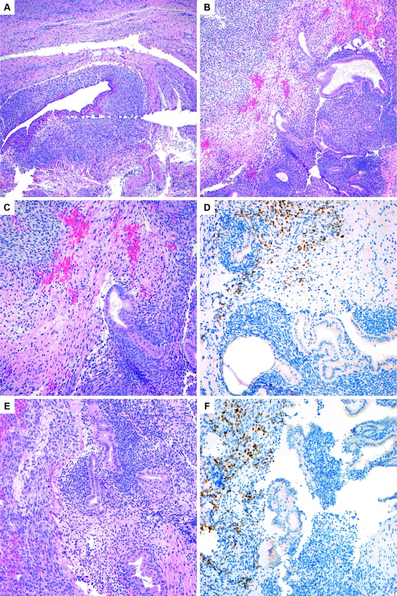
Figure 7.
A, Most of the material in the resection consisted of conventional appearing endometriosis, as pictured here in the ovary (H&E, 10×). B, The right aspect of the image shows benign endometrial glands and stroma of endometriosis, and the left upper corner shows stroma with paler cytoplasm (H&E, 10×). C, Higher-power image of the same area to contrast the benign endometrial-type stroma on the bottom right with the paler stroma in the upper left (H&E, 20×). D, Positive PAX-7 immunohistochemical staining in the paler stroma performed retrospectively confirms the presence of focal rhabdomyosarcoma (20×). E, Another area on the same slide to contrast the morphology of the benign endometrial-type stroma in the endometriosis on the right with the paler rhabdomyosarcomatous stroma on the left. In this image, there is slight nuclear enlargement and apoptotic debris noted in the sarcomatous area (H&E, 20×). F, Positive staining for PAX-7 in the stroma on the left confirms the diagnosis of rhabdomyosarcoma (20×).

