

ABSTRACT
Bouveret syndrome, a rare cause of intestinal obstruction, occurs by passage of a gallstone through a cholecystoduodenal fistula into the intestinal lumen. Presenting symptoms are nausea, vomiting, and abdominal pain. In some cases, chronic symptoms result in weight loss. Typically, the syndrome is diagnosed via x-ray, ultrasound, or computed tomography. Treatment options are endoscopic or surgical. Endoscopic approaches include mechanical lithotripsy, electrohydraulic lithotripsy, stone extraction, laser lithotripsy, extracorporeal shockwave lithotripsy, and/or duodenal stenting. When stone fragments migrate distally, surgical removal becomes necessary. We describe a distinct endoscopic treatment via stone breakage, followed by pushing the fragments of the stone into the jejunum, resolving the intestinal obstruction.
INTRODUCTION
Beaussier published the first case of Bouveret syndrome in 1770 and was followed by Léon Bouveret, a French internist, who published 2 cases in 1896.1,2 Presenting symptoms are nausea, vomiting, abdominal pain, and sometimes weight loss.1 Gallstone ileus is a rare diagnosis and is known to have high clinical and treatment-related mortality and morbidity rates, especially in elderly patients with multiple comorbidities. The syndrome is more common in women, and onset is at a mean age of 60–80 years.3,4 A number of endoscopic and sometimes surgical techniques are used to remove the gallstones.5 Although several cases of Bouveret syndrome have been reported in the literature, we present a rare case of complete resolution of intestinal obstruction by fragmentation of the gallstone and pushing of the stone into the jejunum.
CASE REPORT
A 78-year-old woman with a history of gallstones, diabetes, and chronic kidney disease presented with a complaint of diffuse abdominal pain associated with heartburn, nausea, and vomiting for 1 day. Other reviews of systems were negative. On physical examination, her vitals were stable and the abdomen was soft, nontender, and nondistended. Laboratory examination demonstrated leukocytosis (white blood cells 13,200/μL), normal liver chemistry, serum creatinine 2.49 mg/dL, blood urea nitrogen 43 mg/dL, and lactate 2.7 mmol/L. An abdominal computed tomography (CT) revealed gas in the in common bile duct, pneumobilia, and a 4.7 × 2.8-cm calcified gallstone in the duodenal bulb (Figure 1). Ultrasonography was not performed.
Figure 1.
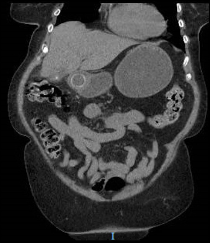
Computed tomography showing large gallstones within an area of inflammation where the gallbladder is near the duodenum.
Esophagogastroduodenoscopy showed a large, 4 cm, obstructing stone adherent to a large ulcer/fistula at the posterior wall of the duodenal bulb with normal anterior and lateral wall mucosa (Figure 2). Using rat tooth forceps, pieces of the outer layer of the stone were broken off because the stone could not pass the pyloric muscle because of its size. Finally, the stone was dislodged from the fistula with gentle pressure with the tip of endoscope (Figure 3). The fistula was then examined and appeared to have fibrotic tissue with no discernible opening into the gallbladder. There was a complication, and the lithotripsy basket was unable to grip the dislodged stone for removal. Further attempts to retrieve the stone past the pylorus were unsuccessful.
Figure 2.

Esophagogastroduodenoscopy showing a large stone obstructing the pylorus in the duodenal bulb after being dislodged from the fistula.
Figure 3.

The stone was advanced beyond the pylorus with a gentle push with the tip of the endoscope.
After discussion with the surgical team, it was decided to advance the stone further into the small bowel so surgical removal could be attempted. Using a pediatric colonoscope, the stone was advanced to the proximal jejunum. Repeat CT of the abdomen showed progression of the stone to the mid-jejunum with resolution of the small bowel obstruction (Figure 4). The patient experienced marked improvement in her symptoms after the procedure although surgical retrieval was planned because of the fear of the stone obstructing at the ileocecal valve because of its size. She underwent a laparoscopic enterotomy with removal of the gallstone and did well postoperation. Her diet was slowly advanced, and she was discharged from the hospital 6 days postoperation.
Figure 4.

Follow-up postendoscopy computed tomography showing the gallstone in the jejunum.
DISCUSSION
Bouveret syndrome is caused by passage of a large gallstone more than 2.5 cm in size through bilio-gastric and gastroduodenal fistulas into various parts of the digestive tract such as the ileocecal valve (50%–90%).6,7 The proximal jejunum and ileum are less common sites (20%–40%), and the colon, stomach, and duodenum are even less common (<5%).5,8 At least 0.3%–0.5% of patients with gallstones develop ileus.5 Morbidity and mortality are high at 15%–33%.9 Factors that lean toward fistula formation include, but are not limited to, large gallstones, extensive history of biliary disease, recurrent episodes of cholecystitis, female sex, age older than 60 years, peptic ulcer, and tumors.5,8,10
The mechanism of gallstone formation includes inflammatory changes that cause gallbladder wall adherence to the adjacent viscera. These stones increase intraluminal pressure, which results in ischemia of the gallbladder wall, and cause gallstones to erode the wall of the gallbladder to pass into the adjacent viscera, usually the intestine, by forming a fistula. Depending on the size of the gallstone, it can either pass into the feces or get lodged into the intestine, causing intestinal obstruction in 15% of cases.5 Most commonly reported symptoms of intestinal obstruction in various case reports and literature reviews are abdominal pain, distention, nausea, vomiting, fever, and hematemesis in rare cases.11
CT scan is the most sensitive diagnostic test for gallstone ileus; however, it can miss 25% of gallstones if they are radiolucent.10 Other options for investigation are ultrasound and x-ray of the abdomen. Isoattenuating gallstones, which are difficult to distinguish from the surrounding bile, are visualized by magnetic resonance cholangiopancreatography in 15%–25% of cases and endoscopy in 69% of cases.1,11 The literature has reported over 300 cases of Bouveret syndrome.4 A PubMed literature review of 24 cases of Bouveret syndrome and a comprehensive review by Cappell and Davis of 128 cases revealed only 2 cases with successful resolution of intestinal obstruction by an endoscopic approach.3,12,13
Treatment of Bouveret syndrome is either endoscopic or surgical. Endoscopic treatments include mechanical, laser lithotripsy, and electrohydraulic. Electrohydraulic is usually the first choice because of its low risk but is only successful in 10% of cases.5,8,9,11 Endoscopic treatment can cause complications such as migration of the gallstone to the intrathoracic cavity via an esophageal tear if patients have concomitant esophagitis or if the gallstone is large in size.8 Surgical methods including enterolithotomy, which is associated with less morbidity, and duodenotomy, occasionally followed by cholecystectomy and fistula repair, are performed when endoscopic approaches are partially or completely unsuccessful.5,8,9,11
This case is unique among cases of Bouveret syndrome because the patient had an endoscopic resolution of intestinal obstruction via fragmentation of a large gallstone, followed by the pushing of the stone into the jejunum, with subsequent prophylactic enterotomy to prevent risk of distal intestinal obstruction. Repositioning the stone via endoscopy made an otherwise difficult surgical procedure less complicated to prevent mortality and morbidity. It highlights an alternative approach in cases in which stones cannot be removed past the pylorus.
DISCLOSURES
Author contributions: S. Khuwaja wrote and edited the manuscript. A. Azeem, J. Afthinos, and S. Guttman edited the manuscript. BA Semkhayev provided the radiologic images. S. Guttmann is the article guarantor.
Financial disclosure: None to report.
Informed consent was obtained for this case report.
REFERENCES
- 1.Al-Habbal Y, Ng M, Bird D, McQuillan T, Al-Khaffaf H. Uncommon presentation of a common disease—Bouveret's syndrome: A case report and systematic literature review. World J Gastrointest Surg. 2017;9(1):25–36. [DOI] [PMC free article] [PubMed] [Google Scholar]
- 2.Iancu C, Bodea R, Al Hajjar N, Todea-Iancu D, Bălă O, Acalovschi I. Bouveret syndrome associated with acute gangrenous cholecystitis. J Gastrointestin Liver Dis. 2008;17:87–90. [PubMed] [Google Scholar]
- 3.Bonam R, Vahora Z, Harvin G, Leland W. Bouveret's syndrome with severe esophagitis and a purulent fistula. ACG Case Reports J. 2014;1(3):158–60. [DOI] [PMC free article] [PubMed] [Google Scholar]
- 4.Nickel F, Müller-Eschner MM, Chu J, von Tengg-Kobligk H, Müller-Stich BP. Bouveret's syndrome: presentation of two cases with review of the literature and development of a surgical treatment strategy. BMC Surg. 2013;13:33. [DOI] [PMC free article] [PubMed] [Google Scholar]
- 5.Shah-Khan S, Vallabh H, Cardinal J, Nasr J. Novel use of an endoscopic suturing device to repair a cholecystoduodenal fistula. ACG Case Rep J. 2017;4:e121. [DOI] [PMC free article] [PubMed] [Google Scholar]
- 6.Gencosmanoglu R, Inceoglu R, Baysal C, Akansel S, Tozun N. Bouveret's syndrome complicated by a distal gallstone ileus. World J Gastroenterol. 2003;9(12):2873–5. [DOI] [PMC free article] [PubMed] [Google Scholar]
- 7.Sağlam F, Sivrikoz E, Alemdar A, Kamalı S, Arslan U, Güven H. Bouveret syndrome: A fatal diagnostic dilemma of gastric outlet obstruction. Ulus Travma Acil Cerrahi Derg. 2015;21(2):157–9. [DOI] [PubMed] [Google Scholar]
- 8.Martin-Cuesta L, Marco De Lucas E, Pellon R, et al. Migrating intrathoracic gallstone imaging findings. J Thorac Imaging. 2008;23(4):272–4. [DOI] [PubMed] [Google Scholar]
- 9.Patel A, Agarwal S. The yellow brick road of Bouveret syndrome. Clin Gastroenterol Hepatol. 2014;12(8):A24. [DOI] [PubMed] [Google Scholar]
- 10.Englert ZP, Love K, Marilley MD, Bower CE. Bouveret syndrome: Gallstone ileus of the duodenum. Surg Laparosc Endosc Percutan Tech. 2012;22(5):e301–3. [DOI] [PubMed] [Google Scholar]
- 11.Smith Z, Totten J, Hughes A, Strote J. Delayed diagnosis of gastric outlet obstruction from bouveret syndrome in a young woman. West J Emerg Med. 2015;16(1):151–3. [DOI] [PMC free article] [PubMed] [Google Scholar]
- 12.Cappell MS, Davis M. Characterization of Bouveret's syndrome: A comprehensive review of 128 cases. Am J Gastroenterol. 2006;101(9):2139–46. [DOI] [PubMed] [Google Scholar]
- 13.Lopes C, Lima F, Hartmann A. Bouveret syndrome and pancreatic acinar cell carcinoma. Endoscopy. 2017;49(S 01):E62–3. [DOI] [PMC free article] [PubMed] [Google Scholar]