

Abstract
Hemangioma of the tendon sheath is an uncommon tumor—often slow-growing and misdiagnosed until final biopsy. We hereby report a 30-year-old woman who presented with pain and swelling of the wrist for 2 years, which was proven to be cavernous hemangioma of extensor tendon sheath.
Keywords: hemangioma, tendon sheath, wrist and hand
Introduction
Hemangioma is a benign hamartoma developing from blood vessels. 1 Synovial hemangiomas are uncommon tumors, arising from surfaces lined by synovium, such as the tendons and joint cavities. The knee joint is the most common intra-articular site followed by the elbow. 2 Until now there have been few reports of these masses arising from tendon in the scientific literature. We report a 30-year-old woman who presented with pain and progressive swelling of the wrist, which was eventually diagnosed as tendon sheath hemangioma of the second extensor compartment.
Case Report
A 30-year-old female housewife, with no comorbidities, presented with pain in the left wrist for 2 years. Insidious in onset and progressive, pain was exacerbated with exertion and lifting heavy weight and was relieved with analgesics. It was mild to moderate with no specific character and no diurnal variation. Pain was followed by appearance of swelling that was progressive over dorsal aspect of the wrist with no restriction of movement. There was no history of fever, rash, other joint involvement, morning stiffness, and any significant loss of weight or appetite. There were no complaints of numbness, tingling, or weakness of the hand. The patient could not recollect any history of mild or significant trauma.
On local physical examination, a 2- × 1.5-cm oval, well-defined, soft, nontender mass was seen over dorsoradial aspect of the left wrist spanning the second and third extensor compartments, with no local rise in temperature or erythema. The mass was well-defined, mobile, and soft to firm in consistency. It did not transilluminate. Plane was deep to subcutaneous. All movements of the wrist were full, except terminal restriction in palmar flexion. Clinically (L) wrist monoarthritis, rheumatoid nodulosis, tubercular arthritis, tenosynovitis, giant cell tumor, ganglion, and lipoma of the wrist were the differentials.
There was mild eosinophilia in hemogram. Erythrocyte sedimentation rate (ESR) was 25 mm/1st hour, C-reactive protein (CRP) was negative, and workup for inflammatory arthritis was negative. Mantoux was 17 mm after 48 hours. X-rays of the local part revealed decreased radiocarpal space and mild osteopenia. Magnetic resonance imaging (MRI) revealed an enhancing, irregular, nodular thickening along the extensor tendons sparing medial two compartments with erosions in the distal end of the radius, scaphoid, and lunate. This appeared consistent with the differential of inflammatory arthritis ( Fig. 1 ).
Fig. 1.

( A–D ) MRI images of the lesion.
A: axial t1 image showing mass arising from extensor compartment encasing tendons.
B: coronal t1 image showing 4- × 2-cm mass at distal end of radius.
C: coronal t2, mass seen enhancing in t2 imaging.
D: axial t2 showing irregularity over radial styloid.
An open excisional biopsy with a dorsal longitudinal incision over the second extensor compartment was performed revealing a red fleshy well-defined mass involving the second extensor compartment, adherent to extensor carpi radialis longus, and brevis tendon sheath was found. There was localized synovial hypertrophy. Articular margin of the distal radius and scaphoid was found to be eroded due to pressure effect. En masse resection was done. Wound was closed in layers and a below-elbow slab was applied. Postoperative course was uneventful. Suture removal was done at 10th day. Wrist and finger active range-of-motion (ROM) exercise was started when the patient was pain free at around 2 weeks. Sections on histologic examination revealed synovial tissue with lymphocytic infiltrate, peripheral dilated ectatic blood vessels suggestive of cavernous hemangioma. On 3- and 12-month follow-up, the patient is symptom free with no restriction in ROM ( Fig. 2 3 4 5 6 ).
Fig. 2.

Intraoperative photograph showing mass arising from extensor carpi radialis longus and brevis.
Fig. 3.

Intraoperative image showing erosion of radial styloid.
Fig. 4.

Mass excised in toto.
Fig. 5.

Hematoxylin and eosin stain section (200X) showing the fibrocollagenous tissue on the right side with adjacent cavernous hemangioma of the left side, having dilated congested vascular spaces and single layer of endothelial lining.
Fig. 6.
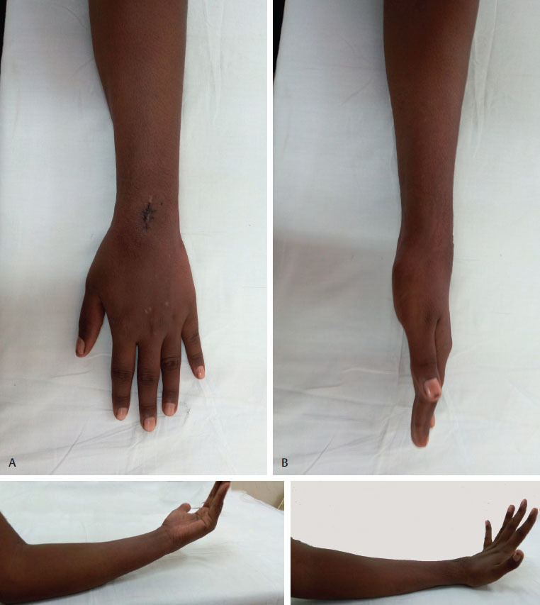
( A–D ) Postoperative images at 6-week follow-up showing complete relief of symptoms and full range of motion.
Discussion
As opposed to infants and pediatric age group in which capillary hemangioma predominates, musculoskeletal hemangiomas are cavernous in nature. 3 Approximately 7 to 10% of all hemangiomas arise in musculoskeletal system, of which 36% are seen in thigh muscles. 4 They rarely arise in tendons and masquerade until final histopathologic report. As seen in this case, symptoms were of 2 years duration. Primary diagnosis was in line of a rheumatoid nodule or tubercular arthritis. Differential diagnosis of a wrist swelling includes ganglion, giant cell tumor of the tendon sheath, bursitis (radial or ulnar bursa), rheumatoid nodule, and tenosynovitis. 1
Most tendon hemangiomas arise in the wrist or hand. Those arising from gracilis, 5 quadriceps and semiten dinosus tendon, 6 and peroneal tendons 7 have also been reported. In the hand and wrist, they can present with myriad of symptoms—painless slow-growing swelling, tenosynovitis, nerve entrapment, or trigger finger (if arising in A1 pulley). 2 Weinzweig et al reported a bizarre case of bilateral cavernous hemangioma arising from the extensor pollicis brevis and was erroneously treated with multiple steroid injection as de Quervain’s disease. 8
Kim et al have reported flexor tendon hemangioma presenting with symptoms of carpal tunnel syndrome in which symptoms were exacerbated after a minor trauma. Trauma as etiology of hemangioma has not been conclusively proven 9 10 11 ( Table 1 ).
Table 1. Studies on hemangioma of tendon sheath of wrist and hand in past century.
| Waddell (1967) 9 | Cavernous hemangioma of extensor pollicis longus and brevis |
| Spinner et al (1983) 12 | Recurrent cavernous hemangioma of 3/4th extensor tendon |
| Weinzweig et al (1996) 8 | Bilateral extensor pollicis brevis hemangioma mimicking as de Quervain’s disease |
| Talwalker et al (2005) 13 | Tenosynovial hemangioma of finger |
| Kim et al (2010) 10 | Flexor tendon sheath hemangioma causing carpal tunnel syndrome |
| Lee et al (2011) 2 | Recurrent synovial hemangioma of ring finger distal to A2 pulley |
| Kim et al (2014) 11 | Flexor digitorum superficialis tendon hemangioma |
Radiographs are normal in more than half of the cases. Sometimes they may present as vague soft tissue mass or localized swelling. Phleboliths, osseous erosions, periosteal new bone, or as arthritic changes are better seen on a CT (computed tomography) scan. MRI (magnetic resonance imaging) demonstrates intermediate signal intensity relative to skeletal muscle on T1-weighted sequences and increased signals on T2-weighted sequences relative to fat. 14 Preoperative diagnosis with imaging is not always conclusive. 5 10 Conclusive evidence is obtained after histopathology study.
The hemangiomatous spectrum ranges from small tumors that regress without sequelae and require no treatment to endangering tumors that require therapy because of neurovascular entrapment. Treatment of middle spectrum is controversial and often surgeon dependent. 3
Treatment includes simple resection open or arthroscopically (if intra-articular). Embolization may be needed preoperatively for diffuse lesions if large feeding vessels are present. Hemangiomas recur if they have been incompletely excised or if feeding vessels have not been ligated. 14 Lee et al reported a recurrent synovial hemangioma of the ring finger distal to A2 pulley. They have discussed need for complete excision of the tendon sheath to prevent recurrence, and because excision of tendon is difficult to manage and given the benign nature of hemangioma, tendon in the hand can be preserved instead of radical excision. 14 Spinner et al in 1983 reported a case of recurrent cavernous tendon hemangioma arising from third and fourth extensor indices tendon 12 years after initial excision. 12
In conclusion, tenosynovial hemangioma is a rare entity with wide spectrum of manifestations. Being vigilant of this can lead to early diagnosis, and complete excision should be the rule.
Footnotes
Conflict of Interest None.
References
- 1.Athanasian E A, Irons G B. 2nd ed. Philadelphia, PA: LWW; 2010. Tumours of the wrist; pp. 1187–1212. [Google Scholar]
- 2.Lee T C, Chein S H, Liu P C, Cheng Y M. A rapidly recurrent synovial hemangioma involving tendon sheath: a rare case in the finger. Formosan Journal of Musculoskeletal Disorders. 2011;2(04):135–138. [Google Scholar]
- 3.Marler J J, Mulliken J B.Vascular anomalies 2nd ed,Vol. 5.Philadelphia, PA: Elsevier; 200619–68. [Google Scholar]
- 4.Wild A T, Raab P, Krauspe R. Hemangioma of skeletal muscle. Arch Orthop Trauma Surg. 2000;120(03)(04):139–143. doi: 10.1007/pl00013761. [DOI] [PubMed] [Google Scholar]
- 5.Nishida Y, Yamada Y, Tsukushi S, Shimoyama Y, Nagasaka T, Ishiguro N. Persistent popliteal pain derived from cavernous hemangioma involving gracilis tendon and tendon sheath. Knee. 2006;13(03):252–254. doi: 10.1016/j.knee.2005.12.001. [DOI] [PubMed] [Google Scholar]
- 6.Burman M S, Milgram J E. Haemangioma of tendon and tendon sheath. Surg Gynecol Obstet. 1930;50:397–406. [Google Scholar]
- 7.Urgüden M, Ozdemir H, Duygulu E, Aydin A T. Cavernous hemangioma behaving like peroneal tenosynovitis. Foot Ankle Int. 2000;21(10):856–859. doi: 10.1177/107110070002101011. [DOI] [PubMed] [Google Scholar]
- 8.Weinzweig J, Watson H K, Wiener B D, Genter B E. Hemangioma of the extensor pollicis brevis in the first dorsal compartment: an unusual cause of bilateral de Quervain’s disease. J Hand Surg Am. 1996;21(02):256–258. doi: 10.1016/S0363-5023(96)80112-2. [DOI] [PubMed] [Google Scholar]
- 9.Waddell G F. A haemangioma involving tendons. J Bone Joint Surg Br. 1967;49(01):138–141. [PubMed] [Google Scholar]
- 10.Kim J Y, Sung J H, Lee S. A haemangioma of the flexor tendon sheath causing carpal tunnel syndrome. J Hand Surg Eur Vol. 2010;35(01):73–74. doi: 10.1177/1753193409347688. [DOI] [PubMed] [Google Scholar]
- 11.Kim D H, Jeong M, Shim S B, Lee J H, Kim C K. Hemangioma of the flexor digitorum superficialis in the hand. J Korean Soc Surg Hand. 2014;19(03):154–158. [Google Scholar]
- 12.Spinner M, Moon S, Young L. Recurrent cavernous haemangioma of the extensor tendons of the hand. Hand. 1983;15(02):223–227. doi: 10.1016/s0072-968x(83)80020-5. [DOI] [PubMed] [Google Scholar]
- 13.Talwalkar S, Hayton M, Stilwell J, Temperley D, Freemont A. Tenosynovial haemangioma of the finger. Acta Orthop Belg. 2005;71(05):618–621. [PubMed] [Google Scholar]
- 14.Garner H W, Bestic J M. Benign synovial tumors and proliferative processes. Semin Musculoskelet Radiol. 2013;17(02):177–178. doi: 10.1055/s-0033-1343095. [DOI] [PubMed] [Google Scholar]