

Abstract
Objectives:
The aim of this study was to compare the shaping ability of three file systems – 2Shape (2S), WaveOne Gold (WOG), and ProTaper Gold – using cone-beam computed tomography (CBCT).
Materials and Methods:
Forty-five freshly extracted mandibular teeth were chosen and instrumented using the following brands of NiTi files: 2S, WOG, and ProTaper Gold. Pre- and post-instrumentation CBCT imaging was performed to measure mesial and distal distance of the dentin walls and calculate the volume of removed dentin, apical transportation, and centering ratio. A statistical analysis of the data was performed using independent t-test. Statistical significance was set at 5%.
Results:
There were no significant differences between the 2S and WOG in terms of the total volume of removed dentin, apical transportation, and centering ratio, whereas ProTaper showed a greater significant difference when compared to 2S and WOG.
Conclusion:
Both 2S and WOG maintained the original canal anatomy better and did not remove excess dentin while chemomechanical preparation as compared to ProTaper Gold. Instruments that use rotary movement achieved an effect similar to that of the reciprocating instruments in relation to change in angle. 2S which is a two-file system and WOG which is a single-file system might perform better clinically and might show enhanced shaping ability with less canal transportation and more centered preparation when compared to ProTaper Gold that is a multiple file-system.
KEYWORDS: 2Shape, apical transportation, centering ratio, ProTaper Gold, volume of removed dentin, WaveOne Gold
INTRODUCTION
The introduction of NiTi rotary instrumentation has revolutionized the endodontics in the past few decades with predictable success. The rotary files have been subjected to constant evaluation in the form of metallurgy, design features, the number of instruments, and the manner in which these instruments are driven (rotary/reciprocation).[1] Various rotary NiTi endodontic file systems have been introduced to improve shaping ability.
It is generally accepted that the excessive removal of dentin compromises the survival of rootfilled teeth and that the strength of endodontically treated teeth is directly related to the amount of remaining sound tooth structure.[2,3]
Recently introduced 2Shape (2S) NiTi rotary file is made of NiTi alloy called T-wire. 2S has a sequence with two instruments: TS1 (#25, 0.04) and TS2 (#25, 0.06).[4]
ProTaper Gold (PG) works on rotating motion and is made of NiTi alloy called M-wire which is considered to feature a progressively tapered design that claimed to improve the cutting efficiency and safety.[5]
Yared proposed a new technique employing reciprocating movements using just one instrument;[6] chemomechanical preparation with reciprocating motion has been postulated to reduce the possibility of unexpected file fractures. WaveOne Gold (WOG) G-wire technology is available in four sizes: small (#20,0.07), primary (#25,0.07), medium (#35,0.06), and large (#45,0.05).[7]
There is paucity of literature comparing the shaping ability of 2S, WOG, and ProTaper Gold. Therefore, this study was planned. The null hypothesis tested is that the new manufacturing methods and type of rotary motion will not have any effect on their shaping ability of root canals.
MATERIALS AND METHODS
In the present study, ethical clearance was obtained from the Ethical Committee of Vyas Dental College and Hospital with reference no. VDCH/IEC/24/2017. Forty-five mesiobuccalroots of extracted mandibular first molar teeth with completely formed apices were collected and stored in saline solution. All teeth were evaluated radiovisiographically to rule out any calcification. Preinstrumentation cone-beam computed tomography (CBCT) scanning was done. Teeth having root curvature ranging from 25° to 30° were selected. Canal curvature was assessed by Schneider’s technique, and the samples were standardized.[8] Access cavities were prepared with Endo-Access bur (Dentsply Maillefer), and the root canals were negotiated using #10 K-file (Dentsply, Maillefer, Switzerland). The distal roots with the respective part of the crown were sectioned at the furcation level using a low-speed diamond bur under water and discarded. The working length was determined by inserting #10 K-file to root canal terminus and subtracting 1 mm from this measurement, which was then confirmed using an electronic apex locator.
The teeth were randomly divided into three experimental groups (n = 15).
In Group 1 (n = 15), teeth were prepared with 2S, in a sequence TS1 (25/0.04)>TS2 (25/0.06) according to the manufacturer’s instructions.
In Group 2 (n = 15), ProTaper Gold in the sequence S1>SX>S1>S2>F1>F2 (25/0.08) was used for shaping according to the manufacturer’s instructions.
In Group 3 (n = 15), WOG was instrumented with the primary file (25/0.07) (Dentsply Maillefer, Switzerland) in reciprocating motion (clockwise 140° and counterclockwise 45°).
The final apical preparation was standardized for all specimens at size 25. Instrumentation was done using Glyde (Dentsply Maillefer) as a lubricating agent. The canals were irrigated with 2 mL of 5% sodium hypochlorite during instrumentation followed by 1 mL of 17% ethylenediaminetetraacetic acid for 3 min and a final irrigation with 2 mL of saline solution. Each instrument was used to prepare three canals, and then, the files were discarded. Teeth were then scanned under the same conditions followed for the initial scan, and the data were analyzed.
CONE-BEAM COMPUTED TOMOGRAPHY ANALYSIS
Pre- and postinstrumentation measurements of MB canals were calculated [Figure 1a–c]. The volume of removed dentine was measured in mm3 for each root canal by subtracting the uninstrumented canal volume from the instrumented canal volume.[8] Canal transportation and centering ratio were calculated at three cross-section levels, i.e., 3, 5, and 7 mm, from the apical end of the root using the following equation:
Figure 1.

(a) Pre- and post-instrumentation cone-beam computed tomography images at 3 mm level. (b) Pre- and post-instrumentation cone-beam computed tomography images at 5 mm level. (c) Pre- and post-instrumentation cone-beam computed tomography images at 7 mm level
Degree of canal transportation: mesiodistally = (m1 − m2) − (d1 − d2)
Canal centering ratio = (m1 − m2 )/(d1 − d2 ) or (d1 − d2)/(m1 − m2)
Mesial (m1) is the shortest distance from the mesial edge of the root to the mesial edge of the uninstrumented canal. Distal (d1) is the shortest distance from the distal edge of the root to the distal edge of the uninstrumented canal.
m2 is the shortest distance from the mesial edge of the root to the mesial edge of the instrumented canal. d2 is the shortest distance from the distal edge of the root to the distal edge of the instrumented canal.
STATISTICAL ANALYSIS
A statistical analysis (SPSS 15.0; SPSS Inc., Chicago, IL, USA) of the data was performed using independent t-test. The statistical significance level was set at P < 0.05.
RESULTS
VOLUME OF REMOVED DENTINE
The significant difference is noted between 2S and ProTaper Gold, WOG, and ProTaper Gold, whereas no statistically significant difference was seen between 2S and WOG [Graph 1].
Graph 1.

Mean ± standard deviation of volume of removed dentine (mm3) for tested groups and statistical analysis
CANAL TRANSPORTATION
The significant difference was seen among the three groups at all the levels: 3 mm, 5 mm, and 7 mm. ProTaper Gold showed maximum canal transportation at all the levels, whereas 2S and WOG produced less canal transportation [Graph 2].
Graph 2.
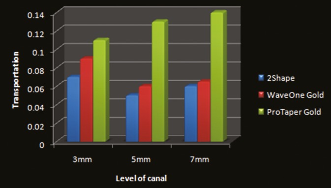
Degree of canal transportation mesiodistally for tested groups
CENTERING RATIO
At all the levels, 3 mm, 5 mm, and 7 mm ProTaper Gold showed the least centered canal preparation as compared to 2S and WOG [Graph 3].
Graph 3.

Centering ratio for tested groups
2S and WOG file system produced better results as compared to ProTaper Gold [Table 1].
Table 1.
The mean and standard deviation values of the canal transportation and centering ratio at the three studied levels (3, 5, and 7 mm) for each tested group
| Level (mm) | Assessment | 2Shape | WaveOne Gold | ProTaper Gold |
|---|---|---|---|---|
| 3 | Transportation | 0.07±0.03 | 0.09±0.02 | 0.11±0.02 |
| Centering ratio | 1.4±0.13 | 1±0.15 | 0.63±0.15 | |
| 5 | Transportation | 0.06±0.03 | 0.06±0.015 | 0.13±0.015 |
| Centering ratio | 1.6±0.15 | 1.35±0.14 | 0.8±0.13 | |
| 7 | Transportation | 0.05±0.035 | 0.065±0.02 | 0.14±0.02 |
| Centering ratio | 1.67±0.13 | 1.55±0.15 | 0.9±0.15 |
DISCUSSION
As there is paucity in literature comparing the shaping ability of recently introduced NiTi rotary instruments 2S, WOG and ProTaper gold, recommended for the preparation of curved root canals, one of which is activated by reciprocating movement (WOG) and other two by continuous rotary movement (2S and ProTaper Gold). The following parameters were evaluated: volume of removed dentin, apical transportation, and centering ratio.
The mesiobuccal root of mandibular first molars was chosen as they typically present with remarkable curvatures. The angle of curvature at 25°–30° (according to Schneider’s technique) was preferred as according to the American Association of Endodontists Endodontic Case Difficulty Assessment, it is considered as moderate curvature to obtain results that cover a large scale of cases.[8] The CBCT imaging technique has been used for shaping ability evaluation of the three files as it provides a accurate, reproducible, three-dimensional analysis of the alterations which might be present in dentin such as dentin thickness. CBCT also provides root canal volume before and after preparation without causing any damage to the specimens.
To allow a proper action of NiTi instruments that use either a rotary or reciprocating motion, creation of a glide path is essential.[9]
In the present study, curvatures with high susceptibility to iatrogenic mishaps usually exist at these three levels: 3, 5, and 7 mm, which represent the apical, middle, and coronal thirds of the root canals, respectively.[10]
The apical preparation was limited to size 25 file in the present study as the amount of canal transportation increases with apical preparation greater than size 25.[11]
In the present study, ProTaper Gold was reported to have aggressive cutting and excessive volume of dentin removal as compared to 2S and WOG [Graph 1].
At all the levels, 3 mm, 5 mm, and 7 mm, mesiodistally, the transportation is more with ProTaper Gold as compared to 2S and WOG, which was statistically significant [Graph 2] (P < 0.05). This could be attributed to the sequence of ProTaper Gold files S1>SX>S1>S2>F1>F2 (25/0.08) used in circumferential brushing motion and the cross-section of the files, convex triangular cross-section of ProTaper Gold file.
WOG works on the principle of reciprocating motion and is claimed to be able to completely shape and clean root canals with only one single-use instrument. Reverse helix, semi-active and modified guiding tip, and offset parallelogram-shaped cross-section limit the engagement zone.[11] Reciprocating movement minimizes torsional and flexural stresses, increases the centering ability of canal, and reduces the taper lock of the instrument within the canal.[12] Hence, WOG reported comparatively less aggressive volume of removed dentin and better centering ratio with less canal transportation.
Ozyurek et al. (2017) conducted an in vitro study on shaping the ability of Reciproc, WOG, and HyFlex EDM single-file systems in simulated S-shaped canals; it was concluded that all of the tested NiTi files caused various levels of resin removal. However, the WOG and HEDM NiTi files were found to cause a lower level of resin removal than the RPC NiTi files.[13] The results for WOG were in accordance with our study.
At all the levels, 3 mm, 5 mm, and 7 mm, 2S and WOG were reported to have a significant difference in centering ratio as compared to ProTaper Gold [Graph 3] (P < 0.05). 2S and WOG were reported to have a better centering ratio as compared to ProTaper Gold. This could be attributed to the metallurgy of 2S NiTi rotary file which works on rotating motion and is made of NiTi alloy called “T-wire” which is a method which allows for an increased resistance to cyclic fatigue (+40%) and a better negotiation of curvatures. The two instruments (TS1 and TS2) return to their original shape after each use. 2S file with the latest generation of the cross-section with triple helix, two primary cutting edges and one secondary cutting edge, aids in perfect compromise between cutting efficiency and debris removal.[4] WOG files are made of a special NiTi alloy called G-Wire which is created by an innovative thermal treatment process. The benefits of this G-wire NiTi are increased flexibility of the instruments and improved resistance to cyclic fatigue.
Abdullah et al. in an in vitro study compared the shaping ability of ProTaper Gold and WOG system in simulated S- and L-shaped canals. The results revealed that there was a highly significant difference noted in preparation; it was concluded that WOG showed better shaping ability with less canal aberrations and faster canal preparation as compared to ProTaper Gold.[14] The results of this study are similar to the results obtained in the current study.
Staffoli et al. conducted a study on simulated teeth with severe curvature and evaluated the centering ability of ProTaper Next and 2S file system. In accordance with the results, it was concluded that there was no significant difference noted in centering ability of ProTaper Next and 2S. Both the file systems showed some degree of canal transportation, especially in the apical third.[15,16]
In an in vitro study, the lower level of resin removal was reported with the use of HEDM and WOG NiTi files when it was compared to the RPC NiTi files. The results for WOG were in accordance with our study that reported less volume of removed dentin.[17]
As there is paucity in literature on shaping ability of 2S file system, the present study was carried out, and it was reported that 2S file system maintained the original canal anatomy better as compared to ProTaper Gold. Further clinical research is needed to conclude the shaping ability of 2S file system.
The results of volume of removed dentin, canal transportation, and centering ratio at all the levels, 3 mm, 5 mm, and 7 mm, revealed that there was no significant difference between 2S and WOG systems (P > 0.05).
CONCLUSION
Within the limitations of this study, it was concluded that 2S file and WOG had better original canal anatomy with less aggressive dentin cutting and minimal canal transportation as compared to ProTaper Gold file system.
FINANCIAL SUPPORT AND SPONSORSHIP
Nil.
CONFLICTS OF INTEREST
There are no conflicts of interest.
REFERENCES
- 1.Krishan S, Aggarwal A, Singh K. Endodontic rotary systems – A review. J Adv Med Dent Sci Res. 2016;4:84–8. [Google Scholar]
- 2.Arora V, Yadav MP, Singh SP, Arora P, Aggarwal A. Comparative evaluation of post obturation materials on reinforcement of PeriCervical Dentin (PCD) – An in vitro study. IJTEEE. 2015;3:39–43. [Google Scholar]
- 3.Grande NM, Plotino G, Lavorgna L, Ioppolo P, Bedini R, Pameijer CH, et al. Influence of different root canal-filling materials on the mechanical properties of root canal dentin. J Endod. 2007;33:859–63. doi: 10.1016/j.joen.2007.02.018. [DOI] [PubMed] [Google Scholar]
- 4.Micro Mega, France. 2Shape. [Last accessed on 2017 Sep 17]. Available from: http://www.micromega.com/shaping/2shape?lang=en .
- 5.Elnaghy AM, Elsaka SE. Shaping ability of ProTaper gold and ProTaper universal files by using cone-beam computed tomography. Indian J Dent Res. 2016;27:37–41. doi: 10.4103/0970-9290.179812. [DOI] [PubMed] [Google Scholar]
- 6.Yared G. Canal preparation using only one Ni-Ti rotary instrument: Preliminary observations. Int Endod J. 2008;41:339–44. doi: 10.1111/j.1365-2591.2007.01351.x. [DOI] [PubMed] [Google Scholar]
- 7.Dentsply Tulsa Dental Specialties. WaveOne Gold. [Last accessed on 2016 Oct 14]. Available from: https://www.Dentsply.com/content/dam/dentsply/pim/manufacturer/Endodontics/Obturation/Gutta_Percha_Points/WaveOne_Gold_Gutta_Percha_Points/W1G_Brochure_EN.pdf .
- 8.Hartmann MS, Barletta FB, Camargo Fontanella VR, Vanni JR. Canal transportation after root canal instrumentation: A comparative study with computed tomography. J Endod. 2007;33:962–5. doi: 10.1016/j.joen.2007.03.019. [DOI] [PubMed] [Google Scholar]
- 9.Schneider SW. A comparison of canal preparations in straight and curved root canals. Oral Surg Oral Med Oral Pathol. 1971;32:271–5. doi: 10.1016/0030-4220(71)90230-1. [DOI] [PubMed] [Google Scholar]
- 10.Michetti J, Maret D, Mallet JP, Diemer F. Validation of cone beam computed tomography as a tool to explore root canal anatomy. J Endod. 2010;36:1187–90. doi: 10.1016/j.joen.2010.03.029. [DOI] [PubMed] [Google Scholar]
- 11.Webber J. Shaping canals with confidence: WaveOne GOLD single-file reciprocating system. Int Dent Afr Ed. 2016;6:6–17. [Google Scholar]
- 12.Guillén RE, Nabeshima CK, Caballero-Flores H, Cayón MR, Mercadé M, Cai S, et al. Evaluation of the waveOne gold and one shape new generation in reducing enterococcus faecalis from root canal. Braz Dent J. 2018;29:249–53. doi: 10.1590/0103-6440201801910. [DOI] [PubMed] [Google Scholar]
- 13.Ozyurek T, Yılmaz K, Uslu G. Shaping ability of reciproc, WaveOne GOLD, and hyFlex EDM single-file systems in simulated S-shaped canals. J Endod. 2017;43:805–9. doi: 10.1016/j.joen.2016.12.010. [DOI] [PubMed] [Google Scholar]
- 14.Abdullah A, Mohammad A, Thomas S, Abdul M. Shaping ability of ProTaper gold and WaveOne gold nickel-titanium rotary file in different canal configurations. Saudi Endod J. 2018;8:202–7. [Google Scholar]
- 15.Staffoli S, Ozyurek T, Hadad A, Lvovsky A, Solomonov M, Azizi H, et al. Comparison of shaping ability of ProTaper next and 2Shape nickel–titanium files in simulated severe curved canals. G Ital Endod. 2018;32:52–6. [Google Scholar]
- 16.Rubio J, Zarzosa JI, Pallarés A. Comparison of shaping ability of 10 rotary and reciprocating systems: An in vitro study with AutoCad. Acta Stomatol Croat. 2017;51:207–16. doi: 10.15644/asc51/3/4. [DOI] [PMC free article] [PubMed] [Google Scholar]
- 17.Surakanti JR, Punna R, Vanapatla A, Vemisetty H, Sajja NP, Nidumukkala TP, et al. Comparative evaluation of cleaning ability of reciproc and waveonegold reciprocating file systems – A scanning electron microscopic study. Indian J Dent Sci. 2018;10:98–101. [Google Scholar]