Abstract
Dermatofibroma (DF) is a benign skin tumor that is well-known among dermatologists. We herein present a rare case of atrophic dermatofibroma presenting linear skin dimpling. The patient was a 25-year-old woman with a history of wild-type recessive dystrophic epidermolysis bullosa who had noticed linear concavity on her right lateral back 1 year before her initial presentation. Anetoderma, atrophic scar, localized morphea, or lupus erythematosus profundus were clinically suspected; however, a biopsy specimen from the dimpling lesion showed the fibrous and histiocytic tumor in the deep dermis. The spindle-to-rhomboid-shaped tumor cells were arranged with irregularly storiform pattern, and immunohistochemistry showed that the tumor cells were positive for factor XIIIa, and negative for CD34 and CD68. Elastica van Gieson staining showed an almost complete loss of elastic fibers, especially at the center of the lesion. The reduction of elastic fibers might have influenced the skin depression in this case. This rare case suggests the need to consider a subtype of DF in the differential diagnosis of dimpling skin lesions.
Keywords: Atrophic dermatofibroma, Rare case, Linear skin dimple, Elastic fiber, Epidermolysis bullosa
Case Report
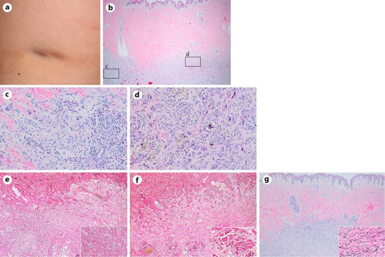
Dermatofibroma (DF) is a benign fibrohistiocytic tumor that is well-known among dermatologists. The typical clinical features of DF include an elevated, painful and hyperpigmented lesion with a smooth surface. Several histopathological variations of the cells have been reported, including - but not limited to - aneurysmal, hemosiderotic, epithelioid, lipidized, cellular, and atypical fibrous variants [1, 2]. We experienced a rare case of atrophic DF in which the patient presented linear dimpling skin lesion of approximately 50 mm in length. The patient was a 25-year-old Japanese woman who had noticed a linear concavity on her right lateral back 1 year prior to her initial presentation. She had initially felt slight pain corresponding to the area of skin dimpling (Fig. 1a). We initially suspected anetoderma, atrophic scar, localized morphea, or lupus erythematosus profundus. The patient had no past history of trauma or treatment with steroid injection. She had a history of wild-type recessive dystrophic epidermolysis bullosa and was observed in our outpatient clinic without medication.
Fig. 1.
a A dimpling and tight skin lesion on the right upper back. b Tumor cells were embedded from the lower dermis to the fatty layer. A slight skin depression was noticed at the center of the lesion. Infiltrating fibroblastic (c) and rhomboid-shaped histiocytic (d) tumor cells are shown in the left and right boxes of b, respectively. Cellular atypia was not evident in either lesion. Elastica van Gieson staining clearly showed the lack and reduction of elastic fibers at the central and peripheral portions of the tumor (e, f), respectively. The inset boxes show high-power views. g H&E staining of a common DF case experienced in our department. Elastica van Gieson staining confirms solid elastic fibers intermingled in the tumor (inset box).
A transverse section of the dimpling lesion was prepared for a histopathological examination. The center of the lesion was slightly depressed without basal melanosis and the fine-to-ovoid tumor cells spread from the reticular dermis to the fatty tissue (Fig. 1b). While Touton giant cells and histiocytic tumor cells with vacuolated cytoplasm infiltrated at the center (Fig. 1c), spindle-shaped tumor cells were predominant at the edge of the lesion (Fig. 1d). The two-dimensional tumor size was histologically estimated as 40 × 15 mm. In addition, Elastica van Gieson staining showed an almost complete loss of elastic fibers at the center (Fig. 1e), while the remaining elastic fibers, which were shortened and degenerated between the collagen fibers, were observed at the edge of the lesion (Fig. 1f). We finally diagnosed the patient with atrophic DF rather than dermatofibrosarcoma protuberans because the tumor cells were positive for factor XIIIa and negative for CD34 and CD68, irrespective of the relatively irregular form and high cellularity.
Requena and Reichel [3] proposed that decreased thickness of the dermis resulted from skin depression above the tumor rather than authentic atrophy and, therefore, that delled DF was a more accurate term than atrophic DF. In fact, because the dermal thinning was not remarkable even at the depressive portion of the tumor in this case, we hypothesize that another mechanism of skin dimpling was involved in the present case. Kiyohara et al. [4] and Ohnishi et al. [5] reported that Elastica van Gieson staining revealed the complete loss of elastic fibers in atrophic DF, while in non-atrophic DF, the expression of elastic fibers within the lesion was markedly decreased. The most common findings of DF in our department were generally the basal pigmentation in the epidermis and increased fibroblast-like cells, with intermingling elastic fibers (Fig. 1g). In contrast, no elastic fiber and basal melanosis was found within the tumor in this case. Decreased and degraded elastic fibers might have induced an imbalance of skin tension and weakened skin tightness, resulting in the clinical feature of skin dimpling. Finally, the present case suggests the need to consider atrophic DF in the differential diagnosis of dimpling skin lesions.
Statement of Ethics
Informed consent was obtained from the patient. The study complied with the Declaration of Helsinki.
Disclosure Statement
The authors declare no conflict of interest.
Author Contributions
The authors are all responsible for and in agreement with the content and writing of the manuscript to which they all contributed significantly.
Acknowledgement
We greatly appreciate Mr. Kenji Nishida and Ms. Eriko Nobuyoshi for an excellent tissue staining.
References
- 1.Beer M, Eckert F, Schmoeckel C. The atrophic dermatofibroma. J Am Acad Dermatol. 1991 Dec;25((6 Pt 1)):1081–2. doi: 10.1016/s0190-9622(08)80444-6. [DOI] [PubMed] [Google Scholar]
- 2.Alves JV, Matos DM, Barreiros HF, Bártolo EA. Variants of dermatofibroma-a histopathological study. An Bras Dermatol. 2014 May-Jun;89((3)):89. doi: 10.1590/abd1806-4841.20142629. 472-7. [DOI] [PMC free article] [PubMed] [Google Scholar]
- 3.Requena L, Reichel M. The atrophic dermatofibroma: a delled dermatofibroma. J Dermatol. 1995 May;22((5)):334–9. doi: 10.1111/j.1346-8138.1995.tb03398.x. [DOI] [PubMed] [Google Scholar]
- 4.Kiyohara T, Kumakiri M, Kobayashi H, Ohkawara A, Lao LM. Atrophic dermatofibroma. Elastophagocytosis by the tumor cells. J Cutan Pathol. 2000 Jul;27((6)):312–5. doi: 10.1034/j.1600-0560.2000.027006312.x. [DOI] [PubMed] [Google Scholar]
- 5.Ohnishi T, Sasaki M, Nakai K, Watanabe S. Atrophic dermatofibroma. J Eur Acad Dermatol Venereol. 2004 Sep;18((5)):580–3. doi: 10.1111/j.1468-3083.2004.00975.x. [DOI] [PubMed] [Google Scholar]


