Abstract
Deafferentation induces rapid plastic changes in the cerebral cortex, probably via unmasking of pre-existent connections. Several mechanisms may contribute, such as changes in neuronal membrane excitability, removal of local inhibition, or various forms of short- or long-term synaptic plasticity. To understand further the mechanisms involved in cortical plasticity, we tested the effects of CNS-active drugs in a plasticity model, in which forearm ischemic nerve block (INB) was combined with low-frequency repetitive transcranial magnetic stimulation (rTMS) of the deafferented human motor cortex. rTMS was used to upregulate the plastic changes caused by INB. We studied six healthy subjects. In two control sessions without drug application, INB plus rTMS increased the motor-evoked potential (MEP) size and decreased intracortical inhibition (ICI) measured with single- and paired-pulse TMS in the biceps brachii muscle proximal to INB. A single oral dose of the benzodiazepine lorazepam (2 mg) or the voltage-gated Na+ and Ca2+ channel blocker lamotrigine (300 mg) abolished these changes. The NMDA receptor blocker dextromethorphan (150 mg) suppressed the reduction in ICI but not the increase in MEP size. With sleep deprivation, used to eliminate sedation as a major factor of these drug effects, INB plus rTMS induced changes similar to that seen in the control sessions. The findings suggest that (1) the INB plus rTMS-induced increase in MEP size involves rapid removal of GABA-related cortical inhibition and short-term changes in synaptic efficacy dependent on Na+ or Ca2+ channels and that (2) the long-lasting (>60 min) reduction in ICI is related to long-term potentiation-like mechanisms given its duration and the involvement of NMDA receptor activation.
Keywords: mechanisms of cortical plasticity, transient ischemic forearm deafferentation, pharmacological blockade of plasticity, GABA, glutamate, paired-pulse inhibition, human motor cortex
The adult mammalian sensorimotor cortex can reorganize rapidly, within minutes to hours, in response to peripheral lesions (Merzenich et al., 1983; Calford and Tweedale, 1988,1991a,b; Sanes et al., 1988; Donoghue et al., 1990; Byrne and Calford, 1991; Kolarik et al., 1994; Silva et al., 1996; Borsook et al., 1998) or as a consequence of motor learning (Grafton et al., 1992; Recanzone et al., 1992; Pascual-Leone et al., 1994, 1995; Karni et al., 1995;Donoghue et al., 1996; Nudo et al., 1996). There is good evidence that these rapid reorganizational processes are mediated by the unmasking of existent but latent corticocortical connections (Schieber and Hibbard, 1993; Donoghue et al., 1996; Huntley, 1997; Sanes and Donoghue, 1997) and that the mechanisms involved include removal of local inhibition (Jacobs and Donoghue, 1991) and changes in synaptic efficacy. One example for short-term synaptic enhancement is post-tetanic potentiation (PTP), which depends on an increase in calcium concentration in presynaptic terminals and decays in the order of minutes (Fisher et al., 1997). An example for a sustained strengthening of synapses is long-term potentiation (LTP), which was first described in the hippocampus (Bliss and Lømo, 1973) but is also present in the mammalian neocortex where it is mediated via NMDA receptors and usually requires the downregulation of local inhibitory circuits (Artola and Singer, 1987; Iriki et al., 1989; Artola et al., 1990;Kirkwood et al., 1993; Hess and Donoghue, 1994; Kirkwood and Bear, 1994; Castro-Alamancos et al., 1995; Hess et al., 1996).
The present experiments used temporary ischemic limb deafferentation, an established experimental model of cortical plasticity in humans (Brasil-Neto et al., 1992, 1993; Ridding and Rothwell, 1995, 1997;Ziemann et al., 1998), combined with low-frequency repetitive transcranial magnetic stimulation (rTMS) of the motor cortex contralateral to ischemia. When tested with single- and paired-pulse TMS, deafferentation leads to a rapid increase in the amplitudes of motor-evoked potentials (MEPs) in muscles proximal to the ischemic nerve block without changes in motor threshold (MT). rTMS upregulates the ischemia-induced MEP increase and, in addition, induces a long-lasting (>60 min) decrease in intracortical inhibition (ICI) (Ziemann et al., 1998). These measures reflect different aspects of motor cortical excitability. MEP size explores in this paradigm excitatory synaptic transmission in the motor cortex (Brasil-Neto et al., 1993), MT probes neuronal membrane excitability (Mavroudakis et al., 1994; Ziemann et al., 1996b; Chen et al., 1997b), and ICI measures strong GABA-dependent inhibitory and weaker NMDA-dependent excitatory interneuronal circuits in the motor cortex (Ziemann et al., 1996a,b; Liepert et al., 1997).
Identification of the mechanisms of plasticity in adult human cortex is an important step toward the development of rationally founded treatment strategies in neurorehabilitation. To understand the mechanisms underlying ischemic nerve block (INB) plus rTMS-induced plasticity better, we tested whether CNS-active drugs, in particular a blocker of NMDA receptors (dextromethorphan), a GABAA receptor agonist (lorazepam), and a blocker of voltage-gated Na+ and Ca2+channels (lamotrigine), suppress this form of plasticity. Suppressive effects by drugs acting via specific mechanisms of action would allow us to identify the involvement of these mechanisms in the plastic process.
MATERIALS AND METHODS
Subjects. We studied six healthy right-handed men (mean age, 25.5 ± 4.3 years). Each subject was tested in six separate sessions. Another three subjects were screened but excluded, because the target muscle [biceps brachii (BB)] was not sufficiently excitable by focal TMS. All subjects gave their written informed consent for the study, and the protocol was approved by the Institutional Review Board.
Measurements of motor excitability. Subjects were seated comfortably in a reclining chair. A surface EMG was recorded from the BB and the abductor pollicis brevis (APB) of the nondominant left arm with silver-silver chloride cup electrodes in a belly-tendon montage. After amplification and bandpass (0.1–2.5 kHz) filtering (Counterpoint electromyograph; Dantec Electronics, Skovlunde, Denmark), the EMG signal was digitized (analog-to-digital rate of 5 kHz) and fed into an IBM 486 AT-compatible laboratory computer for off-line analysis.
Focal TMS was applied to the motor cortex on the right side. A figure eight-shaped coil was connected to two Magstim 200 magnetic stimulators through a BiStim module (Magstim, Whitland, Dyfed, UK). The coil was placed tangentially to the scalp with the handle pointing backward and rotated away from the midline by ∼45°. The current induced in the brain was therefore directed approximately perpendicular to the line of the central sulcus, a condition optimal for activating the corticospinal system trans-synaptically (Kaneko et al., 1996). The coil was moved over the area of the motor cortex on the right side to determine the optimal positions for eliciting MEPs of maximal amplitude in the BB and APB. These positions were marked on the scalp with a pen to ensure an identical coil placement throughout the experiment. The resting MT was determined for both muscles at their optimal stimulation sites to the nearest 1% of the maximum output of the stimulator and was the minimum stimulus intensity that produced MEPs ≥ 50 μV in at least 5 of 10 consecutive trials. MEP size in the BB was obtained in 10 trials each at stimulus intensities of 130 and 150% of the MT of the BB. MEP amplitudes were measured peak-to-peak in the single trials, and averages were then calculated for the two stimulus intensities.
A paired conditioning-test stimulus technique (Kujirai et al., 1993;Ziemann et al., 1996c) was used to study ICI in the BB. The test stimulus was of a suprathreshold intensity to produce a control MEP of 200–500 μV in peak-to-peak amplitude when given alone. The conditioning stimulus was set to 80% of the MT of the APB. This low-intensity stimulus does not produce changes in the excitability of motoneurons at the spinal cord level (Kujirai et al., 1993), so that any changes in the size of the control MEP are probably attributable to intracortical mechanisms, as was recently proved in studies of spinal cord-evoked potentials (Nakamura et al., 1997; Di Lazzaro et al., 1998). Because the MT is usually slightly higher for the BB than for the APB (Brouwer and Ashby, 1990) (also see Table 2), reference to the MT of the APB for setting the conditioning stimulus intensity safely avoids any spinal contribution when measuring the ICI for the BB. Interstimulus intervals (ISIs) of 2 and 4 msec were tested because previous work has shown a clear and consistent inhibitory effect of the conditioning stimulus at these intervals in healthy subjects (Kujirai et al., 1993; Ziemann et al., 1996b). The three conditions (test stimulus alone and paired stimulation at ISIs of 2 and 4 msec) were intermixed in a pseudorandomized order based on single trials and were applied eight times each. The peak-to-peak MEP amplitudes were measured in the single trials, and averages were calculated. ICI was then expressed as the ratio of the average MEP induced by paired stimulation to the control mean (Kujirai et al., 1993). All measurements were taken with the target muscles at rest. Voluntary relaxation was monitored by continuous audiovisual feedback of the EMG signal. The intertrial interval was 5 sec for all measurements.
Table 2.
Resting motor threshold, MEP size, and intracortical inhibition across interventions before the start of forearm ischemia
| Con1 | SLD | DM | LZP | LTG | Con2 | F | p | |
|---|---|---|---|---|---|---|---|---|
| MT(BB) | 51.2 ± 7.1 | 53.5 ± 8.4 | 51.8 ± 7.9 | 52.7 ± 4.4 | 52.5 ± 6.2 | 51.0 ± 8.1 | 0.11 | NS |
| MT(APB) | 49.8 ± 6.9 | 49.0 ± 7.7 | 51.3 ± 8.8 | 52.0 ± 3.6 | 49.8 ± 2.9 | 49.7 ± 6.9 | 0.28 | NS |
| MEP | 0.50 ± 0.30 | 0.50 ± 0.52 | 0.68 ± 0.52 | 0.57 ± 0.38 | 0.52 ± 0.41 | 0.51 ± 0.38 | 0.32 | NS |
| ICI | 41 ± 23 | 53 ± 19 | 49 ± 31 | 64 ± 31 | 49 ± 28 | 51 ± 32 | 1.11 | NS |
Values are means ± SD from six subjects. Resting motor threshold is given as a percentage of maximal stimulator output, MEP size is in millivolts, and ICI is given as a percentage of control MEP size. MT(BB), Motor threshold for biceps brachii muscle; MT(APB), motor threshold for abductor pollicis brevis muscle.
Experimental protocol and interventions. Each of the six sessions followed the same protocol. First, the preischemic measurements of motor excitability (MT, MEP size, and ICI) were obtained. Then a pneumatic tourniquet was placed just below the elbow. The tourniquet was inflated to 220–250 mmHg. Low-frequency rTMS was applied to the optimal scalp position for activating the BB with a water-cooled figure eight-shaped coil and a Cadwell rapid-rate magnetic stimulator (Cadwell Laboratories, Kennewick, WA). The stimulus rate was 0.1 Hz, and the stimulus intensity was 120% of the MT for the BB. rTMS started when the tourniquet was inflated and continued until MEPs in the APB were abolished (on average, after ∼30 min; see Table 1), indicating complete motor block to the hand. Five minutes later, motor excitability was remeasured (“late into ischemia” measurements). Immediately thereafter, the tourniquet was deflated. Another two sets of excitability measurements were taken 20 and 60 min after the tourniquet was released. Low-frequency rTMS at 0.1 Hz does not induce plastic changes in the absence of INB (Chen et al., 1997a; Ziemann et al., 1998). In this study, we used rTMS in combination with INB to enhance the increase in MEP size seen with INB alone and, in addition, to induce a long-lasting decrease in ICI, which likely reflects changes in synaptic efficacy similar to LTP (Ziemann et al., 1998). Therefore, the combination of rTMS and INB allowed us to explore the effects of CNS-active drugs on plastic phenomena that are more pronounced and likely involve a larger number of distinct mechanisms when compared with the plastic changes induced by INB alone.
Table 1.
Time to complete motor block and total time of ischemia across interventions
| Con1 | SLD | DM | LZP | LTG | Con2 | F | p | |
|---|---|---|---|---|---|---|---|---|
| CMB | 28.5 ± 5.3 | 30.6 ± 6.3 | 30.2 ± 5.0 | 29.3 ± 4.4 | 30.2 ± 4.4 | 31.4 ± 7.3 | 0.21 | NS |
| TI | 45.5 ± 6.1 | 46.1 ± 6.7 | 46.4 ± 5.2 | 45.4 ± 4.5 | 46.2 ± 4.3 | 46.5 ± 7.6 | 0.04 | NS |
Values are means ± SD (in minutes) from six subjects. CMB, Time to complete motor block; TI, total time of ischemia.
Sessions 1 and 6 were control sessions (Con1 and Con2, respectively), which meant that there was no further intervention. In sessions 2–5, subjects were treated either with a single oral dose of 150 mg of dextromethorphan (DM), 2 mg of lorazepam (LZP), or 300 mg of lamotrigine (LTG) or with sleep deprivation (SLD). The drugs were taken 2.5–3 hr before the start of an experimental session. Previous TMS experiments (Ziemann et al., 1996a,b) showed that this delay falls into a period in which all of these drugs exhibit significant suppressive effects on motor excitability. SLD was maintained for one full night preceding the experimental session. Each subject was assigned to the four interventions in sessions 2–5 in a pseudorandomized order. The drugs were administered in a double-blind design. One of two figure eight-shaped stimulating coils, which differed in maximum magnetic field strength (difference, ∼30%) but were otherwise identical, was used. Although the coil was the same in one session, the coil was changed between sessions. This introduced some variability across the preischemic measurements so that the known drug effects on motor excitability (Ziemann et al., 1996a,b) could remain hidden to the experimenter, who was unaware of which magnetic coil was used. This was an additional reassurance of the blind status of the investigator toward the intervention. Sessions 2–5 were separated by 1 week each to avoid potential drug interactions between sessions. Each subject was tested at approximately the same time of day in each of the sessions. Side effects of the drugs were generally mild (sedation, ataxia, and nausea) and did not interfere with the ability of the subjects to comply fully with the requirements of the experimental protocol.
The three drugs were selected for their main modes of action. DM is a potent noncompetitive NMDA receptor antagonist (Wong et al., 1988) and in addition blocks voltage-gated Ca2+ and Na+ channels at higher concentrations (Netzer et al., 1993). LZP [7-chloro-5-(o-chlorophenyl)-1,3-dihydro-3-hydroxy-2H-1,4-benzodiazepin-2-one] is a short-acting benzodiazepine that enhances Cl−currents via the GABAA receptor (Macdonald, 1995). LTG [3,5-diamino-6-(2,3-dichlorophenyl)-1,2,4-triazine] is a new antiepileptic drug that principally blocks voltage-gated Na+ and Ca2+ channels (Leach et al., 1995; Stefani et al., 1996; Wang et al., 1996) but also reduces glutamate release (Leach et al., 1986).
Statistical analysis. Each measure of motor excitability (MT, MEP size, and ICI) was analyzed separately. Excitability late into ischemia and 20 and 60 min after ischemia was expressed as a difference from the preischemic value. The two differences in MEP size coming from the two stimulus intensities (130 and 150% of MT) and the two differences for ICI coming from the two ISIs (2 and 4 msec) were averaged, so that for each excitability measure only one value per time point and subject entered the analysis. A two-way factorial ANOVA was used to assess the main effects of intervention and time. Conditional on significant F values, post hoc pairwise comparisons between all interventions were performed with Fisher’s protected least significant difference multiple t statistic. Results were considered significant at the level of p < 0.05.
RESULTS
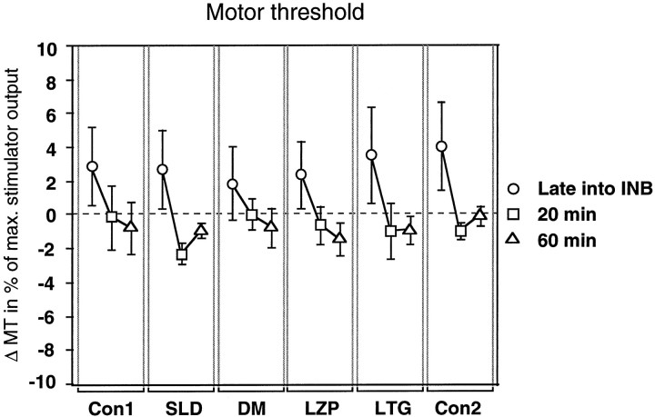
Intervention had no effect on INB plus rTMS-induced changes in MT [Fintervention(5,90) = 0.19], but time was an effective factor [Ftime(2,90) = 10.21; p = 0.0001] (Fig.1). Irrespective of intervention, INB plus rTMS induced a small, nonsignificant increase in MT late into ischemia (post hoc multiple t statistic,p > 0.05), which returned to or was even less than the preischemic MT when measured 20 and 60 min after ischemia (Fig.1). The interaction between intervention and time was not significant [F(10,90) = 0.18].
Fig. 1.
Effect of intervention on the changes in MT induced by ischemic forearm deafferentation and repetitive transcranial magnetic stimulation of the motor cortex contralateral to the side of ischemic nerve block. Changes in MT measured late into ischemia (circles) and 20 min (squares) and 60 min (triangles) after the end of ischemia are shown as differences from measurements obtained before the start of ischemia. These differences are given as a percentage of the maximal stimulator output (y-axis). Data are mean values of six subjects; error bars indicate SEM. Interventions are indicated (x-axis).
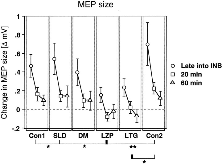
Intervention had a significant effect on the INB plus rTMS-induced changes in MEP size [Fintervention(5,90) = 2.56; p= 0.033]. The effect of time was also significant [Ftime(2,90) = 12.06; p < 0.0001], whereas the interaction between intervention and time was not significant [F(10,90) = 0.27] (Fig.2). A post hoc analysis revealed that the MEP changes under the influence of LZP were different from the changes in the Con1 (p = 0.046), Con2 (p = 0.0042), and SLD (p= 0.023) conditions. Furthermore, the MEP changes under LTG were different from those in the Con2 (p = 0.012) condition. Figure 2 shows that these differences were clearly the result of suppression of the INB plus rTMS-induced increase in MEP size by LZP and LTG.
Fig. 2.
Effect of intervention on the increase in MEP amplitude induced by ischemic forearm deafferentation and repetitive transcranial magnetic stimulation of the motor cortex contralateral to the side of ischemic nerve block. Changes in MEP size measured late into ischemia (circles) and 20 min (squares) and 60 min (triangles) after the end of ischemia are given as differences (Δ mV) from the values obtained before ischemic nerve block, which were assigned a value of 0 (y-axis). Other conventions and arrangements are described in Figure 1. Note that LZP and LTG led to suppression of the ischemia plus transcranial magnetic stimulation-induced increase in MEP size. *p < 0.05; **p < 0.01.
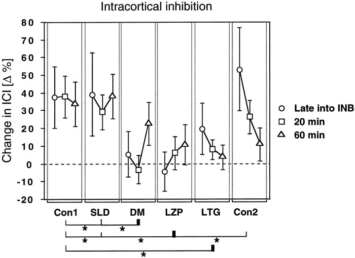
The INB plus rTMS-induced changes in ICI were also significantly affected by intervention [Fintervention(5,90) = 2.60; p = 0.031], whereas the effects of time [Ftime(2,90) = 0.37] and the interaction between intervention and time [F(10,90) = 0.56] were not significant (Fig. 3). Apost hoc analysis demonstrated that the INB plus rTMS-induced changes in ICI under the influence of LZP were significantly different from those in the Con1 (p = 0.014), Con2 (p = 0.048), and SLD (p = 0.017) conditions. Changes in ICI under LTG were different from those in the Con1 (p = 0.049) condition, and changes in ICI under DM were different from those in the Con1 (p = 0.032) and SLD (p = 0.038) conditions. Figure 3shows that these differences were the result of suppression of the INB plus rTMS-induced disinhibition by these drugs.
Fig. 3.
Effect of intervention on the decrease in ICI induced by ischemic forearm deafferentation and repetitive transcranial magnetic stimulation of the motor cortex contralateral to the side of ischemic nerve block. ICI values measured late into ischemia (circles) and 20 min (squares) and 60 min (triangles) after the end of ischemia are given as differences from the values obtained before ischemic nerve block. Because ICI values are percentages of conditioned to control motor-evoked potential amplitudes, the changes in ICI are expressed in Δ% (y-axis). Differences >0 indicate a decrease in ICI. Other conventions and arrangements are described in Figures 1 and 2. Note that DM, LZP, and LTG led to significant suppression of the ischemia plus transcranial magnetic stimulation-induced decrease in intracortical inhibition. *p < 0.05.
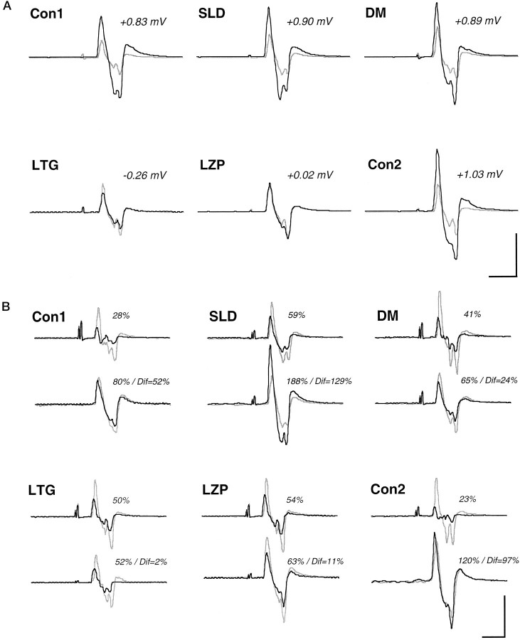
Figure 4 illustrates the effects of intervention on the INB plus rTMS-induced changes in MEP size and in ICI in a single subject. The EMG recordings show that the INB plus rTMS-induced increase in MEP size (Fig.4A) was completely suppressed by LTG and by LZP but not by DM. The INB plus rTMS-induced reduction in ICI (Fig.4B) was abolished by LTG and clearly suppressed by LZP and by DM.
Fig. 4.
EMG recordings from the biceps brachii muscle of one subject, illustrating the effects of intervention on the increase in MEP size (A) and on the decrease in intracortical inhibition (B) induced by ischemic forearm deafferentation and repetitive transcranial magnetic stimulation of the motor cortex contralateral to ischemic nerve block.A, MEPs obtained before ischemic nerve block (gray lines) and late into ischemia (black lines) are superimposed. The numbers (in mV) refer to the deafferentation-induced change in MEP size.B, Control MEPs produced by the test stimulus alone (gray lines) and MEPs elicited by paired stimulation at an interstimulus interval of 2 msec (black lines) are superimposed. For each intervention, thetop and bottom traces refer to MEPs obtained before and late into forearm ischemia, respectively. Thepercentages indicate the amount of intracortical inhibition and the difference (Dif) in intracortical inhibition before and late into ischemia. All MEPs are averages of eight single trials. Calibration bars: A, 20 msec, 0.5 mV; B, 20 msec, 0.25 mV. Note that LTG and LZP led to virtually complete abolition of the deafferentation-induced increase in MEP size in this subject. Furthermore, both of these drugs and DM had a suppressive effect on the deafferentation-induced decrease in intracortical inhibition.
DISCUSSION
Temporary ischemic forearm deafferentation combined with low-frequency rTMS of the motor cortex contralateral to the side of ischemia leads to a rapid increase in the excitability of the motor cortical representation targeting the BB proximal to the ischemic level. The present study shows that a single oral dose of the benzodiazepine LZP, the NMDA receptor antagonist DM, or the voltage-gated Na+ and Ca2+channel blocker LTG affects this form of plasticity to different extents.
Nonspecific factors for these findings were eliminated by the fact that the time to complete motor block, the total time of ischemia (Table1), and the preischemic values for MT, MEP size, and ICI (Table 2) were not different between interventions. Comparison of motor excitability across experimental sessions several days apart and the random use of two different stimulation coils introduced a slightly larger variability in testing than was reported previously (Ziemann et al., 1996a,b). Because preischemic MEP size was slightly, although not significantly, larger and ICI was less prominent in the drug sessions compared with at least one of the control sessions (Table 2), a “floor effect” cannot explain the suppressive drug effects on INB plus rTMS-induced changes in MEP size and in ICI.
The INB-induced increase in MEP size in muscles proximal to the ischemic nerve block (Brasil-Neto et al., 1992, 1993; Ridding and Rothwell, 1995, 1997; Ziemann et al., 1998) occurs at the level of the motor cortex, because subcortical and spinal excitability, tested with transcranial electrical stimulation, spinal electrical stimulation, and Hoffmann reflexes, did not change (Brasil-Neto et al., 1993). The increase in MEP size then reflects enhanced excitability of corticocortical excitatory connections, given that TMS activates corticospinal neurons predominately indirectly (Day et al., 1989;Nakamura et al., 1997). The INB plus rTMS-induced decrease in ICI reflects mainly a reduction in GABA-related motor cortical inhibition (Kujirai et al., 1993; Ziemann et al., 1996c). However, recent evidence showed that ICI consists of weak excitatory in addition to strong inhibitory effects (Ridding et al., 1995; Ziemann et al., 1996b); thus an enhancement of excitatory mechanisms may also contribute to the decrease in ICI. For two reasons, it was proposed that the rTMS-induced reduction in ICI is associated with an LTP-like phenomenon; (1) this reduction lasted for at least 60 min after the end of ischemic deafferentation, and (2) it was dependent on the INB-induced hyperexcitability of the plastic cortex, because in the absence of ischemia, rTMS did not cause any change in ICI (Ziemann et al., 1998). This is reminiscent of the need for sufficient depolarization of the postsynaptic site to allow the occurrence of LTP in the neocortex (Artola and Singer, 1987; Artola et al., 1990;Kirkwood et al., 1993; Hess and Donoghue, 1994; Kirkwood and Bear, 1994; Hess et al., 1996). Depolarization of the postsynaptic site can be achieved by local application of bicuculline, a GABAAreceptor antagonist, before afferent tetanic stimulation is started to induce LTP. The downregulation of GABAergic cortical circuits was proposed to be of crucial importance in deafferentation-induced plasticity (Jones, 1993). This idea is supported by various animal models of deafferentation, which showed a decrease in glutamic acid decarboxylase, the GABA synthetic enzyme (Hendry and Jones, 1986, 1988; Welker et al., 1989; Akhtar and Land, 1991), in GABA (Hendry and Jones, 1986, 1988; Garraghty et al., 1991; Rosier et al., 1995), and in GABA receptors (Hendry et al., 1990). However, it is unclear how soon changes in GABA can occur after the onset of deafferentation. None of these studies started to investigate GABA earlier than 3–5 d after the lesion, at which time the downregulation of GABA was fully expressed (Hendry and Jones, 1988; Welker et al., 1989).
The suppression of the INB plus rTMS-induced increase in MEP size and the decrease in ICI by the GABAA receptor agonist LZP in the present experiments suggest that the rapid removal of GABA-related inhibition is a permissive and necessary step for this form of deafferentation-induced plasticity to occur in the human motor cortex. An intriguing example of the importance of GABA-related circuitry in the mediation of rapid cortical plasticity was the appearance of a “new” forelimb representation in the vibrissae motor cortex of adult rats after GABA receptors in the adjacent forelimb motor cortex were blocked (Jacobs and Donoghue, 1991). These changes were probably caused by unmasking of normally latent excitatory connections from vibrissae to forelimb motor cortex. Other animal studies explored the effect of modulation of GABA-related inhibition on LTP induction in the hippocampus and showed that it was blocked by LZP (Riches and Brown, 1986) and other benzodiazepines (Satoh et al., 1986;del Cerro et al., 1992; Evans and Viola-McCabe, 1996) but facilitated by GABA receptor blockers (Wigström and Gustafsson, 1983).
The main mode of action of DM is a noncompetitive blockade of NMDA receptors (Wong et al., 1988; Netzer et al., 1993). In the present study, DM produced suppression of the INB plus rTMS-induced changes in ICI but had no significant effect on the increase in MEP size, which suggests that the INB plus rTMS-induced reduction in ICI but not the increase in MEP size was dependent on NMDA receptor-mediated plasticity. This further suggests that the decrease in ICI is interrelated with an LTP-like phenomenon, because LTP requires the activation of NMDA receptors (Collingridge et al., 1983;Artola and Singer, 1987; Kirkwood et al., 1993; Kirkwood and Bear, 1994; Hess et al., 1996). The present findings are also compatible with evidence from animal experiments that deafferentation-induced neocortical plasticity can be disrupted by NMDA receptor blockers (Kleinschmidt et al., 1987; Bear et al., 1990; Kano et al., 1991;Garraghty and Muja, 1996).
LTG blocks voltage-gated Na+ channels (Leach et al., 1995) and also high voltage-gated Ca2+ currents (Stefani et al., 1996; Wang et al., 1996) and glutamate release (Leach et al., 1986). In the present experiments, LTG had a suppressive effect on the INB plus rTMS-induced changes in both MEP size and ICI. It is likely that the blockade of Na+ and Ca2+ channels and/or the reduced glutamate release led to a decrease in the deafferentation-induced amount of depolarization in motor cortex output cells. Insufficient depolarization may then have prevented changes in MEP size and ICI from occurring. If a PTP-like mechanism had contributed to the INB plus rTMS-induced short-lasting changes in MEP size, this could explain why LTG but not DM had a suppressive effect, because PTP depends on the accumulation of Ca2+ and probably also Na+ in presynaptic terminals (Fisher et al., 1997) but not on NMDA receptor activation (Malenka, 1991). Although no experimental data are available about the effects of LTG on PTP, drugs with a very similar main mode of action, such as phenytoin, are potent blockers of PTP (Selzer et al., 1985; Griffith and Taylor, 1988). In addition, the changes in ICI may also have been suppressed by the blockade of Ca2+ channels, because a LTP component has been described that is independent of NMDA receptor activation but is dependent on voltage-gated Ca2+ channels (Grover and Teyler, 1990). Accordingly, LTG blocked this component of LTP in animal experiments (Wang et al., 1997).
In conclusion, the effects of CNS-active drugs in the present model of INB plus rTMS-induced plasticity in human sensorimotor cortex provide evidence that the deafferentation-induced increase in MEP size involved rapid downregulation of GABA-related inhibitory circuits and a mechanism mediated via voltage-gated Na+ or Ca2+ channels, either depolarization of motor cortical output cells or a PTP-like mechanism at presynaptic terminals. The rTMS-induced reduction in ICI was long lasting (>60 min), involved NMDA receptor activation, and therefore was likely interrelated with a LTP-like mechanism. The present study introduces a noninvasive experimental model for evaluating mechanisms of plasticity, in this case deafferentation-induced, in intact adult humans. Identification of the specific mechanisms involved in plastic changes in the human cerebral cortex is the necessary first step toward a definition of rationally founded therapeutic strategies in the reorganization of human cortex, that is, the promotion of functionally beneficial (Cohen et al., 1997) but the suppression of probably maladaptive plasticity (Ramachandran et al., 1992; Yang et al., 1994; Flor et al., 1995;Knecht et al., 1995).
Footnotes
This work was supported by Grant Zi 542/1-1 to U.Z. from the Deutsche Forschungsgemeinschaft. We thank Dr. M. A. Rogawski for excellent discussion, B. Corwell for help in some of the experiments, and B. J. Hessie for skillful editing of this manuscript.
Correspondence should be addressed to Dr. Ulf Ziemann or Dr. Leonardo G. Cohen, Building 10, Room 5N234, National Institutes of Health, 10 Center Drive, MSC-1428, Bethesda, MD 20892-1428.
REFERENCES
- 1.Akhtar ND, Land PW. Activity-dependent regulation of glutamic acid decarboxylase in the rat barrel cortex: effects of neonatal versus adult sensory deprivation. J Comp Neurol. 1991;307:200–213. doi: 10.1002/cne.903070204. [DOI] [PubMed] [Google Scholar]
- 2.Artola A, Singer W. Long-term potentiation and NMDA receptors in rat visual cortex. Nature. 1987;330:649–652. doi: 10.1038/330649a0. [DOI] [PubMed] [Google Scholar]
- 3.Artola A, Brocher S, Singer W. Different voltage-dependent thresholds for inducing long-term depression and long-term potentiation in slices of rat visual cortex. Nature. 1990;347:69–72. doi: 10.1038/347069a0. [DOI] [PubMed] [Google Scholar]
- 4.Bear MF, Kleinschmidt A, Gu QA, Singer W. Disruption of experience-dependent synaptic modifications in striate cortex by infusion of an NMDA receptor antagonist. J Neurosci. 1990;10:909–925. doi: 10.1523/JNEUROSCI.10-03-00909.1990. [DOI] [PMC free article] [PubMed] [Google Scholar]
- 5.Bliss TV, Lømo T. Long-lasting potentiation of synaptic transmission in the dentate area of the anaesthetized rabbit following stimulation of the perforant path. J Physiol (Lond) 1973;232:331–356. doi: 10.1113/jphysiol.1973.sp010273. [DOI] [PMC free article] [PubMed] [Google Scholar]
- 6.Borsook D, Becerra L, Fishman S, Edwards A, Jennings CL, Stojanovic M, Papinicolas L, Ramachandran VS, Gonzalez RG, Breiter H. Acute plasticity in the human somatosensory cortex following amputation. NeuroReport. 1998;9:1013–1017. doi: 10.1097/00001756-199804200-00011. [DOI] [PubMed] [Google Scholar]
- 7.Brasil-Neto JP, Cohen LG, Pascual-Leone A, Jabir FK, Wall RT, Hallett M. Rapid reversible modulation of human motor outputs after transient deafferentation of the forearm: a study with transcranial magnetic stimulation. Neurology. 1992;42:1302–1306. doi: 10.1212/wnl.42.7.1302. [DOI] [PubMed] [Google Scholar]
- 8.Brasil-Neto JP, Valls-Sole J, Pascual-Leone A, Cammarota A, Amassian VE, Cracco R, Maccabee P, Cracco J, Hallett M, Cohen LG. Rapid modulation of human cortical motor outputs following ischaemic nerve block. Brain. 1993;116:511–525. doi: 10.1093/brain/116.3.511. [DOI] [PubMed] [Google Scholar]
- 9.Brouwer B, Ashby P. Corticospinal projections to upper and lower limb spinal motoneurons in man. Electroencephalogr Clin Neurophysiol. 1990;76:509–519. doi: 10.1016/0013-4694(90)90002-2. [DOI] [PubMed] [Google Scholar]
- 10.Byrne JA, Calford MB. Short-term expansion of receptive fields in rat primary somatosensory cortex after hindpaw digit denervation. Brain Res. 1991;565:218–224. doi: 10.1016/0006-8993(91)91652-h. [DOI] [PubMed] [Google Scholar]
- 11.Calford MB, Tweedale R. Immediate and chronic changes in responses of somatosensory cortex in adult flying-fox after digit amputation. Nature. 1988;332:446–448. doi: 10.1038/332446a0. [DOI] [PubMed] [Google Scholar]
- 12.Calford MB, Tweedale R. Acute changes in cutaneous receptive fields in primary somatosensory cortex after digit denervation in adult flying fox. J Neurophysiol. 1991a;65:178–187. doi: 10.1152/jn.1991.65.2.178. [DOI] [PubMed] [Google Scholar]
- 13.Calford MB, Tweedale R. Immediate expansion of receptive fields of neurons in area 3b of macaque monkeys after digit denervation. Somatosens Mot Res. 1991b;8:249–260. doi: 10.3109/08990229109144748. [DOI] [PubMed] [Google Scholar]
- 14.Castro-Alamancos MA, Donoghue JP, Connors BW. Different forms of synaptic plasticity in somatosensory and motor areas of the neocortex. J Neurosci. 1995;15:5324–5333. doi: 10.1523/JNEUROSCI.15-07-05324.1995. [DOI] [PMC free article] [PubMed] [Google Scholar]
- 15.Chen R, Classen J, Gerloff C, Celnik P, Wassermann EM, Hallett M, Cohen LG. Depression of motor cortex excitability by low-frequency transcranial magnetic stimulation. Neurology. 1997a;48:1398–1403. doi: 10.1212/wnl.48.5.1398. [DOI] [PubMed] [Google Scholar]
- 16.Chen R, Samii A, Canos M, Wassermann EM, Hallett M. Effects of phenytoin on cortical excitability in humans. Neurology. 1997b;49:881–883. doi: 10.1212/wnl.49.3.881. [DOI] [PubMed] [Google Scholar]
- 17.Cohen LG, Celnik P, Pascual-Leone A, Corwell B, Faiz L, Dambrosia J, Honda M, Sadato N, Gerloff C, Catala MD, Hallett M. Functional relevance of cross-modal plasticity in the blind. Nature. 1997;389:180–183. doi: 10.1038/38278. [DOI] [PubMed] [Google Scholar]
- 18.Collingridge GL, Kehl SJ, McLennan H. Excitatory amino acids in synaptic transmission in the Schaffer collateral-commissural pathway of the rat hippocampus. J Physiol (Lond) 1983;334:33–46. doi: 10.1113/jphysiol.1983.sp014478. [DOI] [PMC free article] [PubMed] [Google Scholar]
- 19.Day BL, Dressler D, Maertens de Noordhout A, Marsden CD, Nakashima K, Rothwell JC, Thompson PD. Electric and magnetic stimulation of the human motor cortex: surface EMG and single motor unit responses. J Physiol (Lond) 1989;412:449–473. doi: 10.1113/jphysiol.1989.sp017626. [DOI] [PMC free article] [PubMed] [Google Scholar]
- 20.del Cerro S, Jung M, Lynch G. Benzodiazepines block long-term potentiation in slices of hippocampus and piriform cortex. Neuroscience. 1992;49:1–6. doi: 10.1016/0306-4522(92)90071-9. [DOI] [PubMed] [Google Scholar]
- 21.Di Lazzaro V, Restuccia D, Oliviero A, Profice P, Ferrara L, Insola A, Mazzone P, Tonali P, Rothwell JC. Magnetic transcranial stimulation at intensities below active motor threshold activates intracortical inhibitory circuits. Exp Brain Res. 1998;119:265–268. doi: 10.1007/s002210050341. [DOI] [PubMed] [Google Scholar]
- 22.Donoghue JP, Suner S, Sanes JN. Dynamic organization of primary motor cortex output to target muscles in adult rats. II. Rapid reorganization following motor nerve lesions. Exp Brain Res. 1990;79:492–503. doi: 10.1007/BF00229319. [DOI] [PubMed] [Google Scholar]
- 23.Donoghue JP, Hess G, Sanes JN. Substrates and mechanisms for learning in motor cortex. In: Bloedel J, Ebner T, Wise SP, editors. Acquisition of motor behavior in vertebrates. MIT; Cambridge, MA: 1996. pp. 363–386. [Google Scholar]
- 24.Evans MS, Viola-McCabe KE. Midazolam inhibits long-term potentiation through modulation of GABAA receptors. Neuropharmacology. 1996;35:347–357. doi: 10.1016/0028-3908(95)00182-4. [DOI] [PubMed] [Google Scholar]
- 25.Fisher SA, Fischer TM, Carew TJ. Multiple overlapping processes underlying short-term synaptic enhancement. Trends Neurosci. 1997;20:170–177. doi: 10.1016/s0166-2236(96)01001-6. [DOI] [PubMed] [Google Scholar]
- 26.Flor H, Elbert T, Knecht S, Wienbruch C, Pantev C, Birbaumer N, Larbig W, Taub E. Phantom-limb pain as a perceptual correlate of cortical reorganization following arm amputation. Nature. 1995;375:482–484. doi: 10.1038/375482a0. [DOI] [PubMed] [Google Scholar]
- 27.Garraghty PE, Muja N. NMDA receptors and plasticity in adult primate somatosensory cortex. J Comp Neurol. 1996;367:319–326. doi: 10.1002/(SICI)1096-9861(19960401)367:2<319::AID-CNE12>3.0.CO;2-L. [DOI] [PubMed] [Google Scholar]
- 28.Garraghty PE, LaChica EA, Kaas JH. Injury-induced reorganization of somatosensory cortex is accompanied by reductions in GABA staining. Somatosens Mot Res. 1991;8:347–354. doi: 10.3109/08990229109144757. [DOI] [PubMed] [Google Scholar]
- 29.Grafton ST, Mazziotta JC, Presty S, Friston KJ, Frackowiak RS, Phelps ME. Functional anatomy of human procedural learning determined with regional cerebral blood flow and PET. J Neurosci. 1992;12:2542–2548. doi: 10.1523/JNEUROSCI.12-07-02542.1992. [DOI] [PMC free article] [PubMed] [Google Scholar]
- 30.Griffith WH, Taylor L. Phenytoin reduces excitatory synaptic transmission and post-tetanic potentiation in the in vitro hippocampus. J Pharmacol Exp Ther. 1988;246:851–858. [PubMed] [Google Scholar]
- 31.Grover LM, Teyler TJ. Two components of long-term potentiation induced by different patterns of afferent activation. Nature. 1990;347:477–479. doi: 10.1038/347477a0. [DOI] [PubMed] [Google Scholar]
- 32.Hendry SH, Jones EG. Reduction in number of immunostained GABAergic neurones in deprived-eye dominance columns of monkey area 17. Nature. 1986;320:750–753. doi: 10.1038/320750a0. [DOI] [PubMed] [Google Scholar]
- 33.Hendry SH, Jones EG. Activity-dependent regulation of GABA expression in the visual cortex of adult monkeys. Neuron. 1988;1:701–712. doi: 10.1016/0896-6273(88)90169-9. [DOI] [PubMed] [Google Scholar]
- 34.Hendry SH, Fuchs J, deBlas AL, Jones EG. Distribution and plasticity of immunocytochemically localized GABAA receptors in adult monkey visual cortex. J Neurosci. 1990;10:2438–2450. doi: 10.1523/JNEUROSCI.10-07-02438.1990. [DOI] [PMC free article] [PubMed] [Google Scholar]
- 35.Hess G, Donoghue JP. Long-term potentiation of horizontal connections provides a mechanism to reorganize cortical motor maps. J Neurophysiol. 1994;71:2543–2547. doi: 10.1152/jn.1994.71.6.2543. [DOI] [PubMed] [Google Scholar]
- 36.Hess G, Aizenman CD, Donoghue JP. Conditions for the induction of long-term potentiation in layer II/III horizontal connections of the rat motor cortex. J Neurophysiol. 1996;75:1765–1778. doi: 10.1152/jn.1996.75.5.1765. [DOI] [PubMed] [Google Scholar]
- 37.Huntley GW. Correlation between patterns of horizontal connectivity and the extent of short-term representational plasticity in rat motor cortex. Cereb Cortex. 1997;7:143–156. doi: 10.1093/cercor/7.2.143. [DOI] [PubMed] [Google Scholar]
- 38.Iriki A, Pavlides C, Keller A, Asanuma H. Long-term potentiation in the motor cortex. Science. 1989;245:1385–1387. doi: 10.1126/science.2551038. [DOI] [PubMed] [Google Scholar]
- 39.Jacobs KM, Donoghue JP. Reshaping the cortical motor map by unmasking latent intracortical connections. Science. 1991;251:944–947. doi: 10.1126/science.2000496. [DOI] [PubMed] [Google Scholar]
- 40.Jones EG. GABAergic neurons and their role in cortical plasticity in primates. Cereb Cortex. 1993;3:361–372. doi: 10.1093/cercor/3.5.361-a. [DOI] [PubMed] [Google Scholar]
- 41.Kaneko K, Kawai S, Fuchigami Y, Morita H, Ofuji A. The effect of current direction induced by transcranial magnetic stimulation on the corticospinal excitability in human brain. Electroencephalogr Clin Neurophysiol. 1996;101:478–482. doi: 10.1016/s0013-4694(96)96021-x. [DOI] [PubMed] [Google Scholar]
- 42.Kano M, Iino K, Kano M. Functional reorganization of adult cat somatosensory cortex is dependent on NMDA receptors. NeuroReport. 1991;2:77–80. doi: 10.1097/00001756-199102000-00003. [DOI] [PubMed] [Google Scholar]
- 43.Karni A, Meyer G, Jezzard P, Adams MM, Turner R, Ungerleider LG. Functional MRI evidence for adult motor cortex plasticity during motor skill learning. Nature. 1995;377:155–158. doi: 10.1038/377155a0. [DOI] [PubMed] [Google Scholar]
- 44.Kirkwood A, Bear MF. Hebbian synapses in visual cortex. J Neurosci. 1994;14:1634–1645. doi: 10.1523/JNEUROSCI.14-03-01634.1994. [DOI] [PMC free article] [PubMed] [Google Scholar]
- 45.Kirkwood A, Dudek SD, Gold JT, Aizenman CD, Bear MF. Common forms of synaptic plasticity in hippocampus and neocortex. Science. 1993;260:1518–1521. doi: 10.1126/science.8502997. [DOI] [PubMed] [Google Scholar]
- 46.Kleinschmidt A, Bear MF, Singer W. Blockade of “NMDA” receptors disrupts experience-dependent plasticity of kitten striate cortex. Science. 1987;238:355–358. doi: 10.1126/science.2443978. [DOI] [PubMed] [Google Scholar]
- 47.Knecht S, Henningsen H, Elbert T, Flor H, Hohling C, Pantev C, Birbaumer N, Taub E. Cortical reorganization in human amputees and mislocalization of painful stimuli to the phantom limb. Neurosci Lett. 1995;201:262–264. doi: 10.1016/0304-3940(95)12186-2. [DOI] [PubMed] [Google Scholar]
- 48.Kolarik RC, Rasey SK, Wall JT. The consistency, extent, and locations of early-onset changes in cortical nerve dominance aggregates following injury of nerves to primate hands. J Neurosci. 1994;14:4269–4288. doi: 10.1523/JNEUROSCI.14-07-04269.1994. [DOI] [PMC free article] [PubMed] [Google Scholar]
- 49.Kujirai T, Caramia MD, Rothwell JC, Day BL, Thompson PD, Ferbert A, Wroe S, Asselman P, Marsden CD. Corticocortical inhibition in human motor cortex. J Physiol (Lond) 1993;471:501–519. doi: 10.1113/jphysiol.1993.sp019912. [DOI] [PMC free article] [PubMed] [Google Scholar]
- 50.Leach MJ, Marden CM, Miller AA. Pharmacological studies on lamotrigine, a novel potential antiepileptic drug: II. Neurochemical studies on the mechanism of action. Epilepsia. 1986;27:490–497. doi: 10.1111/j.1528-1157.1986.tb03573.x. [DOI] [PubMed] [Google Scholar]
- 51.Leach MJ, Lees G, Riddall DR. Lamotrigine. Mechanisms of action. In: Levy RH, Mattson RH, Meldrum BS, editors. Antiepileptic drugs. Raven; New York: 1995. pp. 861–869. [Google Scholar]
- 52.Liepert J, Schwenkreis P, Tegenthoff M, Malin J-P. The glutamate antagonist Riluzole suppresses intracortical facilitation. J Neural Transm. 1997;104:1207–1214. doi: 10.1007/BF01294721. [DOI] [PubMed] [Google Scholar]
- 53.Macdonald RL. Benzodiazepines. Mechanisms of action. In: Levy RH, Mattson RH, Meldrum BS, editors. Antiepileptic drugs. Raven; New York: 1995. pp. 695–703. [Google Scholar]
- 54.Malenka RC. Postsynaptic factors control the duration of synaptic enhancement in area CA1 of the hippocampus. Neuron. 1991;6:53–60. doi: 10.1016/0896-6273(91)90121-f. [DOI] [PubMed] [Google Scholar]
- 55.Mavroudakis N, Caroyer JM, Brunko E, Zegers de Beyl D. Effects of diphenylhydantoin on motor potentials evoked with magnetic stimulation. Electroencephalogr Clin Neurophysiol. 1994;93:428–433. doi: 10.1016/0168-5597(94)90149-x. [DOI] [PubMed] [Google Scholar]
- 56.Merzenich MM, Kaas JH, Wall JT, Sur M, Nelson RJ, Felleman DJ. Progression of change following median nerve section in the cortical representation of the hand in areas 3b and 1 in adult owl and squirrel monkeys. Neuroscience. 1983;10:639–665. doi: 10.1016/0306-4522(83)90208-7. [DOI] [PubMed] [Google Scholar]
- 57.Nakamura H, Kitagawa H, Kawaguchi Y, Tsuji H. Intracortical facilitation and inhibition after transcranial magnetic stimulation in conscious humans. J Physiol (Lond) 1997;498:817–823. doi: 10.1113/jphysiol.1997.sp021905. [DOI] [PMC free article] [PubMed] [Google Scholar]
- 58.Netzer R, Pflimlin P, Trube G. Dextromethorphan blocks N-methyl-d-aspartate-induced currents and voltage-operated inward currents in cultured cortical neurons. Eur J Pharmacol. 1993;238:209–216. doi: 10.1016/0014-2999(93)90849-d. [DOI] [PubMed] [Google Scholar]
- 59.Nudo RJ, Milliken GW, Jenkins WM, Merzenich MM. Use-dependent alterations of movement representations in primary motor cortex of adult squirrel monkeys. J Neurosci. 1996;16:785–807. doi: 10.1523/JNEUROSCI.16-02-00785.1996. [DOI] [PMC free article] [PubMed] [Google Scholar]
- 60.Pascual-Leone A, Grafman J, Hallett M. Modulation of cortical motor output maps during development of implicit and explicit knowledge. Science. 1994;263:1287–1289. doi: 10.1126/science.8122113. [DOI] [PubMed] [Google Scholar]
- 61.Pascual-Leone A, Nguyet D, Cohen LG, Brasil-Neto JP, Cammarota A, Hallett M. Modulation of muscle responses evoked by transcranial magnetic stimulation during the acquisition of new fine motor skills. J Neurophysiol. 1995;74:1037–1045. doi: 10.1152/jn.1995.74.3.1037. [DOI] [PubMed] [Google Scholar]
- 62.Ramachandran VS, Stewart M, Rogers-Ramachandran DC. Perceptual correlates of massive cortical reorganization. NeuroReport. 1992;3:583–586. doi: 10.1097/00001756-199207000-00009. [DOI] [PubMed] [Google Scholar]
- 63.Recanzone GH, Merzenich MM, Jenkins WM, Grajski KA, Dinse HR. Topographic reorganization of the hand representation in cortical area 3b of owl monkeys trained in a frequency-discrimination task. J Neurophysiol. 1992;67:1031–1056. doi: 10.1152/jn.1992.67.5.1031. [DOI] [PubMed] [Google Scholar]
- 64.Riches IP, Brown MW. The effect of lorazepam upon hippocampal long-term potentiation. Neurosci Lett [Suppl] 1986;24:S42. [Google Scholar]
- 65.Ridding MC, Rothwell JC. Reorganisation in human motor cortex. Can J Physiol Pharmacol. 1995;73:218–222. doi: 10.1139/y95-032. [DOI] [PubMed] [Google Scholar]
- 66.Ridding MC, Rothwell JC. Stimulus/response curves as a method of measuring motor cortical excitability in man. Electroencephalogr Clin Neurophysiol. 1997;105:340–344. doi: 10.1016/s0924-980x(97)00041-6. [DOI] [PubMed] [Google Scholar]
- 67.Ridding MC, Inzelberg R, Rothwell JC. Changes in excitability of motor cortical circuitry in patients with Parkinson’s disease. Ann Neurol. 1995;37:181–188. doi: 10.1002/ana.410370208. [DOI] [PubMed] [Google Scholar]
- 68.Rosier AM, Arckens L, Demeulemeester H, Orban GA, Eysel UT, Wu YJ, Vandesande F. Effect of sensory deafferentation on immunoreactivity of GABAergic cells and on GABA receptors in the adult cat visual cortex. J Comp Neurol. 1995;359:476–489. doi: 10.1002/cne.903590309. [DOI] [PubMed] [Google Scholar]
- 69.Sanes JN, Donoghue JP. Static and dynamic organization of motor cortex. Adv Neurol. 1997;73:277–296. [PubMed] [Google Scholar]
- 70.Sanes JN, Suner S, Lando JF, Donoghue JP. Rapid reorganization of adult rat motor cortex somatic representation patterns after motor nerve injury. Proc Natl Acad Sci USA. 1988;85:2003–2007. doi: 10.1073/pnas.85.6.2003. [DOI] [PMC free article] [PubMed] [Google Scholar]
- 71.Satoh M, Ishihara K, Iwama T, Takagi H. Aniracetam augments, and midazolam inhibits, the long-term potentiation in guinea-pig hippocampal slices. Neurosci Lett. 1986;68:216–220. doi: 10.1016/0304-3940(86)90145-x. [DOI] [PubMed] [Google Scholar]
- 72.Schieber MH, Hibbard LS. How somatotopic is the motor cortex hand area? Science. 1993;261:489–492. doi: 10.1126/science.8332915. [DOI] [PubMed] [Google Scholar]
- 73.Selzer ME, David G, Yaari Y. On the mechanisms by which phenytoin blocks post-tetanic potentiation at the frog neuromuscular junction. J Neurosci. 1985;5:2894–2899. doi: 10.1523/JNEUROSCI.05-11-02894.1985. [DOI] [PMC free article] [PubMed] [Google Scholar]
- 74.Silva AC, Rasey SK, Wu X, Wall JT. Initial cortical reactions to injury of the median and radial nerves to the hands of adult primates. J Comp Neurol. 1996;366:700–716. doi: 10.1002/(SICI)1096-9861(19960318)366:4<700::AID-CNE9>3.0.CO;2-8. [DOI] [PubMed] [Google Scholar]
- 75.Stefani A, Spadoni F, Siniscalchi A, Bernardi G. Lamotrigine inhibits Ca2+ currents in cortical neurons: functional implications. Eur J Pharmacol. 1996;307:113–116. doi: 10.1016/0014-2999(96)00265-8. [DOI] [PubMed] [Google Scholar]
- 76.Wang SJ, Huang CC, Hsu KS, Tsai JJ, Gean PW. Inhibition of N-type calcium currents by lamotrigine in rat amygdalar neurones. NeuroReport. 1996;7:3037–3040. doi: 10.1097/00001756-199611250-00048. [DOI] [PubMed] [Google Scholar]
- 77.Wang SJ, Tsai JJ, Gean PW. Lamotrigine inhibits tetraethylammonium-induced synaptic plasticity in the rat amygdala. Neuroscience. 1997;81:667–671. doi: 10.1016/s0306-4522(97)00216-9. [DOI] [PubMed] [Google Scholar]
- 78.Welker E, Soriano E, Van der Loos H. Plasticity in the barrel cortex of the adult mouse: effects of peripheral deprivation on GAD-immunoreactivity. Exp Brain Res. 1989;74:441–452. doi: 10.1007/BF00247346. [DOI] [PubMed] [Google Scholar]
- 79.Wigström H, Gustafsson B. Facilitated induction of hippocampal long-lasting potentiation during blockade of inhibition. Nature. 1983;301:603–604. doi: 10.1038/301603a0. [DOI] [PubMed] [Google Scholar]
- 80.Wong BY, Coulter DA, Choi DW, Prince DA. Dextrorphan and dextromethorphan, common antitussives, are antiepileptic and antagonize N-methyl-d-aspartate in brain slices. Neurosci Lett. 1988;85:261–266. doi: 10.1016/0304-3940(88)90362-x. [DOI] [PubMed] [Google Scholar]
- 81.Yang TT, Gallen CC, Ramachandran VS, Cobb S, Schwartz BJ, Bloom FE. Noninvasive detection of cerebral plasticity in adult human somatosensory cortex. NeuroReport. 1994;5:701–704. doi: 10.1097/00001756-199402000-00010. [DOI] [PubMed] [Google Scholar]
- 82.Ziemann U, Lönnecker S, Steinhoff BJ, Paulus W. The effect of lorazepam on the motor cortical excitability in man. Exp Brain Res. 1996a;109:127–135. doi: 10.1007/BF00228633. [DOI] [PubMed] [Google Scholar]
- 83.Ziemann U, Lönnecker S, Steinhoff BJ, Paulus W. Effects of antiepileptic drugs on motor cortex excitability in humans: a transcranial magnetic stimulation study. Ann Neurol. 1996b;40:367–378. doi: 10.1002/ana.410400306. [DOI] [PubMed] [Google Scholar]
- 84.Ziemann U, Rothwell JC, Ridding MC. Interaction between intracortical inhibition and facilitation in human motor cortex. J Physiol (Lond) 1996c;496:873–881. doi: 10.1113/jphysiol.1996.sp021734. [DOI] [PMC free article] [PubMed] [Google Scholar]
- 85.Ziemann U, Corwell B, Cohen LG. Modulation of plasticity in human motor cortex after forearm ischemic nerve block. J Neurosci. 1998;18:1115–1123. doi: 10.1523/JNEUROSCI.18-03-01115.1998. [DOI] [PMC free article] [PubMed] [Google Scholar]


