Abstract
A 59-year-old woman with end-stage renal disease presented for suspected Stevens-Johnson syndrome that was ultimately diagnosed as generalized bullous fixed drug eruption (GBFDE) secondary to the administration of iodinated nonpolar radiocontrast. The patient had three previous episodes of a generalized bullous eruption after a thrombectomy, fistulogram, and an arteriovenous fistula revision, all requiring radiocontrast administration. Biopsies taken after previous eruptions demonstrated full-thickness epidermal necrosis, and she was diagnosed with Stevens-Johnson syndrome thought to be due to allopurinol. However, against medical advice she continued taking allopurinol and remained asymptomatic until the current presentation. Based on the clinical appearance and time frame of the eruptions, the patient was diagnosed with GBFDE due to radiocontrast. GBFDE, a rare variant of a fixed drug eruption, can be misdiagnosed as Stevens-Johnson syndrome due to their overlapping features of drug-induced whole-body blisters and variable degrees of epidermal necrosis.
Keywords: Generalized bullous fixed drug eruption, radiocontrast
We present the rare case of generalized bullous fixed drug eruption (GBFDE) induced by iodinated nonpolar radiocontrast media (iohexol). Fixed drug eruption (FDE), a cutaneous drug reaction, is characterized by well-demarcated, dusky, circular plaques that reappear at the same location upon re-exposure to the offending agent. GBFDE has a clinical and pathological presentation similar to that of Stevens-Johnson syndrome (SJS).
CASE PRESENTATION
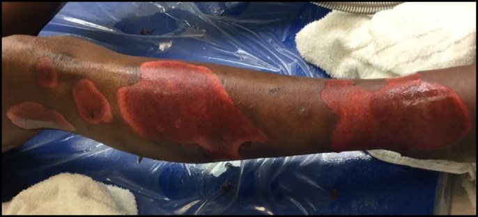
A 59-year-old black woman with end-stage renal disease on hemodialysis presented to the emergency department with painful, dusky brown-red targetoid plaques with superimposed flaccid bullae on her bilateral upper and lower extremities (Figure 1), trunk, and face. Approximately 20% of her total body surface area was affected. Two days prior, the patient underwent a computed tomography angiogram. During administration of the radiocontrast, she experienced severe discomfort and pruritus. That evening, she noticed a painful, pruritic rash on her lower legs that worsened the following day.
Figure 1.
Generalized bullous fixed drug eruption after administration of radiocontrast. Painful, large, dusky, well-demarcated, irregular, and annular plaques after surgical debridement of large flaccid bullae on the lower extremity. Lesions were present on the extremities, trunk, lips, and face. Approximately 20% of total body surface area was involved.
Upon chart review, it was discovered that the patient had three previous episodes of a generalized bullous eruption within 12 hours of contrast administration: after a thrombectomy, fistulogram, and an arteriovenous fistula revision. Biopsies taken after previous eruptions demonstrated pauci-inflammatory full-thickness epidermal necrosis. Based on her biopsy results and medication history of allopurinol use, the patient was diagnosed with SJS. However, against medical advice she continued taking allopurinol and was asymptomatic until the current presentation. Based on the clinical appearance and time frame of the eruption, the patient was diagnosed with GBFDE due to radiocontrast.
DISCUSSION
FDE, a rare variant of drug-induced dermatoses, is characterized by the recurrence of distinct, dusky-red patches and edematous plaques at similar locations upon re-exposure to the offending agent. Within minutes to hours upon re-exposure, the acute episode of FDE presents with pruritus and an intensely painful burning sensation.1 FDE classically involves the limbs, hands, and feet. The different morphological patterns include nonpigmenting and bullous, with our case corresponding to the very rare variant of GBFDE. Rarely, repeated exposure to the offending drug may cause an undifferentiated FDE to evolve into GBFDE.2
GBFDE, the rarest variant of FDE, can be misdiagnosed as SJS due to their overlapping features of drug-induced generalized bullae and erosions.1 There are a few clinical and pathological differences that can help clinicians discern between GBFDE and SJS (Table 1). On physical examination, GBFDE generally presents with well-demarcated, erythematous lesions reoccurring at previously affected sites with sparse mucosal involvement.3 In contrast, SJS generally presents with targetoid lesions with mucosal involvement.4 On histopathological examination, variable degrees of epidermal necrosis and inflammatory infiltrate at the dermoepidermal junction can be seen in both GBFDE and SJS. A lichenoid infiltrate with scattered epidermal necrosis is a common finding in GBFDE, whereas SJS is more pauci-inflammatory with greater epidermal necrosis.5 To differentiate GBFDE from SJS, the biopsy specimen should be of an acute lesion, <24 hours old, because older bullae can display epidermal necrosis observed in SJS.6 The previous biopsies from our patient were all performed several days after contrast administration and were therefore more consistent with the histopathology of SJS. Our case denotes the importance of early biopsy in bullous eruptions for accurate diagnosis.
Table 1.
Distinguishing features of generalized bullous fixed drug eruption and Stevens-Johnson syndrome
| Characteristic | Generalized bullous fixed drug eruption | Stevens-Johnson syndrome |
|---|---|---|
| Implicated drugs | Antibiotics, nonsteroidal anti-inflammatory drugs, acetaminophen, antiepileptic drugs, antimalarials2 | Allopurinol, antiepileptic drugs, antibiotics, nonsteroidal anti-inflammatory drugs, nevirapine2 |
| Onset after inciting agent | Onset within 30 minutes to 12 hours3 | Onset within 6 to 21 days4 |
| Site specificity | Reoccurs at same site: trunk, extremities; spares mucosa or mild mucosal involvement3 | Mucosal involvement in >90%: oral, ocular, urogenital4 |
| Cutaneous lesions | Well-circumscribed annular red/brown macules or plaques with overlying flaccid bullae1 | Painful, ill-defined, coalescing, erythematous macules with purpuric centers progressing to vesicles and bullae1 |
| Histopathology | Scattered apoptotic keratinocytes, lichenoid inflammatory infiltrate with eosinophils, dermal macrophages, and pigment incontinence5 | Partial to full-thickness epidermal necrosis, pauci-inflammatory infiltrate with variable number of eosinophils5 |
Our patient’s clinical and histopathologic findings provide an important example of GBFDE initially mistaken for SJS. Our case was complicated by a well-known SJS inducer: allopurinol. GBFDE can be mistaken for SJS, because both can involve whole-body blisters and variable degrees of epidermal necrosis.
References
- 1.Cho YT, Lin JW, Chen YC, et al. Generalized bullous fixed drug eruption is distinct from Stevens-Johnson syndrome/toxic epidermal necrolysis by immunohistopathological features. J Am Acad Dermatol. 2014;70:539–548. [DOI] [PubMed] [Google Scholar]
- 2.Mockenhaupt M. Epidemiology of cutaneous adverse drug reactions. Chem Immunol Allergy. 2017;1:96–108. doi: 10.5414/ALX01508E. [DOI] [PubMed] [Google Scholar]
- 3.Böhm I, Medina J, Prieto P, Block W, Schild HH. Fixed drug eruption induced by an iodinated non-ionic x-ray contrast medium: a practical approach to identify the causative agent and to prevent its recurrence. Eur Radiol. 2007;17:485–489. doi: 10.1007/s00330-006-0371-6. [DOI] [PubMed] [Google Scholar]
- 4.Fouchard N, Bertocchi M, Roujeau J-C, Revuz J, Wolkenstein P, Bastuji-Garin S. SCORTEN: a severity-of-illness score for toxic epidermal necrolysis. J Invest Dermatol. 2000;115:149–153. doi: 10.1046/j.1523-1747.2000.00061.x. [DOI] [PubMed] [Google Scholar]
- 5.Weyers W, Metze D. Histopathology of drug eruptions—general criteria, common patterns, and differential diagnosis. Dermatol Pract Concept. 2011;1:33–47. doi: 10.5826/dpc.0101a09. [DOI] [PMC free article] [PubMed] [Google Scholar]
- 6.Elston DM, Stratman EJ, Miller SJ. Skin biopsy. J Am Acad Dermatol. 2016;74:1–16. doi: 10.1016/j.jaad.2015.06.033. [DOI] [PubMed] [Google Scholar]


