The fungal cell wall is an essential organelle whose components provide the first line of defense against host-induced antifungal activity. Chitosan is one of the carbohydrate polymers in the cell wall that significantly affects the outcome of host-pathogen interaction. Chitosan-deficient strains are avirulent, implicating chitosan as a critical virulence factor. C. gattii R265 is an important fungal pathogen of concern due to its ability to cause infections in individuals with no apparent immune dysfunction and an increasing geographical distribution. Characterization of the fungal cell wall and understanding the contribution of individual molecules of the cell wall matrix to fungal pathogenesis offer new therapeutic avenues for intervention. In this report, we show that the C. gattii R265 strain has evolved alternate regulation of chitosan biosynthesis under both laboratory growth conditions and during mammalian infection compared to that of C. neoformans.
KEYWORDS: Cryptococcus gattii, R265, chitin, chitosan, chitosan regulation, protection, vaccine, virulence
ABSTRACT
Cryptococcus gattii R265 is a hypervirulent fungal strain responsible for the recent outbreak of cryptococcosis in Vancouver Island of British Columbia in Canada. It differs significantly from Cryptococcus neoformans in its natural environment, its preferred site in the mammalian host, and its pathogenesis. Our previous studies of C. neoformans have shown that the presence of chitosan, the deacetylated form of chitin, in the cell wall attenuates inflammatory responses in the host, while its absence induces robust immune responses, which in turn facilitate clearance of the fungus and induces a protective response. The results of the present investigation reveal that the cell wall of C. gattii R265 contains a two- to threefold larger amount of chitosan than that of C. neoformans. The genes responsible for the biosynthesis of chitosan are highly conserved in the R265 genome; the roles of the three chitin deacetylases (CDAs) have, however, been modified. To deduce their roles, single and double CDA deletion strains and a triple CDA deletion strain were constructed in a R265 background and were subjected to mammalian infection studies. Unlike C. neoformans where Cda1 has a discernible role in fungal pathogenesis, in strain R265, Cda3 is critical for virulence. Deletion of either CDA3 alone or in combination with another CDA (cda1Δ3Δ or cda2Δ3Δ) or both (cda1Δ2Δ3Δ) rendered the fungus avirulent and cleared from the infected host. Moreover, the cda1Δ2Δ3Δ strain of R265 induced a protective response to a subsequent infection with R265. These studies begin to illuminate the regulation of chitosan biosynthesis of C. gattii and its subsequent effect on fungal virulence.
IMPORTANCE The fungal cell wall is an essential organelle whose components provide the first line of defense against host-induced antifungal activity. Chitosan is one of the carbohydrate polymers in the cell wall that significantly affects the outcome of host-pathogen interaction. Chitosan-deficient strains are avirulent, implicating chitosan as a critical virulence factor. C. gattii R265 is an important fungal pathogen of concern due to its ability to cause infections in individuals with no apparent immune dysfunction and an increasing geographical distribution. Characterization of the fungal cell wall and understanding the contribution of individual molecules of the cell wall matrix to fungal pathogenesis offer new therapeutic avenues for intervention. In this report, we show that the C. gattii R265 strain has evolved alternate regulation of chitosan biosynthesis under both laboratory growth conditions and during mammalian infection compared to that of C. neoformans.
INTRODUCTION
Cryptococcosis is an invasive fungal infection caused mainly by the Cryptococcus neoformans and Cryptococcus gattii species. Cryptococcus is ubiquitous with worldwide distribution and causes infections in a wide variety of host species, such as plants, birds, and mammals (1, 2). Infections caused by Cryptococcus may lead to cryptococcal meningitis and is estimated to cause more than 200,000 deaths annually (3). C. neoformans (including serotypes A and D) is an opportunistic pathogen and mainly causes disease in immunocompromised patients (4, 5). However, cryptococcal infections have also been reported even in healthy individuals (non-HIV) with or without underlying risk factors (6, 7). Unlike C. neoformans, its sibling species C. gattii (serotypes B and C) is recognized as a primary pathogen, as it predominately causes infections in immunocompetent individuals (5, 8–10). C. gattii was initially considered to be endemic to tropical and subtropical regions, especially Australia until it attracted attention with a major outbreak on Vancouver Island, British Columbia, Canada, in 1999 (11). Based on the global molecular epidemiologic survey employing a wide variety of molecular techniques, five distinct genetic groups (VGI/AFLP4, VGII/AFLP6, VGIII/AFLP5, VGIV/AFLP7, and VGIV/AFLP10) within the C. gattii species complex were described (12). The C. gattii R265 strain belongs to the VGII subtype and was the strain associated with the outbreak in British Columbia in 1999 (13). The fungus subsequently spread to the Pacific Northwest of the United States (14–16). Infections due to C. gattii R265 are associated with an 8 to 20% mortality rate in spite of antifungal therapies. With advances in genotyping, C. gattii distribution has been revised. It is predominantly isolated from environmental sources and is currently found in a variety of climates, including humid and arid conditions and associated with 53 different tree species across six continents (10). It has also been isolated from diverse groups of organisms, including cats, dogs, marine mammals, koalas, deer, ferrets, llamas, horses, birds, and insects (10). This distribution to diverse conditions of environment and host species demonstrates its adaptability.
Significant differences in the ecological, morphological, biochemical, molecular, pathological, and clinical features exist between C. neoformans and C. gattii species complex (12, 17). For example, the difference in the assimilation of nitrogen and carbon sources between C. neoformans and C. gattii species has been exploited to formulate a one-step diagnostic media for their identification (18). Several differences in the nature of the host immune response elicited by C. gattii R265 compared to C. neoformans have been attributed to its capacity to cause disease in persons with apparently normal immune systems. C. gattii R265 has been shown to proliferate better in macrophages compared to C. neoformans due to their increased resistance to reactive oxygen species (ROS) inside macrophages and to their tubular mitochondrial morphology (19). At the genomic level, strain R265 has shown to evolve by expanding stress-related heat shock proteins, which may offer better fitness inside the host (20). C. gattii R265 readily proliferates in the lung and disseminates poorly to the brain, suggesting that unlike C. neoformans, the major target organ of C. gattii R265 is the host lung (19, 21). C. gattii R265 has been shown to trigger a dampened immune response in the lung with reduced infiltration of macrophages, neutrophils, and Th1/Th17 lymphocytes, and it inhibits host dendritic cell maturity (22–25). Both C. neoformans and C. gattii R265 share a similar suite of virulence factors, yet they differ in the nature of pathogenesis in mammalian hosts.
Chitosan is one of the critical components of the cell wall of C. neoformans and has been shown to be essential for its virulence (26). The genes coding for enzymes that are responsible for the production of chitosan in C. neoformans have been identified and characterized (27). Out of the eight potential chitin synthase genes in the genome, chitin synthase 3 (Chs3) coded by CHS3 and chitin synthase regulator 2 (Csr2) coded by the CSR2 gene are critical for the production of chitosan (27). There are four potential chitin deacetylase (CDA) genes in the genome of C. neoformans; three have been shown to possess chitin deacetylase activity in vegetatively growing cells (28). Chitosan-deficient strains of C. neoformans induce significant host immune responses during mammalian infection and clearance of the fungus (26, 29). These results suggest that the presence of chitosan may influence the cell wall architecture, thereby shielding pathogen-associated molecular patterns (PAMPs) from being recognized by host immune cells. When grown in yeast extract-peptone-dextrose (YPD) culture medium, all three Cda proteins appear to be functionally redundant. However, they are differentially regulated in the lungs of the infected host with Cda1 being preferentially expressed (30). Accordingly, either cda1Δ or the cda mutant with abolished chitin deacetylase activity were found to be avirulent, suggesting that Cda1 alone with its chitin deacetylase activity is sufficient to render the yeast cells fully virulent during a murine infection of CBA/J mice. Deletion of either Cda2 or Cda3 did not affect the virulence of the yeast strains (30). These results indicate that in C. neoformans, chitosan and the mechanisms of its production are the critical mediators of fungal pathogenesis and virulence.
The mechanisms responsible for differences in the pathogenicity and virulence of C. neoformans and C. gattii are not understood. We sought to determine whether there are any differences in either the level of cell wall chitosan or in the regulation of its biosynthesis between these two species. Here, we report the identification of genes present in C. gattii R265 genome that are either responsible for the production of chitosan during growth under vegetative conditions or that contribute to chitosan biosynthesis during mammalian infection. We found significant differences in the amount and regulation of chitosan biosynthesis between C. gattii R265 and C. neoformans. First, the cell walls of C. gattii R265 had more than double the amount of chitosan compared to C. neoformans when either grown in culture or in infected mice. We targeted homologs of C. neoformans chitosan biosynthetic genes in the C. gattii R265 genome for gene deletion and subjected the respective deletion strains to various in vitro phenotypic assays and to mouse virulence studies. For C. neoformans, Cda1 played an important role in the synthesis of chitosan, while Cda3 was found to be dispensable during murine infection. Interestingly for C. gattii R265, we found that Cda3 plays a critical role in fungal virulence, while the deletion of Cda1 did not affect fungal virulence across different mouse strains. The results of these studies will provide a framework to further design strategies to dissect the molecular mechanisms of chitosan in fungus-induced host immune response and virulence.
RESULTS
Identification of C. gattii R265 genes potentially involved in the synthesis of chitosan.
In C. neoformans, the conversion of chitin to chitosan is catalyzed by Cda1, Cda2, and Cda3 (28, 30). We utilized BLASTp homology with C. neoformans Cda1, Cda2, and Cda3 to identify the C. gattii chitin deacetylases in the C. gattii R265 genome. This search yielded CNBG_1745 (Cda1), CNBG_9064 (Cda2), and CNBG_0806 (Cda3) as the C. gattii homologs of C. neoformans (Table 1). At the protein level, Cda1, Cda2, and Cda3 are 85%, 83%, and 85% identical and 92%, 90%, and 90% similar, respectively, between the two species. All three Cda proteins of C. gattii have similar predicted sequence features to those of C. neoformans Cda proteins in having N-terminal signal sequences, S/T-rich regions, and glycosylphosphatidylinositol (GPI) anchor sites, as well as conserved amino acids for catalysis. Pairwise protein sequence alignments are shown in Fig. S1 in the supplemental material.
TABLE 1.
Identification of chitin deacetylases in C. gattii R265 genome
| Locus tag | C. gattii protein | E value for the following query: |
||
|---|---|---|---|---|
| C. neoformans Cda1p | C. neoformans Cda2p | C. neoformans Cda3p | ||
| CNBG_1745 | Cda1 | 0.0 | 4.54E−37 | 4.58E−36 |
| CNBG_0964 | Cda2 | 2.07E−35 | 0.0 | 3.63E−29 |
| CNBG_0806 | Cda3 | 1.01E−37 | 2.34E−34 | 0.0 |
Pair-wise alignment of CDA protein sequences of C. neoformans and C. gattii R265. Download FIG S1, PDF file, 0.2 MB (240.1KB, pdf) .
Copyright © 2019 Lam et al.
This content is distributed under the terms of the Creative Commons Attribution 4.0 International license.
C. gattii R265 cells produce significantly larger amount of chitosan in the cell wall compared to C. neoformans under YPD growth conditions.
We have shown for C. neoformans that the amount of cell wall chitosan significantly influences host immune response during infection (26, 29). Various studies have demonstrated that strain R265 elicits different types of immune response either in the host or when incubated with immune cells under in vitro conditions compared to the response induced by C. neoformans cells (22–25). Therefore, we were curious to see whether there is a difference in the amount of chitosan between KN99, a hypervirulent strain of C. neoformans (31), and R265. To determine the amount of chitosan, we grew both KN99 and R265 cells in YPD at 30°C. We have previously reported the specific affinity of an anionic dye Eosin Y to chitosan in C. neoformans (28). Therefore, we stained wild-type C. neoformans KN99 and C. gattii R265 cells after 5 days of culture. As shown in Fig. 1A, we observed a dramatic increase in the binding of Eosin Y to R265 cells compared to KN99 cells. This difference was further quantified by measuring the mean fluorescence intensity (MFI) per cell using ImageJ (Fig. 1B). The MFI/cell of strain KN99 was 32.9 compared to 62.9 for strain R265. We then measured the total amount of chitin and chitosan biochemically employing the 3-methyl-2-benzothiazolinone hydrazone (MBTH) assay as described in Materials and Methods. Wild-type KN99 and R265 cells were grown for 1 to 5 days. At different days of growth, the cellular chitosan was quantified by the MBTH assay. As shown in Fig. 1C and D, at day 2, R265 cells started to show increased amount of chitosan compared to KN99, and this increase peaked at day 3 of growth before it started to decrease. At its peak, the amount of chitosan as expressed as nanomoles of glucosamine per milligram (dry weight) of the cell wall material in the R265 cells had increased by threefold compared to the amount in the KN99 cells. The levels of chitin in the R265 cells remained almost constant throughout the growth period, and their levels were comparable to those in the KN99 cells. The sharp increase in the amount of chitosan observed in R265 on day 3 was not observed in KN99 cells which showed a slight increase in the chitosan amount (Fig. 1C and D). These data indicate that outbreak R265 cells produce significantly larger amounts of chitosan compared to those produced by KN99 cells under YPD growth conditions.
FIG 1.
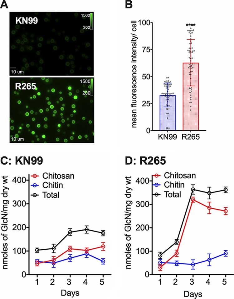
C. gattii R265 cells produce significantly larger amounts of chitosan in the cell wall compared to C. neoformans under YPD growth conditions. (A) Wild-type R265 and KN99 strains were grown in YPD for 5 days at 30°C and stained with Eosin Y to detect cell wall chitosan. Staining intensity was assessed using epifluorescence microscopy with identical exposures for all images. (B) Fluorescent levels for 60 individual cells (represented in panel A) were quantified using ImageJ (Fiji). The two-tailed unpaired t test with Welch’s correction was used to compare mean values of the wild type. Means represent the fluorescence intensity levels from three independent experiments (n = 3). ****, P < 0.0001. Error bars represent standard errors of the means. (C) Quantitative determination of cell wall chitosan and chitin of strain KN99 by the MBTH assay. Cells were grown in YPD for 1 to 5 days, collected, washed, and used for the assay. Data represents the averages of three biological experiments and are expressed as nanomoles of glucosamine per milligram (dry weight) of yeast cells. (D) Quantitative determination of cell wall chitosan and chitin of R265 were determined as in panel C.
C. gattii R265 cells produce significantly larger amount of chitosan compared to C. neoformans under host conditions.
To determine whether the increased amount of chitosan was also seen under host conditions, we first measured chitosan of KN99 and R265 cells cultured in tissue culture conditions and then for Cryptococcus isolated from infected mouse lungs. Wild-type KN99 and R265 cells were first grown in YPD medium at 30°C and then transferred to RPMI 1640 medium containing 10% fetal bovine serum (FBS), 5% CO2 at 37°C for 5 days. The cells were then harvested, and the MBTH assay was used to quantify the chitosan content. As shown in Fig. 2A, R265 cells have a mean value of 63.9 versus 30.2 of KN99 cells as expressed as nanomoles of glucosamine sugar per milligram (dry weight) of the cell wall material. Next, chitosan was determined after isolating the yeast cells from infected mouse lungs. Similar to YPD and RPMI 1640 culture conditions, R265 cells showed significantly higher cell wall chitosan compared to KN99 cells with mean values of 213.9 and 78.3, respectively, as expressed as nanomoles of glucosamine sugar per 108 cells (Fig. 2B). These results suggest that the increase in cell wall chitosan of C. gattii R265 over C. neoformans KN99 is a phenotype that is preserved when these strains are grown in culture and during mouse infection.
FIG 2.
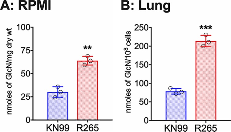
C. gattii R265 cells produce significantly larger amount of chitosan in the cell wall compared to C. neoformans under host conditions. (A) Chitosan levels of strains grown in RPMI 1640 containing 10% FBS and 5% CO2 at 37°C for 5 days. Strains were grown in YPD for 2 days. Yeast cells were harvested, washed with PBS, inoculated at 500 cells/μl in RPMI 1640 containing 10% FBS, and incubated for 5 days at 37°C in the presence of 5% CO2. At the end of incubation, chitosan was measured by the MBTH assay and expressed as nanomoles of glucosamine per milligram (dry weight) of cells. Data represent the averages from three biological experiments. (B) Chitosan levels of strains growing in the murine lung. Mice (CBA/J) (three mice per group) were intranasally inoculated with 107 CFU of each strain. On day 7 postinfection (PI), the lungs were excised and homogenized, and the lung tissue was removed by alkaline extraction, leaving the fungal cells to be harvested, counted, and subjected to the MBTH assay. Data are expressed as nanomoles of glucosamine per 108 cells. Significant differences between the groups were compared by two-tailed unpaired t test with Welch’s correction. Error bars represent standard errors of the means. ***, P < 0.0062; **, P < 0.001.
The deletion of CDA1 in C. gattii R265 causes a decrease in cell wall chitosan under YPD growth conditions.
We generated single CDA deletion mutants of C. gattii R265 by biolistic transformation. The strains generated and used in this study are listed in Table 2. The deletion cassettes for each gene deletion were generated by overlap PCR and biolistically transformed into R265 cells. The primers used to generate these deletion cassettes are listed in Table S1 in the supplemental material. All the isolates were characterized by diagnostic PCR screening and Southern blot hybridization. After growing the strains in YPD for 5 days, we measured chitosan by MBTH assay. We found that deletion of the CDA1 gene decreased the chitosan amount by 33% compared to that in wild-type R265. However, deletion of either CDA2 or CDA3 did not affect the total amount of chitosan, as shown in Fig. 3. This is different from what we have previously described for C. neoformans, where there was no significant difference in chitosan in any of the single CDA deletion strains (28, 30). This suggests that there are differences in the regulation of chitin deacetylation between the two species, with R265 Cda1 having a more significant role in chitosan production when the cells were cultured in YPD.
TABLE 2.
Strains used and generated in this study
| Strain | Resistance marker(s) | Background | Designation |
|---|---|---|---|
| R265 | JLCG924 | ||
| cda1Δ | G418 | JLCG924 | WLCG1156 |
| cda1Δ–2 | G418 | JLCG924 | WLCG1157 |
| cda2Δ | NAT | JLCG924 | WLCG1159 |
| cda3Δ | HYG | JLCG924 | WLCG1162 |
| cda3Δ::CDA3 | G418 | WLCG1162 | JLCG951 |
| cda1Δ2Δ | G418/NAT | WLCG1156 | WLCG1169 |
| cda1Δ2Δ–2 | G418/HYG | WLCG1156 | WLCG1171 |
| cda1Δ3Δ | G418/HYG | WLCG1162 | WLCG1197 |
| cda1Δ3Δ–2 | G418/HYG | WLCG1163 | WLCG1198 |
| cda2Δ3Δ | NAT/HYG | WLCG1162 | WLCG1166 |
| cda2Δ3Δ–2 | NAT/HYG | WLCG1162 | WLCG1167 |
| cda1Δ2Δ3Δ | G418/NAT/HYG | WLCG1166 | WLCG1190 |
| cda1Δ2Δ3Δ–2 | G418/NAT/HYG | WLCG1166 | WLCG1194 |
FIG 3.
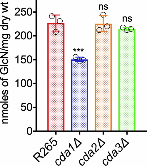
The deletion of CDA1 in strain R265 displays a decrease in cell wall chitosan in cells grown in YPD. Chitosan levels of strains grown in YPD for 5 days were determined. The amount of chitosan in the cell wall of the strains was quantified by the MBTH assay. Data represent the averages from three biological experiments with two technical replicates and are expressed as nanomoles of glucosamine per milligram (dry weight) of yeast cells. Significant differences between the groups were compared by one-way ANOVA followed by Dunnett’s multiple-comparison test. ***, P < 0.0002 comparing wild-type R265 with any other strain. ns, not significant.
Primers used in this study. Download Table S1, PDF file, 0.2 MB (195.1KB, pdf) .
Copyright © 2019 Lam et al.
This content is distributed under the terms of the Creative Commons Attribution 4.0 International license.
R265 Cda3 is critical for fungal virulence.
Next, we assessed the CDA single-deletion mutant strains for virulence by employing the murine intranasal infection model. We infected CBA/J mice with 105 wild-type cells or with cells of the corresponding single CDA deletion strains. The virulence was assayed as described in Materials and Methods. We found that deletion of either CDA1 or CDA2 did not affect virulence (Fig. 4A). The virulence of the cda1Δ mutant was reproduced using a second isolate and is shown in Fig. S2 to further confirm the absence of a role for Cda1 in the virulence of C. gattii R265. Interestingly, we found that R265 Cda3 is essential for fungal virulence. The virulence defect of the cda3Δ mutant was completely restored in a CDA3 complemented strain (Fig. 4A). Since different mouse models show various degrees of sensitivity to Cryptococcus infection, we wanted to verify whether the associations between the absence of CDA genes, amount of cell wall chitosan, and virulence of the specific CDA deletion strain can be recapitulated in C57BL/6 mice, which are routinely employed for diverse immunological studies using readily available mutants. Similar to the results obtained with CBA/J mice, we found that only the R265 cda3Δ mutant is specifically avirulent when tested following orotracheal inoculation of 10e4 CFU of yeast (Fig. 4B). The avirulent phenotype in both mouse strains was accompanied by the complete clearance of the mutant strain from the infected host at the endpoint of the survival study (Fig. S3). This was rather surprising because we have previously seen that for strain KN99, Cda1 plays a major role in fungal virulence without the contribution of Cda3 to pathogenesis and that C. neoformans cda1Δ mutant cells persist in the mouse lungs at low levels, even though the strain does not cause disease (30).
FIG 4.
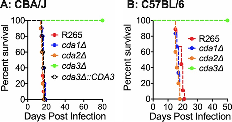
C. gattii cda3Δ displays severely attenuated virulence in CBA/J and C57BL/6 mouse models of infection. (A) CBA/J mice (6 to 8 weeks old, female) were infected intranasally with 105 CFU of each strain. Survival of the animals was recorded as mortality of mice for 80 days PI. Mice that lost 20% of the body weight at the time of inoculation were considered ill and sacrificed. Data are representative of two independent experiments with five animals for each strain. (B) C57BL/6 mice (4 to 6 weeks old, female) were infected with 104 CFU of each strain by intratracheal inoculation. Survival of the animals was recorded as mortality of mice for 50 days PI. Mice that lost 20% of the body weight at the time of inoculation were considered ill and sacrificed. Data are representative of one experiment with 10 animals for each strain. Virulence was determined using Mantel-Cox curve comparison with statistical significance determined by log rank test. P < 0.0001 comparing strain KN99 with cda3Δ strain.
Deletion of CDA1 in C. gattii R265 does not affect fungal virulence. Mice were inoculated with 50,000 CFU of either wild-type R265 or the second isolate of the cda1Δ strain. Survival of the animals was recorded as mortality. Mice that lost 20% of the body weight at the time of inoculation were considered ill and sacrificed. Download FIG S2, PDF file, 0.04 MB (40.5KB, pdf) .
Copyright © 2019 Lam et al.
This content is distributed under the terms of the Creative Commons Attribution 4.0 International license.
Fungal burden in the lungs of the mice at the endpoint of the survival experiment. Fungal burden in the lungs of the mice at the end point of the survival experiment for both CBA/J and C57BL/6 mice. The dashed line indicates the CFU of the initial inoculum for each mouse strain. Download FIG S3, PDF file, 0.04 MB (36.4KB, pdf) .
Copyright © 2019 Lam et al.
This content is distributed under the terms of the Creative Commons Attribution 4.0 International license.
The virulence phenotype of the single CDA deletion strains followed their ability to grow in the infected lung. As shown in Fig. 5, we found that CBA/J mice infected with either cda1Δ or cda2Δ strains had lung fungal burdens similar to those of R265 on day 14 and 21 postinfection (PI). On the other hand, mice infected with the cda3Δ strain showed slow and gradual clearance of the mutant strain. The fungal growth in the lungs of the mice infected with the cda3Δ strain was restored to wild-type R265 levels in a CDA3 complemented strain (Fig. 5, cda3Δ::CDA3).
FIG 5.
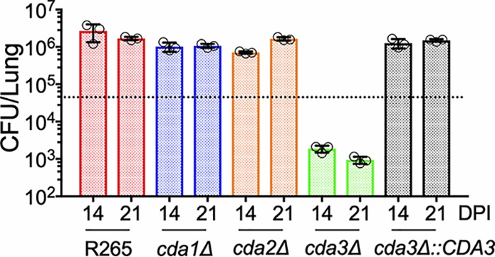
There is a slow but gradual clearance of C. gattii cda3Δ in CBA/J mice. Fungal burden in the lungs of CBA/J mice infected with indicated strains at different days postinfection (DPI). Data are from three mice per group at each time point. The dotted line indicates the CFU of the initial inoculum for each strain.
Fungal virulence of different CDA deletion strains is directly correlated with their ability to produce chitosan under host-mimicking growth conditions.
Recently, we have shown that in C. neoformans, Cda1 is essential for fungal virulence, and this avirulent phenotype is associated with the inability of the mutant to produce wild-type levels of chitosan when grown under host-mimicking conditions, such as RPMI 1640 medium containing 10% FBS, 5% CO2, and 37°C (30). Therefore, we wanted to test whether such defects are also responsible for the avirulent phenotype of R265 mutants that do not harbor the CDA3 gene in the genome. As shown in Fig. 6, the loss of Cda3 resulted in a 77% decrease in the amount of chitosan produced compared to wild-type R265. Even though the amount of chitosan in the cda1Δ strain showed 33% reduction compared to wild-type R265 (Fig. 6), this decrease was similar to what we observed when the cells were grown in YPD culture conditions as well (Fig. 3). These data indicate that in strain R265, Cda1-mediated deacetylation of chitin is not influenced by growth conditions and is not critical for virulence. However, Cda3 is responsible for the majority of the deacetylation either in RPMI 1640 culture conditions or in infected mice and thus contributes significantly to fungal proliferation in the host. Taken together, these results confirmed the importance of cell wall chitosan to fungal virulence and suggests that a certain threshold amount of chitosan needs to be maintained in the cell wall to sustain infection.
FIG 6.
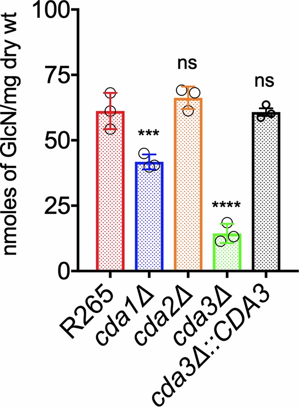
C. gattii Cda3 plays a major role in the synthesis of chitosan under host infection conditions. Chitosan levels of strains grown in RPMI 1640 containing 10% FBS and 5% CO2 at 37°C for 5 days. The indicated strains were grown in YPD for 48 h. Yeast cells were harvested, washed with PBS, and inoculated at 500 cells/μl in RPMI 1640 containing 10% FBS and incubated for 5 days at 37°C in the presence of 5% CO2. At the end of incubation, chitosan was measured by the MBTH assay and expressed as nanomoles of glucosamine per milligram (dry weight) of cells. Data represent the averages from three biological experiments. Significant differences between the groups were compared by one-way ANOVA, followed by Dunnett’s multiple-comparison test (***, P < 0.0002, *, P < 0.0280 comparing KN99 with any other strain; ns, not significant).
Double and triple CDA mutants of C. gattii R265 displayed varied amounts of chitosan under YPD and host-mimicking conditions.
We generated three double CDA deletion strains and a triple CDA gene deletion strain in C. gattii R265 by biolistic transformations as indicated in Table 2. We subjected these strains to chitosan quantification after growing them either in YPD or in host-mimicking conditions of RPMI 1640 medium containing 10% FBS in the presence of 5% CO2 at 37°C. When we compared the chitosan amounts among the CDA double deletion strains, we found that the cell wall chitosan amount was significantly decreased in all the double deletion strains grown under YPD culture conditions (cda1Δ2Δ, cda1Δ3Δ, and cda2Δ3Δ strains in Fig. 7A). The decrease in the chitosan amount was more pronounced in the absence of Cda1 in combination with either Cda2 or Cda3: 63% reduction for the cda1Δ2Δ strain and 54% reduction for the cda1Δ3Δ strain compared to wild-type R265. The amount of chitosan in the cda2Δ3Δ strain showed a reduction of only 26% compared to wild-type R265. These results are consistent with the major role of Cda1 in chitosan production in strain R265 when the cells were grown under YPD conditions (Fig. 3). When we deleted all three CDAs to generate a cda1Δ2Δ3Δ strain, the chitosan amount decreased to almost negligible amounts, suggesting that in spite of differences in the roles of individual CDAs in strains KN99 and R265, the deletion of all three CDAs was sufficient to render the mutant chitosan deficient (Fig. 7A). When the strains were grown under host-mimicking conditions (RPMI 1640 plus 10% FBS grown with CO2 at 37°C), any strain in which CDA3 was deleted in combination with either CDA1 or CDA2 (cda1Δ3Δ or cda2Δ3Δ strain in Fig. 7B) produced negligible amounts of chitosan similar to the cda1Δ2Δ3Δ strain, further indicating the importance of R265 Cda3 in the production of chitosan in the host and its subsequent effect on fungal virulence.
FIG 7.
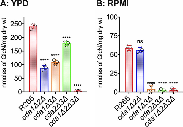
The deletion of C. gattii CDA1, CDA2, and CDA3 results in a strain that is completely chitosan deficient. C. gattii Cda1 in combination with Cda2 or Cda3 plays a major role in chitosan synthesis in vegetative growing conditions. While C. gattii Cda3 in combination with Cda1 or Cda2 results in a stain that is completely chitosan-deficient. (A) Chitosan levels of strains grown in YPD. The indicated strains were grown in YPD for 5 days. The amount of chitosan in the cell wall of the strains was quantified by the MBTH assay. Data are the averages for three biological experiments and are expressed as nanomoles of glucosamine per milligram (dry weight) of yeast cells. (B) Chitosan levels of strains grown in RPMI 1640 containing 10% FBS and 5% CO2 at 37°C for 5 days. The indicated strains were grown in YPD for 48 h. Yeast cells were harvested, washed with PBS, and inoculated at 500 cells/μl in RPMI 1640 containing 10% FBS and incubated for 5 days at 37°C in the presence of 5% CO2. At the end of incubation, chitosan was measured by the MBTH assay and expressed as nanomoles of glucosamine per milligram (dry weight) of cells. Data represent the averages for three biological experiments. Significant differences between the groups were compared by one-way ANOVA, followed by Dunnett’s multiple-comparison test (****, P < 0.0001, comparing KN99 with any other strain; ns, not significant).
Chitosan-deficient R265 cells were sensitive to cell wall stressors and had normal capsule- and melanin-producing abilities.
We subjected the single CDA deletion strains and the triple CDA deletion strain of C. gattii R265 to a panel of cell wall stressors (0.005% SDS, 1 mg/ml Calcofluor white, 0.5 mg/ml caffeine, 0.4% Congo red) added to YPD agar medium; these stressors are routinely employed to determine cell wall integrity. As shown in Fig. S4, only the chitosan-deficient cda1Δ2Δ3Δ strain was sensitive to the various cell wall stressors. None of the CDA deletion mutants were sensitive to temperature when their growth at 30°C was compared to growth at 37°C on YPD agar medium. The deletion of CDA genes in strain R265 either individually or in combination did not affect their ability to produce either capsule (Fig. S5) or melanin as shown in Fig. S6. In contrast to C. neoformans mutants (26), none of the CDA mutants of R265 displayed “leaky melanin” phenotype.
Sensitivity of chitin deacetylase mutants to cell wall inhibitors and temperature. Cultures were grown overnight in YPD and then diluted to an OD650 of 1.0. Tenfold serial dilutions were made in PBS, and 5 μl of each strain was plated. The plates were grown for 5 days at 30°C for the inhibitor plates and at the indicated temperature for all others. The wild-type (R265) and deletion strains are labeled on the left, and the conditions are noted at the top. (A) Sensitivity of mutants to temperature. (B) Sensitivity to cell wall inhibitors. Abbreviations: CFW, Calcofluor white; SDS, sodium dodecyl sulfate; Caff, caffeine; CR, Congo red. Download FIG S4, PDF file, 0.05 MB (53.2KB, pdf) .
Copyright © 2019 Lam et al.
This content is distributed under the terms of the Creative Commons Attribution 4.0 International license.
C. gattii R265 cda1Δ, cda2Δ, or cda3Δ mutants display normal levels of capsule under capsule-inducing conditions. Cells were incubated under capsule-inducing conditions for 5 days. Capsule size was assessed by staining with India ink and visualizing the zone of exclusion at a magnification of ×60. Download FIG S5, PDF file, 0.05 MB (53.6KB, pdf) .
Copyright © 2019 Lam et al.
This content is distributed under the terms of the Creative Commons Attribution 4.0 International license.
R265 CDA1 and CDA2 are dispensable for virulence even when both genes were deleted.
We next subjected the double and triple CDA gene deletion strains to tests of fungal virulence either by intranasal infection of CBA/J mice or by orotracheal inoculation of C57BL/6 mice. We found that deletion of CDA1 and CDA2 did not affect the virulence in either CBA/J or C57BL/6 mice (cda1Δ2Δ strain in Fig. 8A and B). Consistent with the role of C. gattii R265 Cda3 in virulence, double CDA deletion strains in which CDA3 is deleted in combination with either CDA1 or CDA2 showed a major defect in virulence (cda1Δ3Δ and cda2Δ3Δ strains in Fig. 8A and B). These results further point to the importance of just Cda3 in fungal virulence for strain R265. The mutant strain devoid of all three CDA genes (cda1Δ2Δ3Δ strain) was completely avirulent (Fig. 8A and B). For all mutants, the avirulent phenotype of the mutant strains was accompanied by the inability of the different CDA mutant strains to proliferate or maintain in the infected murine lung as revealed by their gradual clearance (data not shown). To further ascertain these results, we subjected a second independent isolate of each mutant to fungal virulence studies and obtained nearly identical results (Fig. S7).
FIG 8.
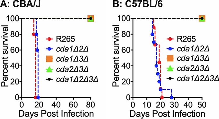
Deletion of C. gattii CDA3 in combination with any of the other two CDAs results in severe attenuation of virulence in CBA/J and C57BL/6 mouse models of infection. (A) CBA/J mice (6 to 8 weeks old, female) were infected intranasally with 105 CFU of each strain. Survival of the animals was recorded as mortality of mice for 80 days postinfection (PI). Mice that lost 20% of the body weight at the time of inoculation were considered ill and sacrificed. Data are representative of two independent experiments with five animals for each strain. (B) C57BL/6 mice (4 to 6 weeks old, female) were infected intratracheally with 104 CFU of each strain. Survival of the animals was recorded as mortality of mice for 50 days PI. Mice that lost 20% of the body weight at the time of inoculation were considered ill and sacrificed.
Melanin phenotype of chitin deacetylase mutants of C. gattii. Chitin deacetylase mutants displayed no difference in melanin production compared to the wild type (R265). Strains were grown overnight in 2 ml YPD medium at 30°C with shaking to saturation. Cells were collected and washed in 1× PBS. Then, 1 × 10⁸ (5 × 10⁷/ml) of each mutant was added to 4 ml of Asn plus l--DOPA for 7 days at 300 rpm and 30°C in the dark. Samples were then spun down and photographed. Black pellets indicate the presence of melanin in the strain. The clear supernatants indicate that there was not a “leaky melanin” phenotype (27, 28). Download FIG S6, PDF file, 0.04 MB (37.8KB, pdf) .
Copyright © 2019 Lam et al.
This content is distributed under the terms of the Creative Commons Attribution 4.0 International license.
Vaccination with the chitosan-deficient R265 cells confers partial protection to subsequent challenge with the virulent wild-type R265.
For C. neoformans, we observed that mice infected with 107 CFU of the chitosan-deficient cda1Δ2Δ3Δ strain resulted in the clearance of the mutant. That clearance was accompanied by the induction of a robust protective response to a subsequent infection with wild-type, fully virulent KN99 (29). This protective response was observed only when mice were vaccinated with 107 CFU of the cda1Δ2Δ3Δ strain, while vaccination with either 106 or 105 CFU did not generate a protective response (29). Therefore, since the complete clearance of the chitosan-deficient cda1Δ2Δ3Δ strain is also observed for mutants generated in the R265 background, we wanted to determine whether this clearance of the mutant strain induces protective immunity to R265 infection. For this, we vaccinated naive mice (CBA/J) with either 105, 106, or 107 CFU of live preparations of the cda1Δ2Δ3Δ strain by intranasal inhalation. After 40 days PI, we challenged them with 105 CFU of strain R265. Two independent isolates of the cda1Δ2Δ3Δ strain (cda1Δ2Δ3Δ–1 and cda1Δ2Δ3Δ-2) were used for this study. As shown in Fig. 9A, vaccination with either one of the chitosan-deficient cda1Δ2Δ3Δ isolates conferred only partial protection to subsequent infection with R265. The control mice that received phosphate-buffered saline (PBS) alone succumbed to R265 infection with a median survival time of 20 days PI. However, the mice vaccinated with a live preparation of the cda1Δ2Δ3Δ strain had a median survival time of 30 to 40 days PI upon secondary challenge infection with strain R265. This protective immunity required a minimal dose of 107 CFU of the cda1Δ2Δ3Δ strain for vaccination (Fig. 9A), as observed for the cda1Δ2Δ3Δ mutant of strain KN99 (29). Next, we wanted to determine whether a heat-killed (HK) preparation of the cda1Δ2Δ3Δ strain induces protective immunity. We confirmed that incubating live R265 (either the wild-type strain or the corresponding cda1Δ2Δ3Δ mutant) at 70°C for 15 min was sufficient to kill both strains by plating them for CFU onto YPD agar. Then we vaccinated mice intranasally with 107 CFU of HK preparation of either strain R265 or the cda1Δ2Δ3Δ mutant, waited 40 days, and challenged them with 105 CFU of R265. The mice vaccinated with either PBS or with HK wild-type R265 succumbed to R265 infection with a median survival time of 20 days PI. However, the mice vaccinated with the HK cda1Δ2Δ3Δ strain had a median survival time of 33 days PI (Fig. 9B), similar to that observed for vaccination with the live cda1Δ2Δ3Δ strain (Fig. 9A). When the vaccination and protection experiment was done in C57BL/6 mice using the HK cda1Δ2Δ3Δ strain as a vaccine, mice had a median survival time of 41.5 days compared to 26 days for unvaccinated mice (Fig. S8).
FIG 9.
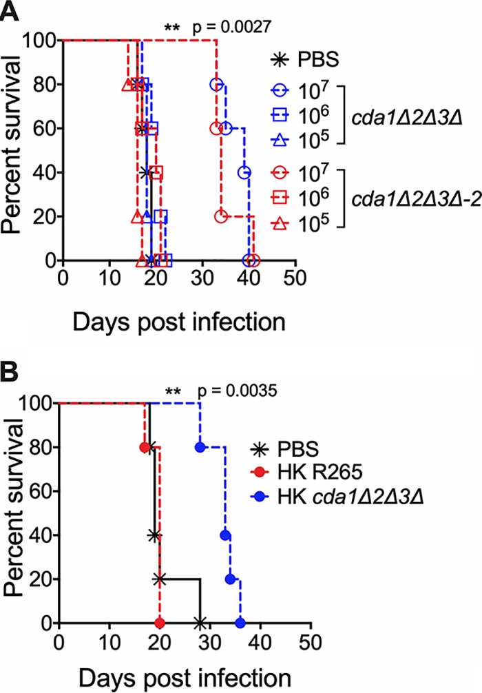
Vaccination of CBA/J mice with 107 CFU of live or heat-killed (HK) cda1Δ2Δ3Δ cells conferred attenuated protective immunity to subsequent infection with wild-type R265 C. gattii cells. (A) Mice were immunized with either 107, 106, or 105 live CFU of cda1Δ2Δ3Δ cells through inhalation. PBS-inoculated mice served as control. Animals were left for 40 days to resolve the infection. Subsequently, both groups of mice were challenged with 50,000 CFU of virulent R265 cells. Virulence was recorded as mortality of mice. Mice that lost 20% of starting body weight at the time of inoculation were considered to be moribund and sacrificed. The percentage of mice that survived was plotted against the day postinfection. Each survival curve is the average of two independent experiments that had five mice per experimental group. (B) Mice were immunized with an inoculum of HK cells with a dose equivalent to 107 CFU of either the wild-type R265 or the cda1Δ2Δ3Δ strain. Control mice were inoculated with PBS. After 40 days, mice were challenged with 50,000 CFU of wild-type R265 cells. Survival of the animals was recorded as described above. The data shown are from a single experiment with five mice per experimental group. A second experiment showed similar results.
Deletion of C. gattii CDA3 in combination with any of the other two CDAs results in severe attenuation of virulence in mouse model of infection. CBA/J mice (6 to 8 weeks old, female) were infected intranasally with 105 CFU of second independent isolate of each strain. Survival of the animals was recorded as mortality of mice for 80 days PI. Mice that lost 20% of the body weight at the time of inoculation were considered ill and sacrificed. Data are representative of two independent experiments with five animals for each strain. Download FIG S7, PDF file, 0.04 MB (43.2KB, pdf) .
Copyright © 2019 Lam et al.
This content is distributed under the terms of the Creative Commons Attribution 4.0 International license.
DISCUSSION
Chitosan is one of the principal components of the C. neoformans cell wall. In its absence, yeast cells display a severe budding defect resulting in irregular shaped, often clumped cells with significant sensitivity to various cell wall-perturbing agents (28). More importantly, the absence of chitosan makes the yeast cells completely avirulent in a mammalian host, as the chitosan-deficient cells stimulate robust host immune responses, which in turn leads to rapid clearance of infection (26, 29). The outbreak strain of C. gattii, R265, is a hypervirulent strain that has been reported to possess several key features that enabled it to cause symptomatic infection, even in immunocompetent individuals (10, 32, 33). Its higher rate of intracellular proliferation with increased resistance to oxidative stress, its ability to dampen host immune response, and its potential to inhibit the maturation of specific immune cells all may contribute to its virulence (19, 22–25, 34, 35). A larger amount of chitosan in C. gattii R265 than in C. neoformans KN99 may efficiently shield surface-exposed PAMPs from being recognized by the host immune system, thereby limiting the intensity and complexity of the host immune response. C. neoformans and C. gattii share diverse ecological niches. Moreover, C. gattii R265 is predominantly detected in the environment and strictly associated with plants where they encounter chitinases from soil microbes and plant hosts, respectively (32). Extensive deacetylation of chitin to form chitosan may provide greater resistance to environmental chitinases, thereby increasing its fitness in the environment.
C. neoformans Cda1 plays an important role in the deacetylation of chitin especially during host infection (30). Among the individual C. neoformans CDA deletion strains, we did not observe phenotypic differences when the strains were cultured in YPD medium, suggesting the redundancy in their function (28). However, deletion of Cda1 alone caused a dramatic avirulent phenotype in virulence studies using CBA/J mice (30). Unlike C. neoformans, Cda1 of C. gattii seems to play an important role in deacetylation when grown in YPD medium, since its deletion caused a 34% reduction in the amount of cell wall chitosan (Fig. 3). The deletion of either CDA2 or CDA3 did not significantly reduce the chitosan amount when the strain was grown in YPD. However, when grown under host-mimicking conditions of RPMI 1640 medium with 10% FBS, 5% CO2, and at 37°C, C. gattii Cda3 played an important role in the deacetylation of chitin, since its deletion in the cda3Δ strain caused a 76% reduction in the amount of chitosan. On the other hand, the decrease in the amount of chitosan in the cda1Δ strain when grown in RPMI 1640 conditions was around 32%, which is similar to what we observed for YPD culture conditions, suggesting a unique role of R265 Cda3 in deacetylating chitin in host-mimicking conditions. For our initial animal virulence experiments, we chose two independent isolates of the cda1Δ mutant strain, expecting that they will mimic the avirulent phenotype observed in C. neoformans (30). However, we were surprised to see that for C. gattii R265, deacetylation of chitin during host infection was even more dependent on Cda3 than Cda1. The avirulent phenotype of the cda3Δ mutant was further confirmed by the CDA3 complemented strain in CBA/J mice. In spite of its role in chitosan production in YPD and host-mimicking media, the decreased levels of chitosan in the cda1Δ strain did not affect its virulence (Fig. 4). It may be that the levels of chitosan present in the cda1Δ strain during infection are still sufficient to promote pathogenesis, or the pattern of deacetylation in the chitosan produced by Cda1 may be different from that of Cda3. This difference in the molecular structure of chitosan may have contributed to the observed differences in the virulence potential of different CDA deletion mutant strains.
All three CDAs contribute to chitosan production in C. gattii R265 while growing in YPD, since deletion of two CDA genes in combination significantly reduced cell wall chitosan (Fig. 7). This again was reflected in the virulence phenotype of the double and triple CDA deletion strains. When CDA3 was deleted in combination with any one or both of the other CDA genes, the resulting strains had substantially reduced chitosan when grown in host-mimicking conditions and were avirulent. Any strain in which CDA3 is deleted was avirulent in both CBA/J and C57BL/6 mouse strains and was cleared from the infected lung. Deletion of all three CDAs made the strain chitosan deficient similar to the cda1Δ2Δ3Δ strain of C. neoformans. The majority of the in vitro phenotypes of the cda1Δ2Δ3Δ strain of R265 were similar to the cda1Δ2Δ3Δ strain made in C. neoformans. However, cda1Δ2Δ3Δ of C. gattii R265 grew better in YPD (with markedly fewer morphological abnormalities) than the cda1Δ2Δ3Δ strain of C. neoformans (data not shown). C. neoformans cda1Δ2Δ3 exhibited a leaky melanin phenotype when incubated in medium containing l--DOPA (28). However, the cda1Δ2Δ3Δ strain of R265 did not show this phenotype (Fig. S6) suggesting that there are differences in the cell wall architecture between cda1Δ2Δ3Δ cells of C. neoformans and C. gattii.
In C. neoformans, CDA1 seems to be the most critical deacetylase gene during infection—the CDA1 gene is much more highly upregulated during the growth of wild-type KN99 yeast cells in the infected lung than CDA2 or CDA3, and a cda1Δ strain is avirulent, consistent with the hypothesis that differential regulation of CDA1 is the driving force behind its importance during infection (30). Unlike C. neoformans, CDA3 is the critical deacetylase gene in C. gattii R265 based on our genetic analysis, but the available data from genome-wide studies of transcriptional regulation are less clear, and may be different for different C. gattii strains. One study that looked only at transcript abundance of genes from strain R265 recovered through bronchoalveolar lavage, found that R265 CDA3 was among the highly expressed genes during mammalian infection (36). Recent whole-genome transcriptome studies of different strains of C. gattii have shown multiple strain-specific differences in gene expression that include chitin and chitosan biosynthesis genes (37, 38). One of these studies (37) examined strains from the four lineages of C. gattii, and it showed that the CDA2 gene of strain R265 was substantially downregulated after incubation of the R265 cells with bone marrow-derived macrophages. The results from these studies suggest that transcriptional regulation of chitin and chitosan biosynthesis in C. gattii is likely divergent from regulation in C. neoformans, is different among different C. gattii strains, may play a role in the virulence of the different strains, and could be a factor in the importance of CDA3 in strain R265.
We have previously shown that vaccination with the chitosan-deficient cda1Δ2Δ3Δ strain of C. neoformans at the optimal concentration confers robust protective immunity to subsequent challenge infection with the wild-type virulent KN99 strain (29). Even though deletion of all three CDA genes of C. gattii produced a chitosan-deficient strain, vaccination with either the live strain or a heat-killed preparation of it induced only partial protection to subsequent infection with strain R265. This may be due to the fact that R265 has the inherent ability to dampen host-induced immune responses (22–25).
In summary, the hypervirulent C. gattii strain R265 has evolved with a distinct transcriptional profile with altered expression of components of chitin deacetylation that may have enabled it to adapt more efficiently to various environmental conditions with ramifications on its virulence. Of the several reported differences in the virulence-related traits between C. neoformans and C. gattii R265, the difference in the regulation of chitosan biosynthesis as revealed from our studies may significantly contribute to the mechanisms of its unique and distinct nature of pathogenesis, since chitosan is one of the macromolecules that resides at the host-pathogen interface.
MATERIALS AND METHODS
Fungal strains and media.
R265, C. gattii strain of VGII subtype linked to the 1999 outbreak in British Columbia, Canada (11), was used as the wild-type strain and as progenitor of mutant strains. This strain was kindly provided by Joseph Heitman (Duke University Medical Center, NC). All the strains used in this study are listed in Table 1. KN99α, a strain of C. neoformans, was used as the wild-type strain for serotype A (31). Strains were grown on YPD (1% yeast extract, 2% Bacto peptone, and 2% dextrose). Solid media contained 2% Bacto agar. Selective YPD medium contained 100 μg/ml nourseothricin (NAT) (Werner BioAgents, Germany) and/or 200 μg/ml G418 (Geneticin; Gibco Life Technologies, USA). RPMI 1640 medium (catalog no. 10-040-CM; Corning) contained 10% fetal bovine serum (FBS) (catalog no. 26140; Gibco-Thermo Fisher Technologies).
Generation of deletion constructs of C. gattii.
Gene-specific deletion constructs of the chitin deacetylases were generated using overlap PCR gene technology described previously (39, 40) and included either the hygromycin resistance, Geneticin resistance cassette (41), or nourseothricin resistance cassette (42). The primers used to disrupt the genes are shown in Table S1 in the supplemental material. The Cda1 deletion cassette contained the Geneticin resistance cassette, resulting in a 1,539-bp replacement of the genomic sequence between regions of primers 3-Cda1 and 6-Cda1 shown in upper case in Table S1. The Cda2 deletion cassette contained the hygromycin resistance cassette, resulting in a 1,587-bp replacement of the genomic sequence, and the Cda3 deletion cassette contained the hygromycin resistance cassette, resulting in a 1,494-bp replacement of the genomic sequence. Constructs were introduced into the R265 strain using biolistic techniques (43).
Transformation and characterization of C. gattii mutants.
Recipient strains of C. gattii were transformed biolistically following the protocol described earlier (41, 43). Drug-resistant transformants that formed colonies in 3 to 5 days were passaged four times in liquid YPD medium before reselection of drug resistance on agar. Transformants were further screened by diagnostic PCR of their genomic DNA using primers at the 5′ and 3′ junction of the integration site of the transforming DNA. Southern blot hybridizations were done to verify the absence of random DNA integrations, as described previously employing digoxigenin (DIG)-labeled DNA probes (44, 45).
Cellular chitosan measurement.
As previously described, MBTH (3-methyl-2-benzothiazolinone hydrazone)-based chemical method was used to determine the chitin and chitosan content of C. gattii or C. neoformans (31). In brief, cells were collected after growing them in appropriate media and growth conditions by centrifugation. The cell pellets were washed two times with phosphate-buffered saline (PBS), pH 7.4, and lyophilized. The dried samples were resuspended in water first before adding KOH to a final concentration of 6% KOH (wt/vol). The alkali-suspended material was incubated at 80°C for 30 min with vortexing in between to eliminate nonspecific MBTH-reactive molecules from the cells. Alkali-treated material was then washed several times with PBS, pH 7.4, to make sure that the pH of the cell suspension was brought back to neutral pH. In the case of the cells grown in RPMI 1640 medium, alkali-treated material was sonicated as described previously to generate a uniform suspension (30). Finally, the cell material was resuspended in PBS, pH 7.4, to a concentration of 10 mg/ml in PBS (by dry weight), and a 0.1-ml aliquot of each sample was used in the MBTH assay (46).
Virulence and fungal burden assays.
C. gattii strains were grown at 30°C and 300 rpm for 48 h in 50 ml YPD. The cells were centrifuged, washed in endotoxin-free 1× PBS, and suspended in 5 ml of the same PBS solution. The cells were counted with a hemocytometer and diluted to 2 × 10⁶ cells/ml. CBA/J female mice (Jackson Laboratories) were anesthetized with an intraperitoneal injection (200 μl) of ketamine (8 mg/ml)-dexmedetomidine (0.05 mg/ml) mixture, which was reversed by an intraperitoneal injection of 200 μl of antipamezole (0.25 mg/ml). Mice were allowed to inhale 1 × 10⁵ cells in 50 μl, which were dripped into the nares. For virulence assays, mice were weighed before and during the course of infection. Mice were euthanized by CO2 asphyxiation if they reached 80% of their original body weight. At this point, the mice appeared morbidly ill, displaying a ruffled coat, lethargy, hunched posture, unstable gait, and loss of appetite. For the determination of CFU, lung or brain from each mouse was placed in 2.0 ml of 1× PBS (pH 7), homogenized, serially diluted, plated onto YPD agar supplemented with 100 μg/ml streptomycin and 100 μg/ml ampicillin, and incubated for 2 days at 30°C. Total CFU per organ were calculated. The infection protocol was reviewed and approved by the Washington University School of Medicine Animal Care and Use Committee (IACUC).
Evaluation of C. gattii to stress under in vitro conditions.
Solid YPD medium was made with the desired amount of either SDS, NaCl, Calcofluor white, or Congo red. For plating, wild-type and mutant strains were grown in liquid YPD for 24 h at 30°C. Cells were diluted to an optical density at 650 nm (OD650) of 1.0, and 10-fold serial dilutions were made. Five microliters of each dilution was spotted on the plate, and the plates were incubated for 2 or 3 days at appropriate temperatures and photographed. Eosin Y staining was conducted as described earlier (29).
Analysis of melanin production.
Strains were grown overnight in 2 ml YPD medium at 30°C with shaking to saturation. Cells were collected and washed in 1× PBS. Then, 1 × 10⁸ (5 × 10⁷/ml) of each mutant was added to 4 ml of glucose-free asparagine medium (1 g/liter l-asparagine, 0.5 g/liter MgSO4·7H2O, 3 g/liter KH2PO4, and 1 mg/liter thiamine, plus 1 mM l-3,4-dihydroxyphenylalanine [l-DOPA]) for 7 days at 300 rpm and 30°C in the dark. Samples were then spun down at >600 × g for 10 min. The cells’ ability to produce pigment was assessed visually.
Capsule analysis.
Cells were grown in YPD at 30°C. After 48 h for growth, cells were collected, washed once with PBS, inoculated into RPMI 1640 medium containing 10% fetal bovine serum (FBS) at a concentration of 500 cells/μl, and incubated for 5 days at 37°C in the presence of 5% CO2. The capsule-induced strains were resuspended in a 1:4 India ink-H2O solution and photographed on an Olympus BX61 microscope. The capsule diameter was measured and averaged for a minimum of 100 cells per strain using SlideBook 5.0 (Intelligent Imaging Innovations, Inc. CO, USA).
Statistics.
Data were analyzed using GraphPad Prism, version 7.0 (GraphPad Software, Inc., La Jolla, CA). The unpaired two-tailed t test with Welch’s correction was used for comparisons of two groups. The one-way analysis of variance (ANOVA) with the Dunnett’s multiple-correction test was used to compare more than two groups. Kaplan-Meier survival curves were compared using the Mantel-Cox log rank test.
Vaccination with a heat-killed preparation of cda1Δ2Δ3Δ strain of C. gattii confers attenuated protection to a subsequent challenge infection with virulent R265. Mice (C57BL/6) were immunized with a heat-killed (HK) preparation of the cda1Δ2Δ3Δ strain with a dose equivalent to 107 CFU. Control mice were inoculated with PBS. After 40 days, mice were challenged with 10,000 CFU of wild-type R265 cells. Survival of the animals was recorded as described above. Download FIG S8, PDF file, 0.04 MB (41KB, pdf) .
Copyright © 2019 Lam et al.
This content is distributed under the terms of the Creative Commons Attribution 4.0 International license.
ACKNOWLEDGMENTS
This work was supported by NIH grant R01AI072195 to J.K.L., NIH grant R01AI125045 to J.K.L., C.A.S., and S.M.L., and NIH grant R01AI025780 to C.A.S. and S.M.L.
REFERENCES
- 1.Spickler AR. 2013. Cryptococcosis. The Center for Food Security and Public Health, Iowa State University, Ames, Iowa. [Google Scholar]
- 2.Barnett JA. 2010. A history of research on yeasts 14: medical yeasts part 2, Cryptococcus neoformans. Yeast 27:875–904. doi: 10.1002/yea.1786. [DOI] [PubMed] [Google Scholar]
- 3.Rajasingham R, Smith RM, Park BJ, Jarvis JN, Govender NP, Chiller TM, Denning DW, Loyse A, Boulware DR. 2017. Global burden of disease of HIV-associated cryptococcal meningitis: an updated analysis. Lancet Infect Dis 17:873–881. doi: 10.1016/S1473-3099(17)30243-8. [DOI] [PMC free article] [PubMed] [Google Scholar]
- 4.Idnurm A, Bahn YS, Nielsen K, Lin X, Fraser JA, Heitman J. 2005. Deciphering the model pathogenic fungus Cryptococcus neoformans. Nat Rev Microbiol 3:753–764. doi: 10.1038/nrmicro1245. [DOI] [PubMed] [Google Scholar]
- 5.Kronstad JW, Attarian R, Cadieux B, Choi J, D’Souza CA, Griffiths EJ, Geddes JMH, Hu G, Jung WH, Kretschmer M, Saikia S, Wang J. 2011. Expanding fungal pathogenesis: Cryptococcus breaks out of the opportunistic box. Nat Rev Microbiol 9:193–203. doi: 10.1038/nrmicro2522. [DOI] [PMC free article] [PubMed] [Google Scholar]
- 6.Chen J, Varma A, Diaz MR, Litvintseva AP, Wollenberg KK, Kwon-Chung KJ. 2008. Cryptococcus neoformans strains and infection in apparently immunocompetent patients. Emerg Infect Dis 14:755–762. doi: 10.3201/eid1405.071312. [DOI] [PMC free article] [PubMed] [Google Scholar]
- 7.Choi YH, Ngamskulrungroj P, Varma A, Sionov E, Hwang SM, Carriconde F, Meyer W, Litvintseva AP, Lee WG, Shin JH, Kim EC, Lee KW, Choi TY, Lee YS, Kwon-Chung KJ. 2010. Prevalence of the VNIc genotype of Cryptococcus neoformans in non-HIV-associated cryptococcosis in the Republic of Korea. FEMS Yeast Res 10:769–778. doi: 10.1111/j.1567-1364.2010.00648.x. [DOI] [PMC free article] [PubMed] [Google Scholar]
- 8.Byrnes EJ III, Bartlett KH, Perfect JR, Heitman J. 2011. Cryptococcus gattii: an emerging fungal pathogen infecting humans and animals. Microbes Infect 13:895–907. doi: 10.1016/j.micinf.2011.05.009. [DOI] [PMC free article] [PubMed] [Google Scholar]
- 9.Harris JR, Lockhart SR, Debess E, Marsden-Haug N, Goldoft M, Wohrle R, Lee S, Smelser C, Park B, Chiller T. 2011. Cryptococcus gattii in the United States: clinical aspects of infection with an emerging pathogen. Clin Infect Dis 53:1188–1195. doi: 10.1093/cid/cir723. [DOI] [PubMed] [Google Scholar]
- 10.Chen SC, Meyer W, Sorrell TC. 2014. Cryptococcus gattii infections. Clin Microbiol Rev 27:980–1024. doi: 10.1128/CMR.00126-13. [DOI] [PMC free article] [PubMed] [Google Scholar]
- 11.Kidd SE, Hagen F, Tscharke RL, Huynh M, Bartlett KH, Fyfe M, Macdougall L, Boekhout T, Kwon-Chung KJ, Meyer W. 2004. A rare genotype of Cryptococcus gattii caused the cryptococcosis outbreak on Vancouver Island (British Columbia, Canada). Proc Natl Acad Sci U S A 101:17258–17263. doi: 10.1073/pnas.0402981101. [DOI] [PMC free article] [PubMed] [Google Scholar]
- 12.Kwon-Chung KJ, Bennett JE, Wickes BL, Meyer W, Cuomo CA, Wollenburg KR, Bicanic TA, Castaneda E, Chang YC, Chen J, Cogliati M, Dromer F, Ellis D, Filler SG, Fisher MC, Harrison TS, Holland SM, Kohno S, Kronstad JW, Lazera M, Levitz SM, Lionakis MS, May RC, Ngamskulrongroj P, Pappas PG, Perfect JR, Rickerts V, Sorrell TC, Walsh TJ, Williamson PR, Xu J, Zelazny AM, Casadevall A. 2017. The case for adopting the “species complex” nomenclature for the etiologic agents of cryptococcosis. mSphere 2:e00357-16. doi: 10.1128/mSphere.00357-16. [DOI] [PMC free article] [PubMed] [Google Scholar]
- 13.Bartlett KH, Kidd SE, Kronstad JW. 2008. The emergence of Cryptococcus gattii in British Columbia and the Pacific Northwest. Curr Infect Dis Rep 10:58–65. doi: 10.1007/s11908-008-0011-1. [DOI] [PubMed] [Google Scholar]
- 14.Byrnes EJ III, Bildfell RJ, Dearing PL, Valentine BA, Heitman J. 2009. Cryptococcus gattii with bimorphic colony types in a dog in western Oregon: additional evidence for expansion of the Vancouver Island outbreak. J Vet Diagn Invest 21:133–136. doi: 10.1177/104063870902100122. [DOI] [PMC free article] [PubMed] [Google Scholar]
- 15.Byrnes EJ III, Bildfell RJ, Frank SA, Mitchell TG, Marr KA, Heitman J. 2009. Molecular evidence that the range of the Vancouver Island outbreak of Cryptococcus gattii infection has expanded into the Pacific Northwest in the United States. J Infect Dis 199:1081–1086. doi: 10.1086/597306. [DOI] [PMC free article] [PubMed] [Google Scholar]
- 16.Byrnes EJ III, Li W, Lewit Y, Ma H, Voelz K, Ren P, Carter DA, Chaturvedi V, Bildfell RJ, May RC, Heitman J. 2010. Emergence and pathogenicity of highly virulent Cryptococcus gattii genotypes in the northwest United States. PLoS Pathog 6:e1000850. doi: 10.1371/journal.ppat.1000850. [DOI] [PMC free article] [PubMed] [Google Scholar]
- 17.Hagen F, Khayhan K, Theelen B, Kolecka A, Polacheck I, Sionov E, Falk R, Parnmen S, Lumbsch HT, Boekhout T. 2015. Recognition of seven species in the Cryptococcus gattii/Cryptococcus neoformans species complex. Fungal Genet Biol 78:16–48. doi: 10.1016/j.fgb.2015.02.009. [DOI] [PubMed] [Google Scholar]
- 18.Kwon-Chung KJ, Polacheck I, Bennett JE. 1982. Improved diagnostic medium for separation of Cryptococcus neoformans var. neoformans (serotypes A and D) and Cryptococcus neoformans var. gattii (serotypes B and C). J Clin Microbiol 15:535–537. [DOI] [PMC free article] [PubMed] [Google Scholar]
- 19.Ma H, Hagen F, Stekel DJ, Johnston SA, Sionov E, Falk R, Polacheck I, Boekhout T, May RC. 2009. The fatal fungal outbreak on Vancouver Island is characterized by enhanced intracellular parasitism driven by mitochondrial regulation. Proc Natl Acad Sci U S A 106:12980–12985. doi: 10.1073/pnas.0902963106. [DOI] [PMC free article] [PubMed] [Google Scholar]
- 20.Farrer RA, Desjardins CA, Sakthikumar S, Gujja S, Saif S, Zeng Q, Chen Y, Voelz K, Heitman J, May RC, Fisher MC, Cuomo CA. 2015. Genome evolution and innovation across the four major lineages of Cryptococcus gattii. mBio 6:e00868-15. doi: 10.1128/mBio.00868-15. [DOI] [PMC free article] [PubMed] [Google Scholar]
- 21.Ngamskulrungroj P, Chang Y, Sionov E, Kwon-Chung KJ. 2012. The primary target organ of Cryptococcus gattii is different from that of Cryptococcus neoformans in a murine model. mBio 3:e00103-12. doi: 10.1128/mBio.00103-12. [DOI] [PMC free article] [PubMed] [Google Scholar]
- 22.Okubo Y, Wakayama M, Ohno H, Yamamoto S, Tochigi N, Tanabe K, Kaneko Y, Yamagoe S, Umeyama T, Shinozaki M, Nemoto T, Nakayama H, Sasai D, Ishiwatari T, Shimodaira K, Yamamoto Y, Kamei K, Miyazaki Y, Shibuya K. 2013. Histopathological study of murine pulmonary cryptococcosis induced by Cryptococcus gattii and Cryptococcus neoformans. Jpn J Infect Dis 66:216–221. doi: 10.7883/yoken.66.216. [DOI] [PubMed] [Google Scholar]
- 23.Cheng PY, Sham A, Kronstad JW. 2009. Cryptococcus gattii isolates from the British Columbia cryptococcosis outbreak induce less protective inflammation in a murine model of infection than Cryptococcus neoformans. Infect Immun 77:4284–4294. doi: 10.1128/IAI.00628-09. [DOI] [PMC free article] [PubMed] [Google Scholar]
- 24.Angkasekwinai P, Sringkarin N, Supasorn O, Fungkrajai M, Wang YH, Chayakulkeeree M, Ngamskulrungroj P, Angkasekwinai N, Pattanapanyasat K. 2014. Cryptococcus gattii infection dampens Th1 and Th17 responses by attenuating dendritic cell function and pulmonary chemokine expression in the immunocompetent hosts. Infect Immun 82:3880–3890. doi: 10.1128/IAI.01773-14. [DOI] [PMC free article] [PubMed] [Google Scholar]
- 25.Huston SM, Li SS, Stack D, Timm-McCann M, Jones GJ, Islam A, Berenger BM, Xiang RF, Colarusso P, Mody CH. 2013. Cryptococcus gattii is killed by dendritic cells, but evades adaptive immunity by failing to induce dendritic cell maturation. J Immunol 191:249–261. doi: 10.4049/jimmunol.1202707. [DOI] [PubMed] [Google Scholar]
- 26.Baker LG, Specht CA, Lodge JK. 2011. Cell wall chitosan is necessary for virulence in the opportunistic pathogen Cryptococcus neoformans. Eukaryot Cell 10:1264–1268. doi: 10.1128/EC.05138-11. [DOI] [PMC free article] [PubMed] [Google Scholar]
- 27.Banks IR, Specht CA, Donlin MJ, Gerik KJ, Levitz SM, Lodge JK. 2005. A chitin synthase and its regulator protein are critical for chitosan production and growth of the fungal pathogen Cryptococcus neoformans. Eukaryot Cell 4:1902–1912. doi: 10.1128/EC.4.11.1902-1912.2005. [DOI] [PMC free article] [PubMed] [Google Scholar]
- 28.Baker LG, Specht CA, Donlin MJ, Lodge JK. 2007. Chitosan, the deacetylated form of chitin, is necessary for cell wall integrity in Cryptococcus neoformans. Eukaryot Cell 6:855–867. doi: 10.1128/EC.00399-06. [DOI] [PMC free article] [PubMed] [Google Scholar]
- 29.Upadhya R, Lam WC, Maybruck B, Specht CA, Levitz SM, Lodge JK. 2016. Induction of protective immunity to cryptococcal infection in mice by a heat-killed, chitosan-deficient strain of Cryptococcus neoformans. mBio 7:e00547-16. doi: 10.1128/mBio.00547-16. [DOI] [PMC free article] [PubMed] [Google Scholar]
- 30.Upadhya R, Baker LG, Lam WC, Specht CA, Donlin MJ, Lodge JK. 2018. Cryptococcus neoformans Cda1 and its chitin deacetylase activity are required for fungal pathogenesis. mBio 9:e02087-18. doi: 10.1128/mBio.02087-18. [DOI] [PMC free article] [PubMed] [Google Scholar]
- 31.Nielsen K, Cox GM, Wang P, Toffaletti DL, Perfect JR, Heitman J. 2003. Sexual cycle of Cryptococcus neoformans var. grubii and virulence of congenic a and alpha isolates. Infect Immun 71:4831–4841. doi: 10.1128/iai.71.9.4831-4841.2003. [DOI] [PMC free article] [PubMed] [Google Scholar]
- 32.Bielska E, May RC. 2016. What makes Cryptococcus gattii a pathogen? FEMS Yeast Res 16:fov106. doi: 10.1093/femsyr/fov106. [DOI] [PubMed] [Google Scholar]
- 33.Kwon-Chung KJ, Saijo T. 2015. Is Cryptococcus gattii a primary pathogen? J Fungi (Basel) 1:154–167. doi: 10.3390/jof1020154. [DOI] [PMC free article] [PubMed] [Google Scholar]
- 34.Schoffelen T, Illnait-Zaragozi MT, Joosten LA, Netea MG, Boekhout T, Meis JF, Sprong T. 2013. Cryptococcus gattii induces a cytokine pattern that is distinct from other cryptococcal species. PLoS One 8:e55579. doi: 10.1371/journal.pone.0055579. [DOI] [PMC free article] [PubMed] [Google Scholar]
- 35.Vecchiarelli A. 2005. The cellular responses induced by the capsular polysaccharide of Cryptococcus neoformans differ depending on the presence or absence of specific protective antibodies. Curr Mol Med 5:413–420. doi: 10.2174/1566524054022585. [DOI] [PubMed] [Google Scholar]
- 36.Ferrareze PAG, Streit RSA, Santos PRD, Santos FMD, Almeida RMC, Schrank A, Kmetzsch L, Vainstein MH, Staats CC. 2017. Transcriptional analysis allows genome reannotation and reveals that Cryptococcus gattii VGII undergoes nutrient restriction during infection. Microorganisms 5:E49. doi: 10.3390/microorganisms5030049. [DOI] [PMC free article] [PubMed] [Google Scholar]
- 37.Farrer RA, Ford CB, Rhodes J, Delorey T, May RC, Fisher MC, Cloutman-Green E, Balloux F, Cuomo CA. 2018. Transcriptional heterogeneity of Cryptococcus gattii VGII compared with non-VGII lineages underpins key pathogenicity pathways. mSphere 3:e00445-18. doi: 10.1128/mSphere.00445-18. [DOI] [PMC free article] [PubMed] [Google Scholar]
- 38.Ngamskulrungroj P, Price J, Sorrell T, Perfect JR, Meyer W. 2011. Cryptococcus gattii virulence composite: candidate genes revealed by microarray analysis of high and less virulent Vancouver Island outbreak strains. PLoS One 6:e16076. doi: 10.1371/journal.pone.0016076. [DOI] [PMC free article] [PubMed] [Google Scholar]
- 39.Heitman J, de Jesus Berrios M, Davidson RC, Wang P, Kraus PR, D’Souza C, Blankenship JR, Hull CM. 2002. A PCR-based strategy to generate integrative targeting alleles with large regions of homology. Microbiology 148:2607–2615. doi: 10.1099/00221287-148-8-2607. [DOI] [PubMed] [Google Scholar]
- 40.Gerik KJ, Donlin MJ, Soto CE, Banks AM, Banks IR, Maligie MA, Selitrennikoff CP, Lodge JK. 2005. Cell wall integrity is dependent on the PKC1 signal transduction pathway in Cryptococcus neoformans. Mol Microbiol 58:393–408. doi: 10.1111/j.1365-2958.2005.04843.x. [DOI] [PubMed] [Google Scholar]
- 41.Hua J, Meyer JD, Lodge JK. 2000. Development of positive selectable markers for the fungal pathogen Cryptococcus neoformans. Clin Diagn Lab Immunol 7:125–128. doi: 10.1128/cdli.7.1.125-128.2000. [DOI] [PMC free article] [PubMed] [Google Scholar]
- 42.McDade HC, Cox GM. 2001. A new dominant selectable marker for use in Cryptococcus neoformans. Med Mycol 39:151–154. doi: 10.1080/mmy.39.1.151.154. [DOI] [PubMed] [Google Scholar]
- 43.Toffaletti DL, Rude TH, Johnston SA, Durack DT, Perfect JR. 1993. Gene transfer in Cryptococcus neoformans by use of biolistic delivery of DNA. J Bacteriol 175:1405–1411. doi: 10.1128/jb.175.5.1405-1411.1993. [DOI] [PMC free article] [PubMed] [Google Scholar]
- 44.Baker LG, Lodge JK. 2012. Multiple gene deletion in Cryptococcus neoformans using the Cre-lox system. Methods Mol Biol 845:85–98. doi: 10.1007/978-1-61779-539-8_6. [DOI] [PubMed] [Google Scholar]
- 45.Upadhya R, Lam WC, Maybruck BT, Donlin MJ, Chang AL, Kayode S, Ormerod KL, Fraser JA, Doering TL, Lodge JK. 2017. A fluorogenic C. neoformans reporter strain with a robust expression of m-cherry expressed from a safe haven site in the genome. Fungal Genet Biol 108:13–25. doi: 10.1016/j.fgb.2017.08.008. [DOI] [PMC free article] [PubMed] [Google Scholar]
- 46.Smith RL, Gilkerson E. 1979. Quantitation of glycosaminoglycan hexosamine using 3-methyl-2-benzothiazolone hydrazone hydrochloride. Anal Biochem 98:478–480. doi: 10.1016/0003-2697(79)90170-2. [DOI] [PubMed] [Google Scholar]
Associated Data
This section collects any data citations, data availability statements, or supplementary materials included in this article.
Supplementary Materials
Pair-wise alignment of CDA protein sequences of C. neoformans and C. gattii R265. Download FIG S1, PDF file, 0.2 MB (240.1KB, pdf) .
Copyright © 2019 Lam et al.
This content is distributed under the terms of the Creative Commons Attribution 4.0 International license.
Primers used in this study. Download Table S1, PDF file, 0.2 MB (195.1KB, pdf) .
Copyright © 2019 Lam et al.
This content is distributed under the terms of the Creative Commons Attribution 4.0 International license.
Deletion of CDA1 in C. gattii R265 does not affect fungal virulence. Mice were inoculated with 50,000 CFU of either wild-type R265 or the second isolate of the cda1Δ strain. Survival of the animals was recorded as mortality. Mice that lost 20% of the body weight at the time of inoculation were considered ill and sacrificed. Download FIG S2, PDF file, 0.04 MB (40.5KB, pdf) .
Copyright © 2019 Lam et al.
This content is distributed under the terms of the Creative Commons Attribution 4.0 International license.
Fungal burden in the lungs of the mice at the endpoint of the survival experiment. Fungal burden in the lungs of the mice at the end point of the survival experiment for both CBA/J and C57BL/6 mice. The dashed line indicates the CFU of the initial inoculum for each mouse strain. Download FIG S3, PDF file, 0.04 MB (36.4KB, pdf) .
Copyright © 2019 Lam et al.
This content is distributed under the terms of the Creative Commons Attribution 4.0 International license.
Sensitivity of chitin deacetylase mutants to cell wall inhibitors and temperature. Cultures were grown overnight in YPD and then diluted to an OD650 of 1.0. Tenfold serial dilutions were made in PBS, and 5 μl of each strain was plated. The plates were grown for 5 days at 30°C for the inhibitor plates and at the indicated temperature for all others. The wild-type (R265) and deletion strains are labeled on the left, and the conditions are noted at the top. (A) Sensitivity of mutants to temperature. (B) Sensitivity to cell wall inhibitors. Abbreviations: CFW, Calcofluor white; SDS, sodium dodecyl sulfate; Caff, caffeine; CR, Congo red. Download FIG S4, PDF file, 0.05 MB (53.2KB, pdf) .
Copyright © 2019 Lam et al.
This content is distributed under the terms of the Creative Commons Attribution 4.0 International license.
C. gattii R265 cda1Δ, cda2Δ, or cda3Δ mutants display normal levels of capsule under capsule-inducing conditions. Cells were incubated under capsule-inducing conditions for 5 days. Capsule size was assessed by staining with India ink and visualizing the zone of exclusion at a magnification of ×60. Download FIG S5, PDF file, 0.05 MB (53.6KB, pdf) .
Copyright © 2019 Lam et al.
This content is distributed under the terms of the Creative Commons Attribution 4.0 International license.
Melanin phenotype of chitin deacetylase mutants of C. gattii. Chitin deacetylase mutants displayed no difference in melanin production compared to the wild type (R265). Strains were grown overnight in 2 ml YPD medium at 30°C with shaking to saturation. Cells were collected and washed in 1× PBS. Then, 1 × 10⁸ (5 × 10⁷/ml) of each mutant was added to 4 ml of Asn plus l--DOPA for 7 days at 300 rpm and 30°C in the dark. Samples were then spun down and photographed. Black pellets indicate the presence of melanin in the strain. The clear supernatants indicate that there was not a “leaky melanin” phenotype (27, 28). Download FIG S6, PDF file, 0.04 MB (37.8KB, pdf) .
Copyright © 2019 Lam et al.
This content is distributed under the terms of the Creative Commons Attribution 4.0 International license.
Deletion of C. gattii CDA3 in combination with any of the other two CDAs results in severe attenuation of virulence in mouse model of infection. CBA/J mice (6 to 8 weeks old, female) were infected intranasally with 105 CFU of second independent isolate of each strain. Survival of the animals was recorded as mortality of mice for 80 days PI. Mice that lost 20% of the body weight at the time of inoculation were considered ill and sacrificed. Data are representative of two independent experiments with five animals for each strain. Download FIG S7, PDF file, 0.04 MB (43.2KB, pdf) .
Copyright © 2019 Lam et al.
This content is distributed under the terms of the Creative Commons Attribution 4.0 International license.
Vaccination with a heat-killed preparation of cda1Δ2Δ3Δ strain of C. gattii confers attenuated protection to a subsequent challenge infection with virulent R265. Mice (C57BL/6) were immunized with a heat-killed (HK) preparation of the cda1Δ2Δ3Δ strain with a dose equivalent to 107 CFU. Control mice were inoculated with PBS. After 40 days, mice were challenged with 10,000 CFU of wild-type R265 cells. Survival of the animals was recorded as described above. Download FIG S8, PDF file, 0.04 MB (41KB, pdf) .
Copyright © 2019 Lam et al.
This content is distributed under the terms of the Creative Commons Attribution 4.0 International license.


