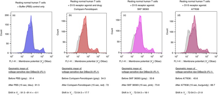
Figure 4.

Three selective agonists of D1‐like receptors (D1R and D5R) induce depolarization of CD3+ normal human T cells, within 15 seconds. (a–d) The fluorescence profiles of the voltage‐sensitive fluorescence dye DiBac3 inside normal human T cells, before (gray) and then 15 seconds after (colored) either: Control buffer (PBS) only (a), Corlopam, the Fenoldopam drug (from its original drug ampoule) (b), SKF 38393 (c) and A77636 (d). All the DR agonists were used in a final concentration of 10−7 M (100 nM), prepared freshly before each experiment, from either their powders or from concentrated stocks stored at −80°. The corresponding intensity of the voltage‐sensitive fluorescence dye DiBac3 was determined by the geometric mean (GM) of the fluorescence (FL1) in each tube before and after addition of each agonist. The GMs and fold shift in each case are shown in the figures below the corresponding fluorescence profiles. As for the depolarization of resting T cells shown in Fig 2, and for activated T cells shown in Fig. 3, here too the results shown are for one representative experiment, of two performed altogether, on T cells of different healthy human participants. In both experiments the same pattern of results was observed, but the actual extent of the depolarization varied. This was not surprising in view of the differences between individuals with regard to the level of DRs on the cell surface of their T cells found in7 and also in the present study (data not shown).
