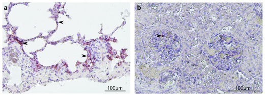
Figure 1. Immunohistochemistry of African green monkey (AGM) tissues after Nipah virus (NiV) infection.
An AGM was infected by NiV via the respiratory route, and necropsy was performed 8 days after infection. Immunostaining of lungs ( a) and kidney ( b) was made by using a polyclonal rabbit antibody specific for NiV nucleoprotein, and hematoxylin was used for the counter-staining. Interstitial pneumonia was found in lungs, inflammatory cells were present in both lungs and kidney, and positive immunostaining for NiV N (arrows) was observed in the alveolar wall and kidney glomerulus.

