Abstract
Background and Aims
C4 plants have higher photosynthetic capacity than C3 plants, but this advantage comes at an energetic cost that is problematic under low light. In the crop canopy, lower leaves first develop under high light but later experience low light because of mutual shading. To explore the re-acclimation of C4 leaves to low light, we investigated the structural and physiological changes of the leaves of maize plants grown in shaded pots.
Methods
Plants were first grown under high light, and then some of them were shaded (20 % of sunlight) for 3 weeks. Four types of leaves were examined: new leaves that developed under low light during shading (L), new leaves that developed under high light (H), mature leaves that developed under high light before shading and were then subjected to low light (H–L) and mature leaves that always experienced high light (H–H).
Key Results
The leaf mass per area, nitrogen and chlorophyll contents per unit leaf area, chlorophyll a/b ratio and activities of C3 and C4 photosynthetic enzymes were lower in H–L than in H–H leaves and in L than in H leaves. Unlike L leaves, H–L leaves maintained the thickness and framework of the Kranz anatomy of H leaves, but chloroplast contents in H–L leaves were reduced. This reduction of chloroplast contents was achieved mainly by reducing the size of chloroplasts. Although grana of mesophyll chloroplasts were more developed in L leaves than in H leaves, there were no differences between H–L and H–H leaves. The light curves of photosynthesis in H–L and L leaves were very similar and showed traits of shade leaves.
Conclusions
Mature maize leaves that developed under high light re-acclimate to low-light environments by adjusting their biochemical traits and chloroplast contents to resemble shade leaves but maintain the anatomical framework of sun leaves.
Keywords: C4 plant, chloroplast, leaf structure, light re-acclimation, maize, photosynthetic traits, shade leaf, sun leaf
INTRODUCTION
Photosynthetic capacity is the major determinant of crop productivity. However, an increase of crop productivity requires improvement of photosynthetic performance at the whole canopy level in the field (Evans, 2013). In a crop canopy, photosynthesis occurs under full sunlight in its upper part, whereas the mutual shading of leaves in the lower canopy results in light-limited conditions for photosynthesis (Long et al., 2006). Based on a modelling study, Long (1993) has shown that light-limited photosynthesis accounts for about half of the carbon fixed in the crop canopy. Because major crops have recently been cultivated at high planting densities (Richards, 2000), acclimation by leaves to low light is one of the critical determinants of crop yields in these systems. Major C4 crops such as maize and sorghum are also grown in dense canopies where most leaves are shaded (Pignon et al., 2017).
The C4 pathway functions as a CO2-concentrating mechanism for the C3 cycle. In leaves of C4 plants, the initial fixation of CO2 by phosphoenolpyruvate carboxylase (PEPC) is performed in the mesophyll (M) cells, whereas the decarboxylation of C4 acids and the re-fixation of CO2 by ribulose 1,5-bisphosphate carboxylase/oxygenase (Rubisco) subsequently occur in the bundle sheath (BS) cells (Hatch, 1987; Kanai and Edwards, 1999). The role of the C4 pathway is to increase the partial pressure of CO2 within BS cells and thereby reduce photorespiration (Hatch, 1987). Under high-light and/or high-temperature conditions, which accelerate photorespiration, C4 plants can therefore exhibit a high photosynthetic efficiency and productivity (Brown, 1999; Ghannoum et al., 2011). However, C4 photosynthesis requires additional ATP to reduce a CO2 molecule (Hatch, 1987). In today’s environment, the light-use efficiency is lower in C4 plants than in C3 plants at leaf temperatures below 25–30 °C, because photorespiration of C3 plants decreases with temperature (Ehleringer and Monson, 1993). Recently, it has been pointed out that CO2 leakiness from BS cells has important implications for C4 performance under low-light conditions (reviewed by Kromdijk et al., 2014).
The acclimation of plants to light conditions via differentiation of sun and shade leaves has been extensively investigated in plant ecophysiology. Previous studies have revealed light acclimation mechanisms mainly in C3 plants (reviewed in Björkman, 1981; Pearcy, 1998; Lambers et al., 2008). However, there have been relatively few studies of light acclimation in C4 plants (Winter et al., 1982; Pearcy and Franceschi, 1986; Ward and Woolhouse, 1986a, b; Sage and Mckown, 2006; Kromdijk et al., 2010; Pengelly et al., 2010; Bellasio and Griffiths, 2014a, b; Sales et al., 2018). In both C3 and C4 plants, shade leaves are thinner, have lower N content per unit leaf area and have lower light compensation and saturation points of photosynthesis than sun leaves. In C3 plants, shade leaves have well-developed grana in chloroplasts. However, the response of chloroplasts to low light is more complex in C4 plants than in C3 plants because C4 leaves have two types of photosynthetic cells. Although several studies have described the ultrastructure of chloroplasts in shaded C4 leaves (Paul and Patterson, 1980; Pearcy and Franceschi, 1986; Ward and Woolhouse, 1986a), precise chloroplast quantification and evaluation of granum development have been very limited (Jiang et al., 2011).
The re-acclimation of mature leaves developed under low light to high light and vice versa is another subject of interest. For example, the re-acclimation of mature shade leaves to a sudden increase of light intensity would be critical for survival of understorey trees (Yamashita et al., 2000). In field crops, however, leaves in the lower canopy develop under high-light conditions at an early stage of canopy formation. The light conditions then gradually shift from sun to shade during canopy growth. Therefore, understanding crop photosynthesis at the field scale requires elucidation of the re-acclimation of mature leaves that developed under high light to progressive shading. Understanding of this phenomenon is particularly important for C4 crops, because they are cultivated in tall, dense canopies (Pignon et al., 2017). The acclimation of C4 plants to low-light environments has been studied with respect to CO2 leakiness from BS cells (Kromdijk et al., 2008, 2014; Tazoe et al., 2008; Pengelly et al., 2010; Bellasio and Griffiths, 2014a; Sharwood et al., 2014). The re-acclimation of mature leaves of C4 plants to low light has not yet been fully explored, although Bellasio and Griffiths (2014b) have recently estimated respiration rates, ATP production rates and CO2 leakiness from BS cells in mature maize leaves. It seems likely that mature leaves do not greatly alter the structure of their photosynthetic tissue in response to changing light conditions. However, previous studies of C3 species have revealed a re-acclimation of photosynthesis to new light environments via adjustments of the structure and quantity of chloroplasts and of the photochemical and biochemical traits of photosynthetic cells (Sebaa et al., 1987; Sims and Pearcy, 1992; Brooks et al., 1996; Oguchi et al., 2003).
In this study, we investigate how mature maize leaves developed under high-light conditions re-acclimate to a low-light environment. We also examined the structural and photosynthetic traits of typical sun and shade leaves. The results of this study will contribute to our understanding of the photosynthetic performance of the canopy of field-grown maize.
MATERIALS AND METHODS
Plant materials and growth conditions
Seeds of maize (Zea mays cv. P1690) were germinated in multi-well nursery boxes filled with loam soil granules and grown for 2 weeks in a greenhouse at the experimental field of Kyushu University. The seedlings were transplanted into 5 L pots (one plant per pot) filled with sandy loam soil mixed with a commercial fertilizer that provided 1 g of nitrogen, phosphorus and potassium for each pot. The plants were then grown in the greenhouse until the start of shade treatment. During the growth of seedlings and the following experimental period, midday maximum photon flux density (PFD) in the greenhouse was about 1200 µmol photons m–2 s–1, and the daytime mean temperature was about 29°C.
At 12 d after transplanting, plants developing the fifth leaf were divided into groups to be grown under high-light (control) or low-light conditions (Fig. 1). Low-light conditions were generated by enclosing a rectangular parallelepiped frame (2.0 m wide × 1.6 m deep ×1.5 m high) with three layers of black shade cloth (1.3 mm net; Hifuku 004, Unitika Co. Ltd, Osaka, Japan). Within the enclosure, light intensity was reduced to 20 % of sunlight in the greenhouse. The pots in the high-light treatment were not shaded, and the light conditions in the high-light treatment were therefore identical to the light conditions in the greenhouse. Before the start of the treatment experiments, the fully expanded uppermost leaves were marked and were considered to represent leaves formed under full sunlight. After the plants had been grown under high-light or low-light conditions for 3 weeks, four types of leaves were investigated (Fig. 1); new leaves developed under high light (H); new leaves developed under low light (L); and mature leaves developed under high light and then exposed to either high light (H–H) or low light (H–L) for 3 weeks. Leaves of 3–5 plants (one leaf per plant) from each treatment were used in the following experiments.
Fig. 1.
Shading experiment for leaves of maize plants. Plants were first grown under high light and then grown for 3 weeks under cloths (shading treatment) or without cloths (control). An H leaf is a new leaf developed under high light. An L leaf is a new leaf developed under low light. An H–H leaf is a mature leaf first developed under high light and then exposed to high light for 3 weeks. An H–L leaf is a mature leaf first developed under high light and then exposed to low light for 3 weeks.
Gas exchange
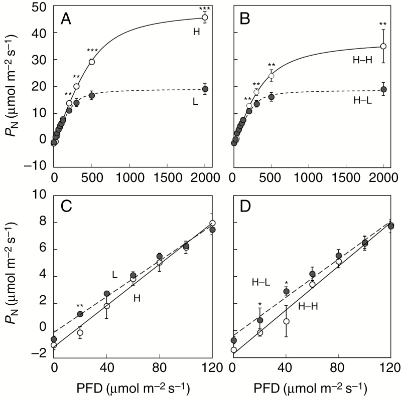
The light response curve of photosynthesis was measured using a portable photosynthesis system (LI-6400XT; LI-COR, Lincoln, NE, USA) at a leaf temperature of 30 °C, a relative humidity of 60 % and a CO2 concentration of 380 µmol mol–1. The light intensity was increased stepwise from 0 to 2000 µmol photons m–2 s–1 (Fig. 2). The net photosynthetic rate (PN) was measured at each light level when the rate had stabilized. From the initial slope of the light response curve, the photosynthetic quantum yield (Φ) was calculated. The dark respiration rate (Rd) and light compensation point (LCP) were calculated from the intercepts of the tangent to the light–response curve in the limit of low light with the ordinate and abscissa, respectively.
Fig. 2.
Response of net photosynthetic rate (PN) to photon flux density (PFD) of maize plants. H, L, H–H and H–L have the same meaning as in Fig. 1. (A, B) Full light curves of PN; (C, D) PN at low irradiances. Means ± s.d. of 3–5 plants in each treatment. Significance at *P < 0.05; **P < 0.01; ***P < 0.001.
Chlorophyll (Chl) and N content, and leaf mass per area
Samples were collected from the same leaves used in the gas exchange measurements. Chl was extracted from each sample (1.1 cm2 from the middle of a leaf) in N,N-dimethylformamide and measured spectrophotometrically according to Porra et al. (1989). The Chl content was expressed on the basis of leaf area. Other leaf samples (2.6 cm2) were air dried for 2 d at 80 °C and used for determination of leaf mass per area (LMA). The remaining leaf parts were also air-dried and milled to a fine powder. The N content of samples was determined by using the Kjeldahl method (Yabiku and Ueno, 2017).
Enzyme assay
Parts of the same leaves used for the gas exchange measurements were sampled, immediately frozen in liquid nitrogen and stored at ‒80 °C. For enzyme assays, leaf samples (0.2 g fresh mass) were ground on ice with a pestle in a mortar containing 1 mL of grinding medium [50 mm HEPES-KOH (pH 7.5), 1 mm EDTA-2Na, 5 mm dithiothreitol, 10 mm MgCl2 and 0.02 % (v/v) Triton X-100] with 0.5 % (w/v) bovine serum albumin, 5 mg of polyvinylpolypyrrolidone and 0.1 g of quartz sand. The homogenates were filtered through two layers of gauze, the filtrates were centrifuged for 5 min at 10 000 g at 4 °C and the supernatants were used for the enzyme assay. An aliquot of the filtrate was taken for determination of Chl content.
Activities of photosynthetic enzymes were assayed spectrophotometrically in 1 mL reaction mixtures at 30 °C. Activities of PEPC and NADP-malic enzyme (NADP-ME) were assayed as described by Ueno and Sentoku (2006). Those of phosphoenolpyruvate carboxykinase (PCK) and Rubisco were measured as described by Yabiku and Ueno (2017). In the assay of Rubisco activity, the supernatant was pre-incubated in the presence of 10 mm NaHCO3 and 10 mm MgCl2 at 25 °C for 10 min. The enzyme activities per Chl content were converted to the enzyme activities per leaf area using the Chl content per leaf area.
Anatomical and ultrastructural traits of leaves
Segments were sampled from the middle of the same leaves used for the gas exchange measurements, fixed in a formalin–acetic acid–alcohol solution for 1 d and cleared according to Ueno et al. (2006). Interveinal distance (IVD) was measured under a light microscope (Eclipse Ci-L, Nikon Instech Co. Ltd, Tokyo, Japan) as described by Yabiku and Ueno (2017). The IVD was the mean of ten measurements of the distance between centres of adjacent small longitudinal veins.
Segments from the middle of the same leaves were also fixed in 3 % (v/v) glutaraldehyde in 50 mm sodium phosphate buffer (pH 6.8) at room temperature for 1.5 h. After washing with phosphate buffer, they were post-fixed in 2 % (w/v) OsO4 in sodium phosphate buffer (pH 6.8) for 2 h, dehydrated through a series of acetone and embedded in Quetol resin (Nisshin-EM Co. Ltd, Tokyo, Japan) at 70 °C. The transverse semi-thin sections (approx. 1 ìm thick) of leaves were sectioned with a glass knife on an ultramicrotome, stained with 1 % toluidine blue O and then observed under a light microscope. Leaf thickness and size (profile area) of M and BS cells were measured on digital images of transverse sections using ImageJ software (Schneider et al., 2012). Leaf thickness was equated to the mean thickness of ten points per leaf. The sizes of M and BS cells were equated to the means of the sizes of 30 and 15 cells per leaf, respectively. The M/BS tissue area ratio was measured between two adjacent vascular bundles.
Ultra-thin transverse sections of leaves were also sectioned with a diamond knife from the same samples used for the semi-thin sections, stained with lead citrate and observed under an electron microscope (JEM-100CX II K, JEOL Ltd, Tokyo, Japan). The number of chloroplasts per cell profile was counted under the electron microscope for 15 M and 15 BS cells per leaf. Other quantitative traits of chloroplasts and the cell wall thickness of M and BS cells were measured on digital images of ultra-thin sections using ImageJ software. The chloroplast sizes (profile areas) were equated to the means of 20 measurements of both the M and BS cells of a leaf. If the chloroplasts accumulated starch grains, the area of the starch grains was subtracted from the estimation of chloroplast size. The chloroplast occupancy (ratio of chloroplast area to cell area) was calculated from the cell size, chloroplast size and number of chloroplasts per cell. Total profile areas of grana per chloroplast and number of grana per chloroplast were equated to the means of 20 measurements of M cells. The granum size of M chloroplasts was calculated from these parameters. The granum occupancy (ratio of granum area to chloroplast area) of M chloroplasts was calculated from the chloroplast size and total profile area of grana per chloroplast. The granum occupancy in BS chloroplasts was not investigated because the granum area was too small. The cell wall thicknesses were measured on the cell walls of M and BS cells exposed to intercellular spaces of M cells and the interface between M and BS cells for four M cells and four BS cells (three sites per cell).
Statistical analysis
The split-plot analysis of variance (ANOVA) was carried out regarding light condition as the main plot and leaf position as the sub-plot by use of PROC GLM by SAS 9.3 (SAS Institute Inc., Cary, NC, USA) software with SAS Studio interface. Fisher’s least significant difference (LSD) test was used to determine differences of the means between H and L leaves and between H–H and H–L leaves.
RESULTS
Gas exchange traits
The light curve of photosynthesis was compared among the four types of leaves (Fig. 2). The PN at PFDs >200 µmol photons m–2 s–1 was lower in L leaves than in H leaves and in H–L leaves than in H–H leaves (Fig. 2A, B; Table 1). In L and H–L leaves, PN was saturated at a PFD >500 µmol photons m–2 s–1. In H and H–H leaves, however, PN increased with increasing PPFD at PFDs of 500–2000 µmol photons m–2 s–1 (Fig. 2A, B). At PFDs below about 120 µmol photons m–2 s–1, however, PN was higher (Fig. 2C, D) and Φ was lower (Table 1) in L leaves than in H leaves and in H–L leaves than in H–H leaves. The Rd and LCP were lower in L leaves than in H leaves and in H–L leaves than in H–H leaves (Fig. 2C, D; Table 1).
Table 1.
Comparison of physiological and biochemical traits in upper and lower leaves of maize plants.
| Traits | Upper leaf | Lower leaf | ANOVA | ||||||||
|---|---|---|---|---|---|---|---|---|---|---|---|
| H | L | L/H | H–H | H–L | H–L/ H–H | Light | Leaf position | Interaction | |||
| P 2000 (µmol m–2 s–1) | 45.6 ± 2.1 | 19.1 ± 2.0 | ** | 0.42 | 34.9 ± 6.1 | 19.0 ± 2.4 | ** | 0.54 | ** | + | + |
| Φ (mol mol–1) | 0.078 ± 0.008 | 0.064 ± 0.002 | * | 0.82 | 0.082 ± 0.005 | 0.06 ± 0.01 | * | 0.73 | * | n.s. | n.s. |
| LCP (µmol m–2 s–1) | 17.2 ± 3.1 | 5.6 ± 1.7 | ** | 0.33 | 22.9 ± 2.5 | 6.8 ± 3.1 | *** | 0.30 | *** | + | n.s. |
| R d (µmol m–2 s–1) | 1.32 ± 0.29 | 0.40 ± 0.13 | ** | 0.31 | 1.77 ± 0.27 | 0.49 ± 0.22 | *** | 0.28 | ** | * | + |
| Leaf N content (mmol m–2) | 98.3 ± 8.5 | 49.8 ± 2.5 | *** | 0.51 | 64.9 ± 5.8 | 42.6 ± 6.0 | ** | 0.66 | *** | *** | *** |
| Chl a + b content (mg m–2) | 432 ± 40 | 224 ± 14 | ** | 0.52 | 398 ± 64 | 275 ± 27 | * | 0.69 | ** | n.s. | * |
| Chl a + b content (mg g–1 d. wt) | 10.4 ± 0.7 | 12.2 ± 1.1 | n.s. | 1.17 | 13.2 ± 2.4 | 17.1 ± 2.0 | * | 1.30 | + | ** | n.s. |
| Chl a/b ratio | 4.83 ± 0.14 | 4.29 ± 0.16 | * | 0.89 | 4.44 ± 0.19 | 3.56 ± 0.25 | ** | 0.80 | ** | ** | n.s. |
| LMA (g m–2) | 41.6 ± 1.1 | 18.4 ± 1.5 | *** | 0.44 | 30.5 ± 2.0 | 16.2 ± 2.2 | *** | 0.53 | *** | *** | ** |
| PEPC activity (µmol m–2 s–1) | 85.3 ± 9.6 | 19.8 ± 5.7 | *** | 0.23 | 44.0 ± 17.5 | 14.4 ± 6.1 | * | 0.33 | ** | * | * |
| Rubisco activity (µmol m–2 s–1) | 27.0 ± 4.2 | 9.4 ± 4.3 | ** | 0.35 | 18.6 ± 4.6 | 7.3 ± 4.9 | * | 0.39 | * | * | n.s. |
| NADP-ME activity (µmol m–2 s–1) | 73.8 ± 6.9 | 24.2 ± 4.2 | *** | 0.33 | 60.4 ± 6.5 | 36.7 ± 10.4 | ** | 0.61 | ** | n.s. | * |
| PCK activity (µmol m–2 s–1) | 15.6 ± 4.7 | 3.9 ± 1.2 | ** | 0.25 | 6.0 ± 1.1 | 2.5 ± 1.0 | n.s. | 0.42 | * | ** | * |
The values are given as the mean ± s.d. of 3–5 plants in each treatment.Significant difference between H and L leaves and between H–H and H–L leaves by Fisher’s LSD test at *P < 0.05; **P < 0.01; ***P < 0.001. n.s., not significant.
Symbols for ANOVA as in the Fisher’s LSD test except + < 0.10.
Chl, chlorophyll; LCP, light compensation point; Rd, dark respiration rate; N, nitrogen; P2000, net photosynthetic rate at PFD = 2000 µmol m–2 s–1; Φ, quantum yield of CO2 fixation; LMA, leaf mass per area.
Physiological and biochemical traits
Leaf N content, Chl content per unit leaf area, Chl a/b ratio and LMA were lower in L leaves than in H leaves and in H–L leaves than in H–H leaves, whereas Chl content per unit leaf mass showed the opposite tendency (Table 1). Activities of PEPC, Rubisco and NADP-ME were lower in L leaves than in H leaves and in H–L leaves than in H–H leaves (Table 1). Although the activity of PCK also showed a similar trend, there was no significant difference between H–H and H–L leaves (Table 1). The activity ratios of L to H leaves were greater in Rubisco than in PEPC and in NADP-ME than in PCK (Table 1). Similar trends were also apparent in the activity ratios of H–L to H–H leaves for these enzymes (Table 1).
Leaf anatomical traits and cell wall thickness
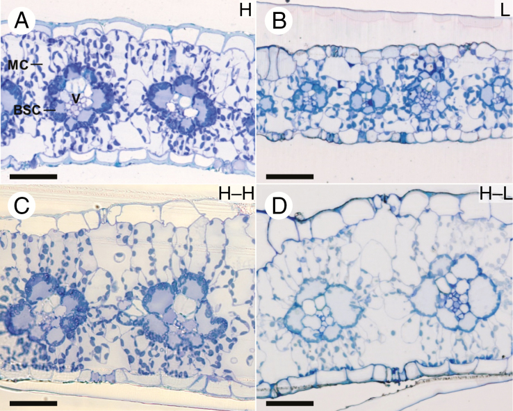
All four types of leaves showed Kranz-type anatomies (Fig. 3). The vascular bundles were surrounded by an outer layer of M cells and an inner layer of BS cells. The chloroplasts in the BS cells were located centrifugally in all leaf types.
Fig. 3.
Transverse sections of upper and lower leaves of maize plants. (A) H leaf; (B) L leaf; (C) H–H leaf; (D) H–L leaf. BSC, bundle sheath cell; MC, mesophyll cell; V, vascular bundle. Scale bars = 50 μm.
The L leaves were thinner and had smaller IVDs than H leaves (Fig. 3A, B; Table 2). The M and BS cells were smaller in L leaves than in H leaves. However, the M/BS tissue area ratio did not differ significantly between L and H leaves (Table 2). There was no significant difference in these parameters between H–L and H–H leaves (Fig. 3C, D; Table 2).
Table 2.
Comparison of structural traits in upper and lower leaves of maize plants
| Traits | Cell | Upper leaf | Lower leaf | ANOVA | ||||||||
|---|---|---|---|---|---|---|---|---|---|---|---|---|
| H | L | L/H | H–H | H–L | H–L/H–H | Light | Leaf position | Interaction | ||||
| Leaf thickness (µm) | 144 ± 5 | 100 ± 13 | ** | 0.69 | 184 ± 16 | 167 ± 17 | n.s. | 0.91 | * | *** | + | |
| IVD (µm) | 103 ± 5 | 63 ± 9 | ** | 0.61 | 122 ± 12 | 106 ± 17 | n.s. | 0.87 | * | ** | + | |
| M/BS tissue area ratio | 2.77 ± 0.08 | 2.97 ± 0.14 | n.s. | 1.07 | 3.32 ± 0.13 | 3.26 ± 0.13 | n.s. | 0.98 | n.s. | ** | + | |
| Cell size (µm2) | MC | 356 ± 35 | 238 ± 13 | * | 0.67 | 567 ± 68 | 503 ± 59 | n.s. | 0.89 | * | *** | n.s. |
| BSC | 412 ± 24 | 196 ± 46 | ** | 0.48 | 563 ± 37 | 523 ± 110 | n.s. | 0.93 | + | *** | * | |
| Cell wall thickness (µm) | MC | 0.083 ± 0.019 | 0.061 ± 0.004 | * | 0.73 | 0.075 ± 0.003 | 0.071 ± 0.009 | n.s. | 0.95 | n.s. | n.s. | n.s. |
| BSC† | 0.491 ± 0.053 | 0.192 ± 0.040 | *** | 0.39 | 0.288 ± 0.032 | 0.213 ± 0.042 | * | 0.74 | ** | ** | ** | |
| MC/BSC interface | 0.448 ± 0.027 | 0.167 ± 0.020 | *** | 0.37 | 0.239 ± 0.037 | 0.194 ± 0.043 | n.s. | 0.81 | *** | ** | ** | |
†The cell wall thickness of BS cells exposed to intercellular space of mesophyll cells.
The values are given as the mean ± s.d. of 3–5 plants in each treatment. Significant difference between H and L leaves and between H–H and H–L leaves by Fisher’s LSD test at *P < 0.05; **P < 0.01; ***P < 0.001. n.s., not significant.
Symbols for ANOVA as in the Fisher’s LSD test except + < 0.10.
BS, bundle sheath; BSC, bundle sheath cell; IVD, interveinal distance; M, mesophyll; MC, mesophyll cell.
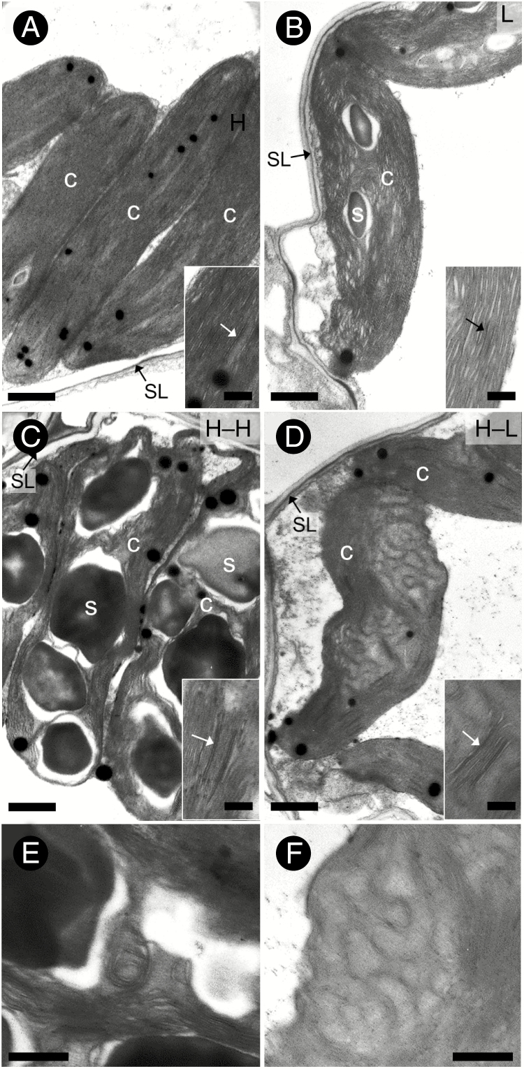
The cell wall thickness of M cells was thinner in L leaves than in H leaves, but it did not differ significantly between H–L and H–H leaves. The walls of BS cells exposed to the intercellular space between M cells were much thinner in L leaves than in H leaves and slightly thinner in H–L leaves than in H–H leaves (Table 2). As a result, the extent of reduction of wall thickness in BS cells was greater in L leaves than in H–L leaves (Table 2). The cell wall thickness at the interface between M and BS cells was thinner in L leaves than in H leaves, but it did not differ significantly between H–L and H–H leaves (Table 2). Suberized lamellae occurred in the cell walls of BS cells of all leaf types (Fig. 4).
Fig. 4.
Ultrastructure of bundle sheath chloroplasts in upper and lower leaves of maize plants. (A) H leaf. (B) L leaf. (C) H–H leaf. (D) H–L leaf. (E, F) Convoluted thylakoid structure of H–H and H–L leaves, respectively. Insets in (A) to (D) show enlarged images of thylakoids, and arrows show rudimentary grana. c, chloroplast; s, starch grain; SL, suberized lamella. Scale bars = 1 μm for (A–D), 0.5 μm for (E) and (F), and 0.25 μm for insets of (A–D).
Structural and quantitative traits of chloroplasts
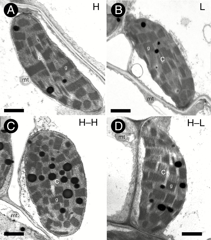
In all leaf types, M chloroplasts had well-developed grana (Fig. 5), whereas BS chloroplasts had only rudimentary grana (Fig. 4). The thylakoids ran mostly parallel in the BS chloroplasts of H and L leaves (Fig. 4A, B). In the BS chloroplasts of H–L and H–H leaves, the thylakoids were partially convoluted (Fig. 4C–F). This conformation was more remarkable in H–L leaves (Fig. 4D, F). In addition, a large accumulation of starch grains was apparent in the BS chloroplasts of H–H leaves (Fig. 4C).
Fig. 5.
Ultrastructure of mesophyll chloroplasts in upper and lower leaves of maize plants. (A) H leaf. (B) L leaf. (C) H–H leaf. (D) H–L leaf. c, chloroplast; g, granum; mt, mitochondrion. Scale bars = 1 μm.
The size of M chloroplasts did not differ in H and L leaves, but it was smaller in H–L leaves than in H–H leaves (Table 3). The BS chloroplasts were smaller in L leaves than in H leaves and in H–L leaves than in H–H leaves (Table 3). The numbers of chloroplasts per cell in M and BS cells were smaller in L than in H leaves (Table 3). Although the number of chloroplasts per cell in M cells was also smaller in H–L leaves than in H–H leaves, there was no significant difference in BS cells (Table 3). In M cells, there was no significant difference in the chloroplast occupancy between H and L leaves and between H–H and H–L leaves (Table 3). In BS cells, the chloroplast occupancy was lower in L leaves than in H leaves and in H–L leaves than in H–H leaves (Table 3). The size of grana in M chloroplasts was greater in L leaves than in H leaves, but it did not differ significantly between H–H and H–L leaves (Table 3). The granum occupancy in M chloroplasts was higher in L leaves than in H leaves, whereas there was no significant difference between H–H and H–L leaves (Table 3).
Table 3.
Comparison of quantitative traits of chloroplasts in upper and lower leaves of maize plants
| Traits | Cell | Upper leaf | Lower leaf | ANOVA | ||||||||
|---|---|---|---|---|---|---|---|---|---|---|---|---|
| H | L | L/H | H–H | H–L | H–L/ H–H | Light | Leaf position | Interaction | ||||
| Chloroplast size (µm2) | MC | 12.1 ± 0.7 | 12.4 ± 1.7 | n.s. | 1.02 | 14.4 ± 0.5 | 10.6 ± 0.2 | *** | 0.74 | ** | n.s. | ** |
| BSC | 13.4 ± 1.4 | 10.0 ± 1.2 | ** | 0.75 | 15.5 ± 1.1 | 9.3 ± 0.4 | *** | 0.60 | *** | n.s. | ** | |
| Chloroplast number (no. per cell) | MC | 6.3 ±0.5 | 4.5 ± 0.3 | ** | 0.71 | 6.8 ± 0.8 | 5.7 ± 1.1 | * | 0.84 | * | * | n.s. |
| BSC | 9.6 ± 1.2 | 5.1 ± 0.5 | ** | 0.53 | 11.1 ± 1.3 | 9.4 ± 2.3 | n.s. | 0.85 | * | ** | n.s. | |
| Chloroplast occupancy (%) | MC | 21.8 ± 3.1 | 21.5 ± 0.3 | n.s. | 0.99 | 17.4 ± 3.4 | 12.1 ± 1.6 | n.s. | 0.70 | + | ** | n.s. |
| BSC | 35.0 ± 3.4 | 26.4 ± 3.0 | ** | 0.75 | 30.5 ± 3.0 | 16.7 ± 2.0 | ** | 0.55 | *** | ** | n.s. | |
| Granum size (µm2) | MC | 0.177 ± 0.022 | 0.213 ± 0.036 | * | 1.20 | 0.206 ± 0.025 | 0.183 ± 0.037 | n.s. | 0.89 | n.s. | n.s. | + |
| Granum occupancy (%) | MC | 38.7 ± 2.2 | 44.6 ± 1.9 | ** | 1.15 | 41.1 ± 1.6 | 41.7 ± 0.5 | n.s. | 1.01 | * | n.s. | * |
The values are given as the mean ± s.d. of 3–5 plants in each treatment.
Significant difference between H and L leaves and between H–H and H–L leaves by Fisher’s LSD test at *P < 0.05; **P < 0.01; ***P < 0.001. n.s., not significant.
Symbols for ANOVA as in the Fisher’s LSD test except + < 0.10.
BSC, bundle sheath cell; IVD, interveinal distance; MC, mesophyll cell.
DISCUSSION
We discovered that the combination of anatomical, biochemical and physiological traits differed greatly between mature maize leaves that developed under high light and were then exposed to low light vs. new leaves that developed under either high or low light.
The L leaves were thinner than H leaves. The smaller photosynthetic cells in the L leaves resulted in a small IVD. These traits did not differ significantly between H–L leaves and H–H leaves (Table 2). The fact that the M/BS tissue area ratio did not differ between H and L leaves (Table 2) suggests that a quantitative balance between the M and BS tissues in the operation of C4 metabolism is important (Hattersley, 1984; Dengler and Nelson, 1999). The M/BS tissue area ratio was quite similar in H–H and H–L leaves. Although L leaves acclimated to low light by changes in leaf thickness and anatomical framework, these results show that H–L leaves maintained these traits. As a result, the anatomical features of H–L leaves differed greatly from those of L leaves. The H–L leaves showed unusual anatomical features because small chloroplasts were distributed in large photosynthetic cells (Fig. 3). The responses of cell wall thickness in M and BS cells to low-light conditions were very complex in L and H–L leaves (Table 2). However, there was an apparent tendency for the cell walls of photosynthetic cells to be thinner in L leaves than in H leaves, whereas the cell wall thicknesses did not differ much between H–L and H–H leaves.
The LMA and N content per unit leaf area were lower in L leaves than in H leaves (Table 1), similar to shade leaves of many C3 species (Björkman, 1981; Pearcy, 1998; Lambers et al., 2008) and several C4 species (Ward and Woolhouse, 1986a; Pengelly et al., 2010). The Chl content per unit leaf area and Chl a/b ratio were also lower in L leaves than in H leaves (Table 1). The response of the Chl a/b ratio is typical of shade leaves (Björkman, 1981; Pearcy, 1998; Lambers et al., 2008). The H–L leaves behaved similarly to the L leaves with respect to these traits, although the extents of the decreases, except for the Chl a/b ratio, were somewhat smaller (Table 1). The H–L leaves had lower contents of N and Chl per unit leaf area than H–H leaves, despite the similarity of the leaf thicknesses. The activities of C3 and C4 photosynthetic enzymes per unit leaf area decreased in L leaves vs. H leaves (Table 1). Such responses of photosynthetic enzymes have also been reported in leaves of maize acclimated to low light (Ward and Woolhouse, 1986b; Sharwood et al., 2014) and a C4 eudicot, Flaveria bidentis (Pengelly et al., 2010). In our study, a greater reduction of PEPC activity relative to that of Rubisco was apparent in L leaves (Table 1). Sharwood et al. (2014) have also found a similar response of the two carboxylases in shade-acclimated maize leaves. Although maize is classified in the NADP-ME type of C4 biochemical groups, both NADP-ME and PCK are involved in the decarboxylation of C4 acids (Wingler et al., 1999; Koteyeva et al., 2015). The reduction of the activities of both of these enzymes in L leaves led to a greater reduction of PCK activity relative to that of NADP-ME activity (Table 1; Sharwood et al., 2014). This differential downregulation of the two enzymes may represent a flexible response of the two decarboxylation pathways to changes of irradiance (Furbank, 2011; Bellasio and Griffiths, 2014c; Sharwood et al., 2014). As expected, activities of all photosynthetic enzymes except PCK decreased in H–L leaves vs. H–H leaves (Table 1).
The decreased activities of photosynthetic enzymes in L leaves explain the lower PN at high PFD of L leaves vs. H leaves (Fig. 2A). The L leaves also showed a higher PN at low PFD and lower LCP and Rd than H leaves (Fig. 2C). This behaviour of the light curve of photosynthesis has been reported in shade-acclimated leaves of C3 species (Björkman, 1981; Lambers et al., 2008) and of C4 species (Winter et al., 1982; Ward and Woolhouse, 1986a; Bellasio and Griffiths, 2014b). The PN at high PFD was also lower in H–L leaves vs. H–H leaves (Fig. 2B). The PN at high PFD was very similar in H–L and L leaves, whereas it was lower in H–H leaves than in H leaves (Fig. 2A, B). The H–L leaves also showed higher PN at low PFD and lower LCP and Rd than did H–H leaves (Fig. 2D; Bellasio and Griffiths, 2014b). These results show that H–L leaves can change to the photosynthetic gas exchange traits of shade leaves by modifying their biochemical components, whereas their anatomical framework maintains that of sun leaves.
The quantitative responses of the chloroplasts in the M and BS cells of maize to shading were complex (Table 3). The sizes of BS chloroplasts decreased in L leaves vs. H leaves, but the sizes of M chloroplasts did not change. The pattern of chloroplast occupancy was similar to that of chloroplast size. The chloroplast number per cell decreased with a reduction of the size of the M and BS cells in L leaves. These results therefore show that the decreases in N and Chl contents per unit leaf area and LMA in L leaves under low light caused the decrease in the number of chloroplasts in the M cells and the decreases in both the size and number of chloroplasts in the BS cells. The physiological basis of this regulatory mechanism is unclear. Chloroplast size decreased in the M and BS cells of H–L leaves, but the chloroplast number in these cells did not differ greatly between H–H and H–L leaves. These results show that the decreases in N and Chl contents and LMA in H–L leaves under low light caused the decrease in the size of their chloroplasts.
Shade leaves have more developed grana in their chloroplasts and lower Chl a/b ratios than sun leaves (Björkman, 1981; Pearcy, 1998; Lambers et al., 2008). A reduction of the Chl a/b ratio is associated with an increase of the light-harvesting Chl–protein complex II (LHCII) that constitutes the antenna protein of photosystem II (PSII) (Björkman, 1981; Anderson et al., 2008). The location of PSII is predominantly in the appressed grana stacks, whereas PSI is enriched in the non-appressed thylakoid membranes (Mustardy and Garab, 2003). In our study, the granum size and occupancy in the M chloroplasts was higher in L leaves than in H leaves. Correspondingly, the Chl a/b ratio decreased in L leaves (Table 1). The presence of rudimentary grana in the BS chloroplasts of both H and L leaves suggests that this thylakoid structure is essential for the operation of NADP-ME-type C4 photosynthesis (Hatch, 1987; Kanai and Edwards, 1999). Similar responses of M and BS chloroplasts in L leaves have been reported in leaves of maize (Ward and Woolhouse, 1986a) and sorghum (Jiang et al., 2011). The granum size and occupancy of H–L leaves did not increase (Table 3). This result suggests that the granum structure is not reorganized under low-light conditions. In H–L leaves, the extent of granum development does not appear to be linked to changes in the Chl a/b ratio because this ratio decreased (Table 1). The reason for this unusual relationship is unclear, and further studies will be required to provide an explanation. However, it has been pointed out that maize chloroplasts have a particular structural organization of their photosynthetic apparatus (Drozak and Romanowska, 2006; Romanowska et al., 2008).
The BS chloroplasts of H–H leaves accumulated numerous starch grains (Fig. 4C). This accumulation may have been caused by an imbalance between photosynthesis and the translocation of assimilates. Makino and Ueno (2018) have reported that nitrogen deficiency results in similar starch accumulation in the BS chloroplasts of sorghum C4 leaves. Correspondingly, H–H leaves showed a 34 % reduction of leaf N content compared with H leaves (Table 1). It is also noteworthy that the BS chloroplasts of H–L leaves had remarkably convoluted thylakoids (Fig. 4D). It is unclear whether the formation of such thylakoids is involved in acclimation to low light, because convoluted thylakoids have also been observed in the BS chloroplasts of many C4 sedges (Ueno et al., 1988) and some C4 species of Portulaca (Kim and Fisher, 1990), both of which are classified as NADP-ME-type plants.
In all four types of leaves examined in this study, the BS cells had suberized lamellae and thicker cell walls than the M cells (Table 2). In general, these structural features are considered to be responsible for the maintenance of high CO2 concentrations within BS cells (Hattersley and Browning, 1981; Dengler and Nelson, 1999; Mertz and Brutnell, 2014). Recent studies on CO2 leakiness from the BS cells of maize have revealed that the CO2 leakiness in leaves that developed under low-light conditions is reduced (Bellasio and Griffiths, 2014a; Sharwood et al., 2014). The leakiness is also reduced in mature sun leaves of maize re-acclimated to low light (Bellasio and Griffiths, 2014b). However, in our study the cell wall of BS cells was thinner in L leaves than in H leaves. In H–L leaves, the thickness of BS cell walls exposed to the intercellular space of M cells was reduced, whereas that of cell walls in the interface between M and BS cells remained unchanged (Table 2). It is therefore unlikely that the cell wall thickness of BS cells is responsible for the lowered CO2 leakiness associated with the acclimation of maize leaves to low light. Balancing C4 and C3 cycle activity may be a more critical factor involved in CO2 leakiness.
The ANOVA shows that the leaf position may also affect the structural and photosynthetic traits of lower leaves, although we focused on the effect of light (Tables 1–3). The maturity and senescence of leaves may also be involved in these effects. On the other hand, there is the possibility that the lower leaves may receive somewhat lower light intensity with a modified spectrum as a result of the presence of upper leaves. Further studies will be required to elucidate the effects of these light factors.
We demonstrated that when mature maize leaves developed under high-light conditions were subjected to low light, their photosynthetic traits re-acclimated to the new light environment by an adjustment of the N and Chl contents, activities of C3 and C4 photosynthetic enzymes and chloroplast size. In contrast, there was little change of anatomical framework such as leaf thickness, IVD, cell size and cell wall thickness. The Chl a/b ratio was also adjusted to the low-light environment, but without a change of granum structure. Our study provides insights that are needed to understand the photosynthetic performance of each leaf in a maize canopy. The responses of the cellular components of mature sun leaves are reasonable for the efficient spatial distribution of N resources to photosynthetic organs as light intensity decreases during canopy growth. Further studies will be required to be certain whether the re-acclimation of mature leaves shown here actually occurs in field-grown maize populations.
FUNDING
This work was supported by a Japan Society for the Promotion of Science KAKENHI grant (JP 16H04868) to O.U.
ACKNOWLEDGEMENTS
We thank Dr T. Hasegawa, Tohoku Agricultural Research Center, for his valuable advice on the statistic analysis, and Professor N. Furuya, Faculty of Agriculture, Kyushu University, for the use of an electron microscope.
LITERATURE CITED
- Anderson JM, Chow WS, Rivas JDL. 2008. Dynamic flexibility in the structure and function of photosystem II in higher plant thylakoid membranes: the grana enigma. Photosynthesis Research 98: 575–587. [DOI] [PubMed] [Google Scholar]
- Bellasio C, Griffiths H. 2014a Acclimation to low light by C4 maize: implications for bundle sheath leakiness. Plant, Cell & Environment 37: 1046–1058. [DOI] [PubMed] [Google Scholar]
- Bellasio C, Griffiths H. 2014b Acclimation of C4 metabolism to low light in mature maize leaves could limit energetic losses during progressive shading in a crop canopy. Journal of Experimental Botany 65: 3725–8736. [DOI] [PMC free article] [PubMed] [Google Scholar]
- Bellasio C, Griffiths H. 2014c The operation of two decarboxylases, transamination, and partitioning of C4 metabolic processes between mesophyll and bundle sheath cells allows light capture to be balanced for the maize C4 pathway. Plant Physiology 164: 466–480. [DOI] [PMC free article] [PubMed] [Google Scholar]
- Björkman O. 1981. Responses to different quantum flux densities. In: Lange OL, Nobel PS, Osmond CB, Zeigler H, eds. Encyclopedia of plant physiology, New Series, 12A. Dordrecht: Springer, 57–107. [Google Scholar]
- Brooks JR, Sprugel DG, Hinckley TM. 1996. The effects of light acclimation during and after foliage expansion on photosynthesis of Abies amabilis foliage within the canopy. Oecologia 107: 21–32. [DOI] [PubMed] [Google Scholar]
- Brown RH. 1999. Agronomic implications of C4 photosynthesis. In: Sage RF, Monson RK, eds. C4plant biology. San Diego: Academic Press, 473–507. [Google Scholar]
- Dengler NG, Nelson T. 1999. Leaf structure and development in C4 plants. In: Sage RF, Monson RK, eds. C4plant biology. San Diego: Academic Press, 133–172. [Google Scholar]
- Drozak A, Romanowska E. 2006. Acclimation of mesophyll and bundle sheath chloroplasts of maize to different irradiances during growth. Biochimica et Biophysica Acta 1757: 1539–1546. [DOI] [PubMed] [Google Scholar]
- Ehleringer JR, Monson RK. 1993. Evolutionary and ecological aspects of photosynthetic pathway variation. Annual Review of Ecology and Systematics 24: 411–439. [Google Scholar]
- Evans JR. 2013. Improving photosynthesis. Plant Physiology 162: 1780–1793. [DOI] [PMC free article] [PubMed] [Google Scholar]
- Furbank RT. 2011. Evolution of the C4 photosynthetic mechanism: are there really three C4 acid decarboxylation types? Journal of Experimental Botany 62: 3103–3108. [DOI] [PubMed] [Google Scholar]
- Ghannoum O, Evans JR, von Caemmerer S. 2011. Nitrogen and water use efficiency of C4 plants. In: Raghavendra AS, Sage RF, eds. C4photosynthesis and related CO2concentrating mechanisms. Dordrecht: Springer, 129–146. [Google Scholar]
- Hatch MD. 1987. C4 photosynthesis: a unique blend of modified biochemistry, anatomy and ultrastructure. Biochimica et Biophysica Acta 895: 81–106. [Google Scholar]
- Hattersley PW. 1984. Characterization of C4 type leaf anatomy in grasses (Poaceae). Mesophyll:bundle sheath area ratios. Annals of Botany 53: 163–179. [Google Scholar]
- Hattersley PW, Browning AJ. 1981. Occurrence of the suberized lamella in leaves of grasses of different photosynthetic types. I. In parenchymatous bundle sheaths and PCR (‘Kranz’) sheaths. Protoplasma 109: 371–401. [Google Scholar]
- Jiang CD, Wang X, Gao HY, Shi L. Chow WS. 2011. Systemic regulation of leaf anatomical structure, photosynthetic performance, and high-light tolerance in sorghum. Plant Physiology 155: 1416–1424. [DOI] [PMC free article] [PubMed] [Google Scholar]
- Kanai R, Edwards GE. 1999. The biochemistry of C4 photosynthesis. In: Sage RF, Monson RK, eds. C4plant biology. San Diego: Academic Press, 49–87. [Google Scholar]
- Kim I, Fisher DG. 1990. Structural aspects of the leaves of seven species of Portulaca growing in Hawaii. Canadian Journal of Botany 68: 1803–1811. [Google Scholar]
- Koteyeva NK, Voznesenskaya EV, Edwards GE. 2015. An assessment of the capacity for phosphoenolpyruvate carboxykinase to contribute to C4 photosynthesis. Plant Science 235: 70–80. [DOI] [PubMed] [Google Scholar]
- Kromdijk J, Schepers HE, Albanito F, et al. 2008. Bundle-sheath leakiness and light limitation during C4 leaf and canopy CO2 uptake. Plant Physiology 148: 2144–2155. [DOI] [PMC free article] [PubMed] [Google Scholar]
- Kromdijk J, Griffiths H, Schepers HE. 2010. Can the progressive increase of C4 bundle sheath leakiness at low PFD be explained by incomplete suppression of photorespiration? Plant, Cell & Environment 33: 1935–1948. [DOI] [PubMed] [Google Scholar]
- Kromdijk J, Ubierna N, Cousins AB, Griffiths H. 2014. Bundle-sheath leakiness in C4 photosynthesis: a careful balancing act between CO2 concentration and assimilation. Journal of Experimental Botany 65: 3443–3457. [DOI] [PubMed] [Google Scholar]
- Lambers H, Chapin FS III, Pons TL. 2008. Plant physiological ecology, 2nd edn. New York: Springer. [Google Scholar]
- Long SP. 1993. The significance of light-limiting photosynthesis to crop canopy carbon gain and productivity: a theoretical analysis. In: Abrol YP, Govindjee PM, eds. Photosynthesis: photoreactions to plant productivity. New Delhi: Oxford & IBH Publishing, 547–560. [Google Scholar]
- Long SP, Zhu XG, Naidu SL, Ort DR. 2006. Can improvement in photosynthesis increase crop yields? Plant, Cell & Environment 29: 315–330. [DOI] [PubMed] [Google Scholar]
- Makino Y, Ueno O. 2018. Structural and physiological responses of the C4 grass Sorghum bicolor to nitrogen limitation. Plant Production Science 21: 39–50. [Google Scholar]
- Mertz RA, Brutnell TP. 2014. Bundle sheath suberization in grass leaves: multiple barriers to characterization. Journal of Experimental Botany 65: 3371–3380. [DOI] [PubMed] [Google Scholar]
- Mustardy L, Garab G. 2003. Granum revised. A three-dimensional model – where things fall into places. Trends in Plant Science 8: 117–122. [DOI] [PubMed] [Google Scholar]
- Oguchi R, Hikosaka T, Hirose T. 2003. Does the photosynthetic light-acclimation need change in leaf anatomy? Plant, Cell & Environment 26: 505–512. [Google Scholar]
- Paul R, Patterson DT. 1980. Effects of shading on the anatomy and ultrastructure of the leaf mesophyll and vascular bundles of itchgrass (Rottboellia exaltata). Weed Science 28: 216–224 [Google Scholar]
- Pearcy RW. 1998. Acclimation to sun and shade. In: Raghavendra AS, ed. Photosynthesis: a comprehensive treatise. Cambridge: Cambridge University Press, 250–263. [Google Scholar]
- Pearcy RW, Franceschi VR. 1986. Photosynthetic characteristics and chloroplast ultrastructure of C3 and C4 tree species grown in high- and low-light environments. Photosynthesis Research 9: 317–331. [DOI] [PubMed] [Google Scholar]
- Pengelly JL, Sirault XRR, Tazoe Y, Evans JR, Furbank RT, von Caemmerer S. 2010. Growth of the C4 dicot Flaveria bidentis: photosynthetic acclimation to low light through shifts in leaf anatomy and biochemistry. Journal of Experimental Botany 61: 4109–4122. [DOI] [PMC free article] [PubMed] [Google Scholar]
- Pignon CP, Jaiswal D, McGrath JM, Long SP. 2017. Loss of photosynthetic efficiency in the shade. An Achilles heel for the dense modern stands of our most productive C4 crops? Journal of Experimental Botany 68: 335–345. [DOI] [PMC free article] [PubMed] [Google Scholar]
- Porra RJ, Thompson WA, Kriedemann PE. 1989. Determination of accurate extinction coefficients and simultaneous equations for assaying chlorophyll a and b extracted in four different solvents. Biochimica et Biophysica Acta 975: 389–394 [Google Scholar]
- Richards RA. 2000. Selectable traits to increase crop photosynthesis and yield of grain crops. Journal of Experimental Botany 51: 447–458. [DOI] [PubMed] [Google Scholar]
- Romanowska E, Kargul J, Powikrowska M, et al. 2008. Structural organization of photosynthetic apparatus in agranal chloroplasts of maize. Journal of Biological Chemistry 283: 26037–26096. [DOI] [PMC free article] [PubMed] [Google Scholar]
- Sage RF, Mckown AD. 2006. Is C4 photosynthesis less phenotypically plastic than C3 photosynthesis? Journal of Experimental Botany 57: 303–317. [DOI] [PubMed] [Google Scholar]
- Sales CRG, Ribeiro RV, Hayashi AH, et al. 2018. Flexibility of C4 decarboxylation and photosynthetic plasticity in sugarcane plants under shading. Environmental and Experimental Botany 149: 34–42. [Google Scholar]
- Schneider CA, Rasband WS, Eliceiri KW. 2012. NIH Image to ImageJ: 25 years of image analysis. Nature Methods 9: 671–675. [DOI] [PMC free article] [PubMed] [Google Scholar]
- Sebaa ED, Prioul JL, Brangeon J. 1987. Acclimation of adult Lolium multiflorum leaves to changes in irradiance: effect on leaf photosynthesis and chloroplast ultrastructure. Journal of Plant Physiology 127: 431–442. [Google Scholar]
- Sharwood RE, Sonawane BV, Ghannoum O. 2014. Photosynthetic flexibility in maize exposed to salinity and shade. Journal of Experimental Botany 65: 3715–3724. [DOI] [PMC free article] [PubMed] [Google Scholar]
- Sims DA, Pearcy RW. 1992. Response of leaf anatomy and photosynthetic capacity in Alocasia macrorrhiza (Araceae) to a transfer from low to high light. American Journal of Botany 79: 449–455. [Google Scholar]
- Tazoe Y, Hanba YT, Furumoto T, Noguchi K, Terashima I. 2008. Relationships between quantum yield for CO2 assimilation, activity of key enzymes and CO2 leakiness in Amaranthus cruentus, a C4 dicot, grown in high or low light. Plant & Cell Physiology 49: 19–29. [DOI] [PubMed] [Google Scholar]
- Ueno O, Sentoku N. 2006. Comparison of leaf structure and photosynthetic characteristics of C3 and C4Alloteropsis semialata subspecies. Plant, Cell and Environment 29: 257–268. [DOI] [PubMed] [Google Scholar]
- Ueno O, Takeda T, Maeda E. 1988. Leaf ultrastructure of C4 species possessing different Kranz anatomical types in the Cyperaceae. Botanical Magazine, Tokyo 101: 141–152. [Google Scholar]
- Ueno O, Kawano Y, Wakayama M, Takeda T. 2006. Leaf vascular systems in C3 and C4 grasses: a two-dimensional analysis. Annals of Botany 97: 611–621. [DOI] [PMC free article] [PubMed] [Google Scholar]
- Ward DA, Woolhouse HW. 1986a Comparative effects of light during growth on the photosynthetic properties of NADP-ME type C4 grasses from open and shaded habitats. I. Gas exchange, leaf anatomy and ultrastructure. Plant, Cell & Environment 9: 261–270. [Google Scholar]
- Ward DA, Woolhouse HW. 1986b Comparative effects of light during growth on the photosynthetic properties of NADP-ME type C4 grasses from open and shaded habitats. II. Photosynthetic enzyme activities and metabolism. Plant, Cell & Environment 9: 271–277. [Google Scholar]
- Wingler A, Walker RP, Chen ZH, Leegood RC. 1999. Phosphoenolpyruvate carboxykinase is involved in the decarboxylation of aspartate in the bundle sheath of maize. Plant Physiology 120: 539–545. [DOI] [PMC free article] [PubMed] [Google Scholar]
- Winter K, Schmitt MR, Edwards GE. 1982. Microstegium vimineum, a shade adapted C4 grass. Plant Science Letters 24: 311–318. [Google Scholar]
- Yabiku T, Ueno O. 2017. Variations in physiological, biochemical, and structural traits of photosynthesis and resource use efficiency in maize and teosintes (NADP-ME-type C4). Plant Production Science 20: 448–458. [Google Scholar]
- Yamashita N, Ishida A, Kushima H, Tanaka N. 2000. Acclimation to sudden increase in light favoring an invasion over native trees in subtropical islands, Japan. Oecologia 125: 412–419. [DOI] [PubMed] [Google Scholar]


