Dear Editor,
Antimicrobial peptides (AMPs) are an ancient class of short polypeptides present in a large number of species in nature with a variety of functions.1 PFR (PFWRIRIRR-NH2) is one kind of AMP identified among the derivatives of lactoferrin.2 Our previous results showed that PFR inhibited the proliferation of human acute myeloid leukemia (AML) HL60 cells potentially without toxicity against normal cells. In addition, PFR induced necrosis by membrane disruption detected using scanning electron microscopy.3 However, the underlying mechanisms of these effects are not clearly understood.
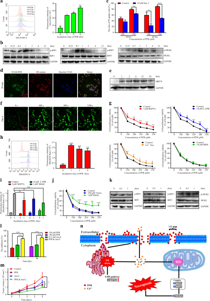
To investigate the mechanisms involved in necrosis4 induced by PFR in HL60 cells (Fig. S1a–d), we found that 5(6)-FAM was taken up by HL60 cells after PFR treatment in a time-dependent manner (Fig. 1a), indicating that PFR induced the formation of permeable pores with open diameters of at least the molecular size of 5(6)-FAM (≈1 nm). In addition, levels of phosphorylated RIP1, RIP3, and MLKL were increased significantly after PFR treatment (Figs. 1b and S1e), indicating that necroptosis had occurred. Furthermore, necrostatin-1 (Nec-1), a specific inhibitor of necroptosis, significantly reduced propidium iodide (PI) uptake induced by PFR (Fig. 1c).
Fig. 1.
PFR induces necroptosis through ER stress and elevated cytoplasmic calcium and mitochondrial ROS levels and cooperates with Ara-C to act against acute myeloid leukemia. Detailed explanations for all subfigures are given in the Supplementary Information
We further synthesized green fluorescent 5-FAM-PFR and traced its dynamic location for up to 6 h (Fig. S2a). The dynamic distribution of PFR on the cytomembrane (~3–10 min) and endoplasmic reticulum (ER) (after 30 min) was clearly indicated by green and bright yellow fluorescence, respectively (Figs. 1d and S2a). The unexpected localization of PFR on the ER prompted us to detect whether PFR induces ER stress because of the fact that ER stress is involved in cell death.5,6 The expression level of the classic ER stress marker GRP78 was increased significantly after PFR treatment (Figs. 1e and S2b).
That PFR can target the ER to induce ER stress was completely unexpected. The ER plays an essential role in regulating Ca2+ homeostasis.7 Thus, we monitored intracellular calcium mobilization in response to PFR treatment. PFR caused a rapid and consistent increase in cytosolic calcium (observed by Fluo-4 staining) followed by a delayed and moderate increase in mitochondrial calcium concentration (observed by Rhod-2 staining) in a dose-dependent manner (Figs. 1f and S3a). The calcium chelator BAPTA; 2-APB, which inhibits the IP3R ER calcium channel; and culture in calcium-free medium could reduce cell death induced by PFR treatment (Fig. 1g). This effect was not observed following treatment with DIDS, which inhibits the voltage-dependent anion channel type 1 calcium channel on the outer mitochondrial membrane, indicating that elevated cytoplasmic calcium from both the influx of extracellular Ca2+ and release of intracellular ER Ca2+ induced by ER stress, but not mitochondrial calcium, mediates the cytotoxicity of PFR in HL60 cells. As ROS production is the executioner and mediator of necroptosis,8 cytosolic ROS were decreased significantly (Fig. S3b), while mitochondrial ROS were increased significantly after PFR treatment (Fig. 1h). Moreover, both BAPTA and 2-APB significantly decreased mitochondrial ROS production induced by PFR treatment (Fig. 1i), indicating that elevated cytoplasmic calcium contributes to elevated mitochondrial ROS. The mitochondrial-targeted antioxidant MitoQ both decreased elevated mitochondrial ROS levels (Fig. 1i) and increased cell proliferation and viability (Fig. 1j) in the presence of PFR, while the cytosolic antioxidant Trolox had no obvious effect on cell proliferation and viability in the presence of PFR (Fig. 1j). Meanwhile, the PFR-induced increase in phosphorylated RIP1, RIP3, and MLKL levels (Figs. 1b and S1e) was blocked by pretreatment with MitoQ (Figs. 1k and S3c), indicating that the elevation of mitochondrial ROS is a major mechanism of necroptosis by RIP1-RIP3-MLKL activation in response to PFR treatment.
Cytosine arabinoside (Ara-C) is a common drug used in the treatment of AML with the drawbacks of drug resistance and drug-related toxicity.9 Cooperation between PFR and Ara-C (Fig. 1l) increased the number of necrotic cells (Fig. S4a). Similarly, PI uptake (Fig. S4b) and LDH release (Fig. S4c) were increased significantly in the group treated with both PFR and Ara-C. Combined treatment with PFR and Ara-C also significantly inhibited the growth of tumors in the HL60 xenograft mouse model (Figs. S5a–c and 1m). In addition, no serious side effects and no difference in weight gain (Fig. S5d) were observed in the combined group, and no toxicity was detected in the liver (Fig. S5e) and kidney (Fig. S5f).
In summary, we found a novel mechanism by which PFR induces necroptosis through ER stress, elevated cytoplasmic calcium, and mitochondrial ROS (Fig. 1n). Furthermore, PFR can also cooperate with Ara-C to enhance the efficacy of Ara-C in vitro and in vivo. The novel molecular mechanisms of PFR used to treat AML and the efficacy of cooperation between PFR and Ara-C may provide new insights into the molecular mechanisms of AMP and a new therapeutic option to treat human AML.
Supplementary information
Acknowledgements
This work was supported by grants from the National Natural Science Foundation of China (No. 81770176), the New Century 151 Talent Project of Zhejiang Province, the 521 Talent Foundation and the Fundamental Research Funds of Zhejiang Sci-Tech University (No. 2019Y001), and the Science Technology Department of Zhejiang Province (No. LGC19H080001).
Competing interests
The authors declare no competing interests.
Footnotes
These authors contributed equally: Yudie Lv, Gang Shao, Qiyu Zhang
Supplementary information
The online version of this article (10.1038/s41392-019-0073-6) contains supplementary material, which is available to authorized users.
References
- 1.Wang GS, Li X, Wang Z. APD3: the antimicrobial peptide database as a tool for research and education. Nucleic Acids Res. 2016;44:D1087–D1093. doi: 10.1093/nar/gkv1278. [DOI] [PMC free article] [PubMed] [Google Scholar]
- 2.Zweytick D, et al. Studies on lactoferricin-derived Escherichia coli membrane-active peptides reveal differences in the mechanism of N-acylated versus nonacylated peptides. J. Biol. Chem. 2011;286:21266–21276. doi: 10.1074/jbc.M110.195412. [DOI] [PMC free article] [PubMed] [Google Scholar]
- 3.Lu Y, et al. PFR peptide, one of the antimicrobial peptides identified from the derivatives of lactoferrin, induces necrosis in leukemia cells. Sci. Rep. 2016;6:20823–20834. doi: 10.1038/srep20823. [DOI] [PMC free article] [PubMed] [Google Scholar]
- 4.Chen Q, Kang J, Fu C. The independence of and associations among apoptosis, autophagy, and necrosis. Signal Transduct. Target Ther. 2018;3:18. doi: 10.1038/s41392-018-0018-5. [DOI] [PMC free article] [PubMed] [Google Scholar]
- 5.Xu C, Bailly-Maitre B, Reed JC. Endoplasmic reticulum stress: cell life and death decisions. J. Clin. Invest. 2005;115:2656–2664. doi: 10.1172/JCI26373. [DOI] [PMC free article] [PubMed] [Google Scholar]
- 6.Ge Chentao, Huang Hemiao, Huang Feiyan, Yang Tianxin, Zhang Tengfei, Wu Hongzhang, Zhou Hanwei, Chen Qi, Shi Yue, Sun Yanfang, Liu Liangjue, Wang Xi, Pearson Richard B., Cao Yihai, Kang Jian, Fu Caiyun. Neurokinin-1 receptor is an effective target for treating leukemia by inducing oxidative stress through mitochondrial calcium overload. Proceedings of the National Academy of Sciences. 2019;116(39):19635–19645. doi: 10.1073/pnas.1908998116. [DOI] [PMC free article] [PubMed] [Google Scholar]
- 7.Krebs J, Agellon LB, Michalak M. Ca2+ homeostasis and endoplasmic reticulum (ER) stress: an integrated view of calcium signaling. Biochem. Biophys. Res. Commun. 2015;460:114–121. doi: 10.1016/j.bbrc.2015.02.004. [DOI] [PubMed] [Google Scholar]
- 8.Fulda S. Regulation of necroptosis signaling and cell death by reactive oxygen species. Biol. Chem. 2016;397:657–660. doi: 10.1515/hsz-2016-0102. [DOI] [PubMed] [Google Scholar]
- 9.Abraham A, et al. Effect of cytosine arabinoside metabolizing enzyme expression on drug toxicity in acute myeloid leukemia. Ann. Hematol. 2015;94:883–885. doi: 10.1007/s00277-014-2254-2. [DOI] [PMC free article] [PubMed] [Google Scholar]
Associated Data
This section collects any data citations, data availability statements, or supplementary materials included in this article.


