Abstract
Background:
The aim of the study was to characterize the clinical profile of patients with anterior communicating artery (ACoA) aneurysms and examine potential correlations between clinical findings, aneurysm morphology, and outcome.
Methods:
A review of medical records and diagnostic neuroimaging reports of patients treated at a neurosurgical service in Porto Alegre, Brazil, between August 2008 and January 2015 was performed.
Results:
During the period, 100 patients underwent surgery for ACoA aneurysms. Fifteen had unruptured aneurysms and 85 had ruptured aneurysms. Ruptured aneurysms had a higher aspect ratio than unruptured ones (2.37 ± 0.71 vs. 1.93 ± 0.51, P = 0.02). Intraoperative rupture occurred in 3%, and temporary clipping was performed in 15%. Clinical vasospasm occurred in 43 patients with ruptured aneurysms (50.6%). Overall, mortality was 26%; 25 patients in the ruptured group (29.4%) and one in the unruptured group (6%). The Glasgow Outcome Scale (GOS) was favorable (GOS 4 or 5) in 54% of patients, significantly more so in those with unruptured aneurysms (P = 0.01). In patients with ruptured aneurysms, mortality was associated with preoperative Hunt and Hess (HH) score (P < 0.001), hydrocephalus (P < 0.001), and clinical complications (P < 0.001). Unfavorable outcomes were associated with HH score (P < 0.001), Fisher grade (P = 0.015), clinical vasospasm (P = 0.012), external ventricular drain (P = 0.015), hydrocephalus (P < 0.001), and presence of clinical complications (P = 0.001). In patients with unruptured aneurysms, presence of clinical complications was the only factor associated with mortality (P < 0.001).
Conclusion:
Despite advances in the management of subarachnoid hemorrhage and surgical treatment of aneurysms, mortality is still high, especially due to clinical complications.
Keywords: Anterior communicating artery aneurysm, Craniotomy, Intracranial aneurysm, Ruptured aneurysm, Subarachnoid hemorrhage

INTRODUCTION
Intracranial aneurysms (IAs) are present in 2%–5% of the population.[9,35] and are more prevalent in women and individuals over the age of 30.[35] Anterior communicating artery (ACoA) aneurysms are the most frequent in several series[13,16] and are, according to some studies, those most likely to rupture.[16,23] Subarachnoid hemorrhage due to rupture of an IA is an extremely serious event, with a mortality rate reaching 25% and permanent sequelae occurring in up to half of those who survive.[6] When considering only subarachnoid hemorrhage due to ACoA aneurysm rupture, mortality can be even higher.[16] Several factors are related to an unfavorable outcome, such as age, large aneurysm size, Fisher grade, and poor neurological status.[28,31]
The ACoA complex often exhibits anatomical variations, such as asymmetry of the A1 segment, lateral rotation of the complex, ACoA aplasia, and hypoplasia. Aneurysms usually arise at the junction of A1 with ACoA. Due to their multiple vascular relationships, deep location, and frequent anatomical variations, they are considered complex aneurysms.[1,13]
Surgery of ACoA aneurysms is usually performed through the pterional approach,[40] which provides direct visualization of the aneurysm while minimizing the necessary cerebral retraction. There is no consensus about the best therapeutic modality for ACoA aneurysms.[25,32] There are no definite results about the best or preferable technique (surgical or endovascular), considering short- and long-run results.[32]
Within this context, the aim of the present study was to characterize the clinical and morphological profile of ACoA aneurysms treated surgically at Hospital Beneficência Portuguesa de Porto Alegre, Brazil, from August 2008 to January 2015. We present the clinical data of this group of patients, the morphological features of the aneurysms; and we try to correlate these data with the clinical outcome, according to the findings of the existing literature.
METHODS
This was a retrospective chart review study of patients with ACoA aneurysms who underwent microsurgical treatment by physicians of the Department of Neurosurgery, Hospital Beneficência Portuguesa de Porto Alegre (Dr. Mario Coutinho Neurosurgical Service), Brazil, from August 2008 to January 2015. Only those patients in whom aneurysm diagnosis was established or confirmed by digital angiography or computed tomography (CT) angiography before the intervention were considered. Patients who underwent endovascular treatment were not included in the sample. Ethics Committee Approval was obtained before data collection (CAAE 79257717.9.1001.5327).
Demographic data (age and sex) and clinical information (risk factors, craniotomy side, and presence of postoperative complications) were obtained from medical records. In patients with ruptured aneurysms, additional data were obtained: initial symptoms, Hunt and Hess (HH) classification at admission and at the time of the procedure, and Glasgow Coma Score (GCS). Clinical vasospasm was defined as the late onset of neurological deficits, including a GCS decline of two or more points, with no other attributable cause, such as fluid-electrolyte disturbance, hydrocephalus, or ventriculitis. The clinical outcome at discharge was assessed using the Glasgow Outcome Scale (GOS).
The following neuroimaging data were obtained through direct analysis of patient scans: aneurysm dome direction in the coronal plane, aneurysm size, aneurysm neck size, presence of A1 dominance, presence of multiple aneurysms, and presence of preferential angiographic filling. The aspect ratio (AR) (i.e., the ratio of aneurysm size to neck size) was calculated on the basis of the aforementioned data.
CT angiography images were obtained in a GE Brightspeed CT scanner, using a specific thin-slice protocol (0.625 mm). Digital angiography images were obtained with GE OEC 9800 Series and Novomédica Radius S/R C-arms, using a bilateral three-view protocol (anteroposterior, lateral, and oblique) for the anterior circulation.
Statistical analysis
Quantitative variables were described as mean and standard deviation or median and interquartile range as appropriate. Categorical variables were expressed as absolute and relative frequencies.
Student’s t-test or the Mann–Whitney U test (in case of asymmetrically distributed data) was used to compare means. Pearson’s Chi-square test or Fisher’s exact test was used to compare proportions. For polytomous variables, a supplemental analysis using adjusted residuals was performed as well.
To control for confounding factors, Poisson regression analysis was carried out to evaluate factors independently associated with mortality and unfavorable outcomes. The level of significance was set at 5% (P ≤ 0.05), and all analyses were performed using SPSS, version 23.0.
RESULTS
From August 2008 to January 2015, 100 patients with ACoA aneurysms underwent surgery at the study facility. The mean age was 53.1 ± 12.1 years. On average, patients with unruptured aneurysms were older (ruptured = 51.4 years and unruptured = 62.3 years). There was a slight female predominance (43 men and 57 women). Among the 100 patients, 85 had ruptured aneurysms and 15 had unruptured aneurysms. Of those with ruptured aneurysms, most had a HH score of 1 or 2 (HH1/2 = 44, HH3 = 32, HH4 = 10, and HH5 = 0). Detailed demographic data are described in Table 1.
Table 1:
Demographic data.
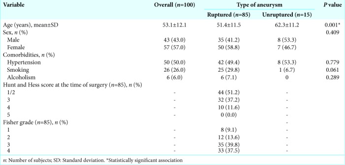
During the study period, only three patients with ACoA aneurysms underwent endovascular treatment: two unruptured and one ruptured aneurysm (HH4). There were no deaths. GOS of the patients was five for the patients with unruptured aneurysms and three for the ruptured aneurysm patient.
The morphological features of the aneurysms are described in Table 2.
Table 2:
Morphological features of aneurysms.
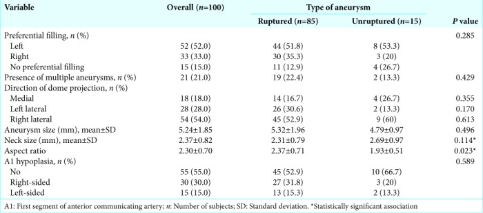
Regarding A1 segment morphology, preferential filling of the left side predominated in the sample (left = 52, right = 33, and bilateral = 15). Multiple aneurysms were present in 21% of patients (n = 21). Hypoplasia of the A1 segment was present in 45% of cases (left = 15, right = 30, and no hypoplasia = 55).
On average, ruptured aneurysms were larger than unruptured ones (5.32 ± 1.96 mm vs. 4.79 ± 0.97 mm), but the difference was not statistically significant (P = 0.49). There was also no significant difference in neck size between ruptured and unruptured aneurysms (2.31 ± 0.79 vs. 2.69 ± 0.97, P = 0.11).
Conversely, the AR differed significantly between ruptured and unruptured aneurysms (2.37 ± 0.71 vs. 1.93 ± 0.51, P = 0.02).
Surgical intervention was performed most often 4 days after the hemorrhagic stroke (range, 2–6 days). In all cases, access was obtained through pterional craniotomy (left-sided in 54 cases and right-sided in the remaining 46). The laterality of the approach was defined by preferential angiographic filling. In cases with no evidence of preferential filling, craniotomy was performed contralateral to the projection of the aneurysm dome. In patients with multiple aneurysms, craniotomy was performed on the side that would allow access to the largest number of lesions. Symmetrically filling single aneurysms with no lateral projection were approached from the right. Hydrocephalus was present in 43 patients (43%).
Intraoperative aneurysm rupture occurred in 3% of cases, and temporary clipping was performed in 15%. The mean duration of temporary clipping was 115 s. An external ventricular drain was placed at some point during hospitalization (either at admission or intraoperatively) in 37% of patients. Patients with mild ventricular enlargement and a normal level of consciousness (HH score 1 or 2) were treated with daily therapeutic lumbar puncture and cerebrospinal fluid (CSF) manometry per routine hospital protocol, obviating the need for ventriculostomy.
Clinical vasospasm occurred in 43 patients (43%). Clinical complications occurred in 41% of patients and are listed in Table 3.
Table 3:
Clinical and surgical data.
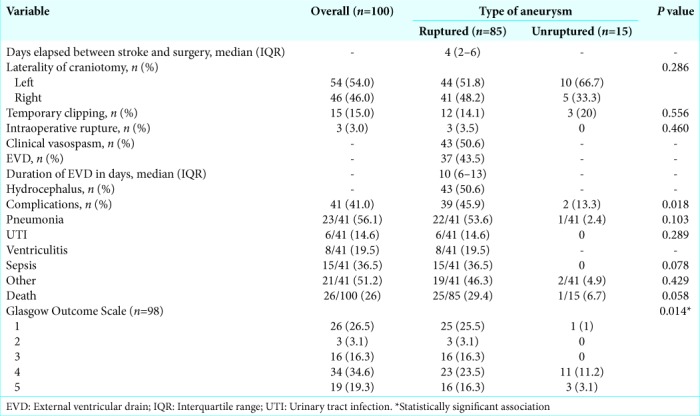
In this case series, 26 patients died (26%). Among patients with ruptured aneurysms, 25 died (29.4%), whereas only one patient in the unruptured group died (6.6%).
Regarding the patients with ruptured aneurysm, the ones who died had a preoperative HH score of 3 or 4 (HH1/2 = 8, HH3 = 11, HH4 = 6, and HH5 = 0); clinical vasospasm occurred in 13 of them. Clinical complications occurred on 22 patients (pneumonia = 14, urinary tract infection = 3, sepsis = 14, and other = 8).
In the unruptured group, the only death occurred due to pulmonary thromboembolism on the third postoperative day, which occurred despite routine prophylactic measures.
Regarding clinical outcome, information on the GOS was available for 98 of the 100 patients. The clinical outcome was favorable (GOS 4 or 5) in 53 patients (54%) and unfavorable (GOS 1, 2, or 3) in 45 (46%). Outcomes were significantly better among patients with unruptured aneurysms than in those with ruptured aneurysms (P = 0.01).
Prognostic factors
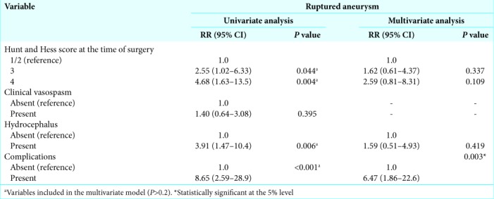
Among the factors of interest, the following were associated with mortality in patients with ruptured aneurysms: HH score in the immediate preoperative period (P < 0.001), hydrocephalus (P < 0.001), and presence of clinical complications (P < 0.001). Detailed data are provided in Table 4. On multivariate analysis to evaluate factors independently associated with death, only the presence of clinical complications (P = 0.003) remained statistically significant [Table 5].
Table 4:
Factors associated with mortality.
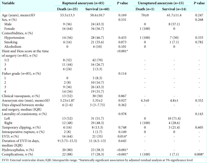
Table 5:
Poisson regression analysis of factors independently associated with death.
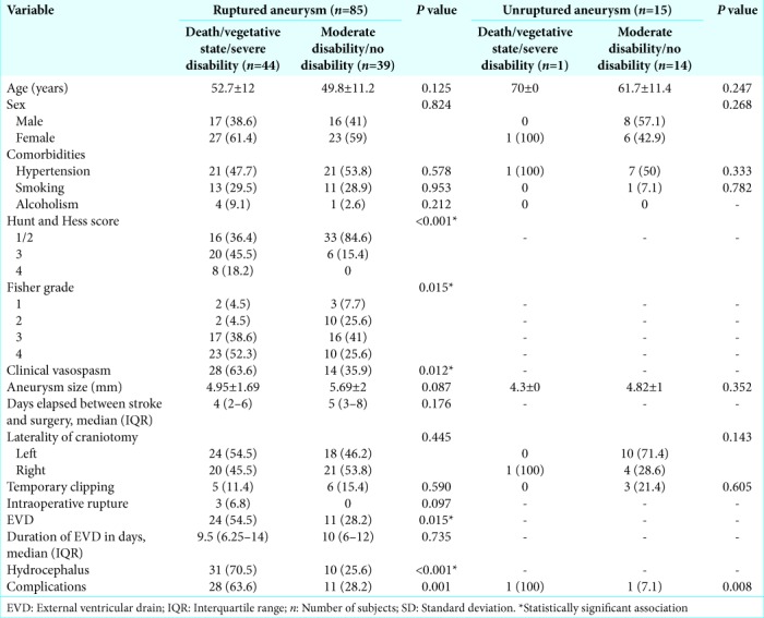
Comparison of patients with favorable versus unfavorable outcomes in the group of ruptured aneurysms revealed that the following factors were associated with an unfavorable outcome: HH score (P < 0.001), Fisher grade (P = 0.015), clinical vasospasm (P = 0.012), external ventricular drainage (P = 0.015), hydrocephalus (P < 0.001), and presence of clinical complications (P = 0.001) [Table 6]. On multivariate analysis, no factor was independently associated with an unfavorable outcome. In the unruptured aneurysms group, presence of clinical complications was the only factor associated with an unfavorable outcome (P = 0.008).
Table 6:
Factors associated with unfavorable outcomes (death, vegetative state, severe disability).
DISCUSSION
There have been a few recent case series of patients undergoing surgical treatment for ACoA aneurysms. With the advent of endovascular techniques, these approaches have become increasingly popular, although they are not necessarily superior to conventional surgical treatment.[25,26,33] In our service, surgical treatment was offered to all patients with anterior circulation aneurysms except when the patient was considered nonoperable due to clinical conditions or, in the case of nonruptured aneurysms, when the patient expressly opted for endovascular treatment and also there were no contraindications.
Petraglia et al. analyzed a series of 28 patients with ACoA aneurysms treated operatively and found two deaths (7%). The authors note that this mortality may be attributable to the low proportion of patients with poor neurological status (HH score 3 or higher) in the series.[29] Diraz et al. reported a mortality rate of 6.8% in their series of 102 ACoA aneurysms. Lin et al., in a series of 65 patients undergoing surgical treatment for ACoA aneurysms, reported mortality of 13.8%.[8,20] Andaluz et al. analyzed 75 patients with ACoA aneurysms and found a poor outcome (severe disability or death) in 10 (17.3%).[2] Nussbaum et al. found a mortality rate of 0.27% in their series of 450 patients (76 with ACoA aneurysms), and Lai et al., in a sample of 103 patients, had no mortality; however, both series were limited to patients with unruptured aneurysms.[18,27] In the present study, we analyzed a series of 100 aneurysms operated over a period of 7 years. The mortality rate was 26%, quite higher than elsewhere in the literature. The presence of clinical complications was the only factor independently correlated with mortality in ruptured aneurysms in our sample.
We observed a high rate of infectious clinical complications, which explains the high mortality. It should be noted that, in our series, most patients (85%) had ruptured aneurysms and among these cases, 48.8% were in severe neurological condition (HH score 3 or higher) preoperatively. The postoperative course of this patient population tends to be worse due to the higher incidence of clinical complications and clinical vasospasm.[21,22]
Ventriculitis was present in 7 patients (18.9% of those who received an external ventricular drain), which is equivalent to 19.1 cases/1000 catheter days. Ramanan, in a meta-analysis of 35 observational studies, found an overall incidence of 11.4/1000 catheter days. When analyzing only smaller studies (<1000 catheter-days), the observed incidence was higher (18.3/1000 catheter-days).[30] Considering that the total number of catheter days in our series is 365, our rate is consistent with that of the smaller studies included in the meta-analysis. Lozier, in a review article, observed that, in most studies, the presence of hemorrhagic CSF is associated with a higher incidence of ventriculitis.[24]
Clinical vasospasm occurred in 50% of patients. Although the occurrence of clinical vasospasm had no direct correlation with mortality, it did correlate with unfavorable outcomes (GOS 1, 2, or 3). However, this statistical association was not maintained on multivariate analysis. Rosengart and Orakdogen, among other authors, have reported an association between clinical vasospasm and mortality.[28,31] Angiographic vasospasm was found in 34% of patients in a case series by Brown, who reported that the incidence of late ischemia was 31% higher in patients with angiographic vasospasm than in patients without it.[3] However, 25% of patients with late ischemia did not exhibit angiographic evidence of vasospasm. There are several possible explanations for the presence of clinical vasospasm and late cerebral ischemia in the absence of detectable angiographic vasospasm. These include initial early damage related to intracranial hypertension in the first 72 h after stroke, which could lead to subsequent global cerebral ischemia; increased concentrations of procoagulant factors in CSF; and cortical spreading depolarization, secondary to dysfunctional cation influx in the neuronal membranes, with subsequent dysfunction and spasm in the cerebral microvasculature.[10]
In our series, there was no significant difference in overall aneurysm size or aneurysm neck size between ruptured and unruptured aneurysms. Aneurysm size has been studied by several authors as a potential predictor of rupture.[5] In 1998, a cooperative analysis of a retrospective cohort (International Study of Unruptured Intracranial Aneurysms) concluded that aneurysms smaller than 10 mm in patients with no history of SAH have a risk of rupture of 0.05% per year.[38] Juvela et al., in their cohort, observed that, although larger size is a risk factor for aneurysm rupture, most ruptured aneurysms were smaller than 7 mm.[15] In a series reported by Weir, 77% of ruptured aneurysms were <10 mm in size.[37] Regarding the aneurysm AR, in our series, it was larger in ruptured than in unruptured aneurysms, which is consistent with the literature. In a retrospective study by Weir, the mean AR of unruptured aneurysms was 1.8 versus 3.4 in ruptured aneurysms. The odds of aneurysm were 20-fold greater when the AR was >3.47 than when the AR was <1.38.[36] Ujiie, in another retrospective study, found that almost 80% of ruptured aneurysms had an AR >1.6, while almost 90% of unruptured aneurysms had an AR <1.6.[34]
In our series, the criterion used to define the laterality of craniotomy was preferential filling, as described by Chemale.[4] Preferential filling corresponds to the side on which the aneurysm is most completely visualized on angiography, if there is a difference. Chemale noted in his series that, in most cases, the dome of the aneurysm is directed to contralateral to the side of preferential filling, even when both A1 segments are symmetrical. When this did not occur, the A1 segment was tortuous and its terminal portion, adjoining the ACoA, was directed contralaterally to preferential filling. Chemale argues that, although the right side is classically preferred when obtaining access to ACoA aneurysms because of the lower risk of morbidity in the nondominant hemisphere,[39] the preferential filling approach allows easier dissection of the aneurysm neck and is associated with a lower risk of intraoperative rupture.[4] Similar findings were observed in our study: nearly 85% of aneurysms exhibited preferential filling on one side; however, A1 segment hypoplasia occurred in only 45%. The aneurysm dome was directed contralateral to the side of preferential filling in 85.8% of cases (73/85). When filling was symmetrical, most aneurysms (10/15) did not project laterally. The laterality of craniotomy did not influence morbidity or mortality in our series.
Intraoperative rupture occurred in 3% of the cases, a lower rate than those reported in the literature. Leipzig et al., in a series of 1694 aneurysms,[19] found moderate or severe intraoperative rupture (disregarding small bleeds that could be controlled immediately using the microsurgical aspirator or clipping) in 3.2% of aneurysms. ACoA aneurysms ruptured intraoperatively in 9.3% of cases, a rate higher than in the present series. In another series of 694 aneurysms, reported by Kheireddin et al.,[17] the overall intraoperative rupture rate was 11.7%, being highest in ACoA aneurysms. Hsu et al.,[14] in a series of 538 surgically treated aneurysms, found that experienced surgeons (more than 300 procedures performed) had a significantly lower intraoperative rupture rate than surgeons with little experience (8% vs. 16%).
We found no association between temporary clippings and worse outcomes in our patients, a finding consistent with the literature. Araújo Jr., in a series of 32 patients with ACoA aneurysm, of whom 21 required temporary clipping, did not find a significant association between duration of temporary clipping and outcome.[7] Griessenauer, in two follow-up studies of patients who underwent temporary clipping during treatment of cerebral aneurysms, also found no association between duration of clipping and outcome, even with an average time as high as 19 min.[11,12]
Study limitations
The limitations of this case series are those inherent to retrospective study designs. Our data were collected from medical records completed by different individuals in a heterogeneous manner over time. Sometimes, specific data were unavailable for a specific patient.
Regarding morbidity and mortality, due to the paucity of data available in outpatient medical records at our facility, it was impossible to evaluate late outcomes in the cohort.
CONCLUSION
The present study reports a series of 100 cases of ACoA aneurysms treated surgically over 7 years at a tertiary care center in Southern Brazil. The overall mortality rate was 26%, demonstrating that, despite advances in the management of subarachnoid hemorrhage, it is still an event that carries high morbidity and mortality rates, especially in patients who present with severe neurological deficit (as did a substantial portion of our sample). The development of clinical complications, especially infectious ones, was the key determinant of mortality, highlighting the importance of adequate neurointensive care in these patients.
Footnotes
How to cite this article: Soares FP, Velho MC, Antunes AC. Clinical and morphological profile of aneurysms of the anterior communicating artery treated at a neurosurgical service in Southern Brazil. Surg Neurol Int 2019;10:193.
Contributor Information
Fabiano Pasqualotto Soares, Email: fabianoslasher@gmail.com.
Maira Cristina Velho, Email: maira.velho@gmail.com.
Apio Claudio Martins Antunes, Email: aantunes@hcpa.edu.br.
Financial support and sponsorship
Nil.
Conflicts of interest
There are no conflicts of interest.
REFERENCES
- 1.Agrawal A, Kato Y, Chen L, Karagiozov K, Yoneda M, Imizu S, et al. Anterior communicating artery aneurysms: An overview. Minim Invasive Neurosurg. 2008;51:131–5. doi: 10.1055/s-2008-1073169. [DOI] [PubMed] [Google Scholar]
- 2.Andaluz N, Zuccarello M. Anterior communicating artery aneurysm surgery through the orbitopterional approach: Long-term follow-up in a series of 75 consecutive patients. Skull Base. 2008;18:265–74. doi: 10.1055/s-2008-1058367. [DOI] [PMC free article] [PubMed] [Google Scholar]
- 3.Brown RJ, Kumar A, Dhar R, Sampson TR, Diringer MN. The relationship between delayed infarcts and angiographic vasospasm after aneurysmal subarachnoid hemorrhage. Neurosurgery. 2013;72:702–7. doi: 10.1227/NEU.0b013e318285c3db. [DOI] [PMC free article] [PubMed] [Google Scholar]
- 4.Chemale IM. Consideraçöes hemodinâmicas sobre abordagem dos aneurismas da artéria comunicante anterior [Haemodynamic considerations regarding the approach to anterior communicating artery aneurysms] J Bras Neurocir. 1996;7:22–30. [Google Scholar]
- 5.Chen PR, Frerichs K, Spetzler R. Natural history and general management of unruptured intracranial aneurysms. Neurosurg Focus. 2004;17:E1. doi: 10.3171/foc.2004.17.5.1. [DOI] [PubMed] [Google Scholar]
- 6.Connolly ES, Jr, Rabinstein AA, Carhuapoma JR, Derdeyn CP, Dion J, Higashida RT, et al. Guidelines for the management of aneurysmal subarachnoid hemorrhage: A guideline for healthcare professionals from the American heart association/ American stroke association. Stroke. 2012;43:1711–37. doi: 10.1161/STR.0b013e3182587839. [DOI] [PubMed] [Google Scholar]
- 7.de Araújo Júnior AS, de Aguiar PH, Fazzito MM, Simm R, Stefani MA, Zicarelli CA, et al. Prospective factors of temporary arterial occlusion during anterior communicating artery aneurysm repair. Acta Neurochir Suppl. 2015;120:231–5. doi: 10.1007/978-3-319-04981-6_39. [DOI] [PubMed] [Google Scholar]
- 8.Diraz A, Kobayashi S, Toriyama T, Ohsawa M, Hokama M, Kitazama K, et al. Surgical approaches to the anterior communicating artery aneurysm and their results. Neurol Res. 1993;15:273–80. doi: 10.1080/01616412.1993.11740148. [DOI] [PubMed] [Google Scholar]
- 9.Etminan N, Beseoglu K, Barrow DL, Bederson J, Brown RD, Jr, Connolly ES, Jr, et al. Multidisciplinary consensus on assessment of unruptured intracranial aneurysms: Proposal of an international research group. Stroke. 2014;45:1523–30. doi: 10.1161/STROKEAHA.114.004519. [DOI] [PubMed] [Google Scholar]
- 10.Flynn L, Andrews P. Advances in the understanding of delayed cerebral ischaemia after aneurysmal subarachnoid haemorrhage. F1000Res. 2015;4:F1000. doi: 10.12688/f1000research.6635.1. [DOI] [PMC free article] [PubMed] [Google Scholar]
- 11.Griessenauer CJ, Poston TL, Shoja MM, Mortazavi MM, Falola M, Tubbs RS, et al. The impact of temporary artery occlusion during intracranial aneurysm surgery on long-term clinical outcome: Part I. Patients with subarachnoid hemorrhage. World Neurosurg. 2014;82:140–8. doi: 10.1016/j.wneu.2013.02.068. [DOI] [PubMed] [Google Scholar]
- 12.Griessenauer CJ, Poston TL, Shoja MM, Mortazavi MM, Falola M, Tubbs RS, et al. The impact of temporary artery occlusion during intracranial aneurysm surgery on long-term clinical outcome: Part II. The patient who undergoes elective clipping. World Neurosurg. 2014;82:402–8. doi: 10.1016/j.wneu.2013.02.067. [DOI] [PubMed] [Google Scholar]
- 13.Hernesniemi J, Dashti R, Lehecka M, Niemelä M, Rinne J, Lehto H, et al. Microneurosurgical management of anterior communicating artery aneurysms. Surg Neurol. 2008;70:8–28. doi: 10.1016/j.surneu.2008.01.056. [DOI] [PubMed] [Google Scholar]
- 14.Hsu CE, Lin TK, Lee MH, Lee ST, Chang CN, Lin CL, et al. The impact of surgical experience on major intraoperative aneurysm rupture and their consequences on outcome: A Multivariate analysis of 538 microsurgical clipping cases. PLoS One. 2016;11:e0151805. doi: 10.1371/journal.pone.0151805. [DOI] [PMC free article] [PubMed] [Google Scholar]
- 15.Juvela S, Porras M, Poussa K. Natural history of unruptured intracranial aneurysms: Probability of and risk factors for aneurysm rupture. J Neurosurg. 2008;108:1052–60. doi: 10.3171/JNS/2008/108/5/1052. [DOI] [PubMed] [Google Scholar]
- 16.Kassell NF, Torner JC, Haley EC, Jr, Jane JA, Adams HP, Kongable GL, et al. The international cooperative study on the timing of aneurysm surgery. Part 1: Overall management results. J Neurosurg. 1990;73:18–36. doi: 10.3171/jns.1990.73.1.0018. [DOI] [PubMed] [Google Scholar]
- 17.Kheĭreddin AS, Filatov IuM, Belousova OB, Pilipenko IuV, Zolotukhin SP, Sazonov IA, et al. Intraoperative rupture of cerebral aneurysm incidence and risk factors. Zh Vopr Neirokhir Im N N Burdenko. 2007;4:33–8. [PubMed] [Google Scholar]
- 18.Lai LT, Gragnaniello C, Morgan MK. Outcomes for a case series of unruptured anterior communicating artery aneurysm surgery. J Clin Neurosci. 2013;20:1688–92. doi: 10.1016/j.jocn.2013.02.015. [DOI] [PubMed] [Google Scholar]
- 19.Leipzig TJ, Morgan J, Horner TG, Payner T, Redelman K, Johnson CS, et al. Analysis of intraoperative rupture in the surgical treatment of 1694 saccular aneurysms. Neurosurgery. 2005;56:455–68. doi: 10.1227/01.neu.0000154697.75300.c2. [DOI] [PubMed] [Google Scholar]
- 20.Lin CL, Kwan AL, Howng SL. Surgical outcome of anterior communicating artery aneurysms. Kaohsiung J Med Sci. 1998;14:561–8. [PubMed] [Google Scholar]
- 21.Lindvall P, Runnerstam M, Birgander R, Koskinen LO. The fisher grading correlated to outcome in patients with subarachnoid haemorrhage. Br J Neurosurg. 2009;23:188–92. doi: 10.1080/02688690802710668. [DOI] [PubMed] [Google Scholar]
- 22.Lo BW, Fukuda H, Nishimura Y, Farrokhyar F, Thabane L, Levine MA, et al. Systematic review of clinical prediction tools and prognostic factors in aneurysmal subarachnoid hemorrhage. Surg Neurol Int. 2015;6:135. doi: 10.4103/2152-7806.162676. [DOI] [PMC free article] [PubMed] [Google Scholar]
- 23.Locksley HB. Natural history of subarachnoid hemorrhage, intracranial aneurysms and arteriovenous malformations. Based on 6368 cases in the cooperative study. J Neurosurg. 1966;25:219–39. doi: 10.3171/jns.1966.25.2.0219. [DOI] [PubMed] [Google Scholar]
- 24.Lozier AP, Sciacca RR, Romagnoli MF, Connolly ES., Jr Ventriculostomy-related infections: A critical review of the literature. Neurosurgery. 2002;51:170–81. doi: 10.1097/00006123-200207000-00024. [DOI] [PubMed] [Google Scholar]
- 25.Molyneux A, Kerr R, Stratton I, Sandercock P, Clarke M, Shrimpton J, et al. International subarachnoid aneurysm trial (ISAT) of neurosurgical clipping versus endovascular coiling in 2143 patients with ruptured intracranial aneurysms: A randomised trial. Lancet. 2002;360:1267–74. doi: 10.1016/s0140-6736(02)11314-6. [DOI] [PubMed] [Google Scholar]
- 26.Molyneux AJ, Kerr RS, Yu LM, Clarke M, Sneade M, Yarnold JA, et al. International subarachnoid aneurysm trial (ISAT) of neurosurgical clipping versus endovascular coiling in 2143 patients with ruptured intracranial aneurysms: A randomised comparison of effects on survival, dependency, seizures, rebleeding, subgroups, and aneurysm occlusion. Lancet. 2005;366:809–17. doi: 10.1016/S0140-6736(05)67214-5. [DOI] [PubMed] [Google Scholar]
- 27.Nussbaum ES, Madison MT, Myers ME, Goddard J. Microsurgical treatment of unruptured intracranial aneurysms. A consecutive surgical experience consisting of 450 aneurysms treated in the endovascular era. Surg Neurol. 2007;67:457–64. doi: 10.1016/j.surneu.2006.08.069. [DOI] [PubMed] [Google Scholar]
- 28.Orakdogen M, Emon ST, Somay H, Engin T, Ates O, Berkman MZ, et al. Prognostic factors in patients who underwent aneurysmal clipping due to spontaneous subarachnoid hemorrhage. Turk Neurosurg. 2016;26:840–8. doi: 10.5137/1019-5149.JTN.13654-14.1. [DOI] [PubMed] [Google Scholar]
- 29.Petraglia AL, Srinivasan V, Moravan MJ, Coriddi M, Jahromi BS, Vates GE, et al. Unilateral subfrontal approach to anterior communicating artery aneurysms: A review of 28 patients. Surg Neurol Int. 2011;2:124. doi: 10.4103/2152-7806.85056. [DOI] [PMC free article] [PubMed] [Google Scholar]
- 30.Ramanan M, Lipman J, Shorr A, Shankar A. A meta-analysis of ventriculostomy-associated cerebrospinal fluid infections. BMC Infect Dis. 2015;15:3. doi: 10.1186/s12879-014-0712-z. [DOI] [PMC free article] [PubMed] [Google Scholar]
- 31.Rosengart AJ, Schultheiss KE, Tolentino J, Macdonald RL. Prognostic factors for outcome in patients with aneurysmal subarachnoid hemorrhage. Stroke. 2007;38:2315–21. doi: 10.1161/STROKEAHA.107.484360. [DOI] [PubMed] [Google Scholar]
- 32.Spetzler RF, McDougall CG, Zabramski JM, Albuquerque FC, Hills NK, Russin JJ, et al. The barrow ruptured aneurysm trial: 6-year results. J Neurosurg. 2015;123:609–17. doi: 10.3171/2014.9.JNS141749. [DOI] [PubMed] [Google Scholar]
- 33.Steklacova A, Bradac O, de Lacy P, Lacman J, Charvat F, Benes V, et al. “Coil mainly” policy in management of intracranial ACoA aneurysms: Single-centre experience with the systematic review of literature and meta-analysis. Neurosurg Rev. 2018;41:825–39. doi: 10.1007/s10143-017-0932-y. [DOI] [PubMed] [Google Scholar]
- 34.Ujiie H, Tamano Y, Sasaki K, Hori T. Is the aspect ratio a reliable index for predicting the rupture of a saccular aneurysm? Neurosurgery. 2001;48:495–502. doi: 10.1097/00006123-200103000-00007. [DOI] [PubMed] [Google Scholar]
- 35.Vlak MH, Algra A, Brandenburg R, Rinkel GJ. Prevalence of unruptured intracranial aneurysms, with emphasis on sex, age, comorbidity, country, and time period: A systematic review and meta-analysis. Lancet Neurol. 2011;10:626–36. doi: 10.1016/S1474-4422(11)70109-0. [DOI] [PubMed] [Google Scholar]
- 36.Weir B, Amidei C, Kongable G, Findlay JM, Kassell NF, Kelly J, et al. The aspect ratio (dome/neck) of ruptured and unruptured aneurysms. J Neurosurg. 2003;99:447–51. doi: 10.3171/jns.2003.99.3.0447. [DOI] [PubMed] [Google Scholar]
- 37.Weir B, Disney L, Karrison T. Sizes of ruptured and unruptured aneurysms in relation to their sites and the ages of patients. J Neurosurg. 2002;96:64–70. doi: 10.3171/jns.2002.96.1.0064. [DOI] [PubMed] [Google Scholar]
- 38.Wiebers DO, Whisnant JP, Huston J, 3rd, Meissner I, Brown RD, Jr, Piepgras DG, et al. Unruptured intracranial aneurysms: Natural history, clinical outcome, and risks of surgical and endovascular treatment. Lancet. 2003;362:103–10. doi: 10.1016/s0140-6736(03)13860-3. [DOI] [PubMed] [Google Scholar]
- 39.Yasargil MG. Anterior cerebral and anterior communicating aneurysms. In: Yasargil MG, editor. Microneurosurgery: Clinical Considerations, Surgery of the Intracranial Aneurysms and Results (Microsurgical Anatomy of Basal Cisterns and Vessels of Brain). II. New York: Georg Thieme/ Thieme-Stratton; 1984. pp. 165–231. [Google Scholar]
- 40.Yasargil MG. General operative techniques. In: Yasargil MG, editor. Microneurosurgery: Microsurgical Anatomy of the Basal Cisterns and Vessels of the Brain, Diagnostic Studies, General Operative Techniques and Pathological Considerations of the Intracranial Aneurysms. I. New York: Georg Thieme/Thieme- Stratton; 1984. pp. 208–71. [Google Scholar]


