Abstract
Background/Aims
Epigallocatechin gallate (EGCG) has established protective actions against myocardial ischemia/reperfusion (I/R) injury by regulating autophagy. However, little is known about the mechanisms of EGCG in posttranscriptional regulation in the process of cardioprotection. Here we studied whether microRNAs play a role in EGCG-induced cardioprotection.
Methods
The myocardial I/R injury in vitro and in vivo model were made, with or without EGCG pretreatment. The upregulation and silencing of microRNA-384-5p (miR-384) and Beclin-1 in H9c2 cell lines were established. Rats were transfected with miR-384 specific shRNA. Dual-luciferase reporter gene assay was conducted to verify the relationship between miR-384 and Beclin-1. TTC staining was performed to analyze the area of myocardial infarct size. Cell viability was monitored by cell counting kit-8 (CCK-8). The release of cardiac troponin-I (cTnI) was examined by ELISA. The levels of autophagy-related genes or proteins expression were evaluated by qRT-PCR or Western blotting. Autophagosomes of myocardial cells were detected by transmission electron microscopy and laser scanning confocal microscope.
Results
I/R increased both autophagosomes and autolysosomes, thereby increasing autophagic flux both in vitro and in vivo. Pretreatment with EGCG attenuated I/R-induced autophagic flux expression, accompanied by an increase in cell viability and a decrease in the size of myocardial infarction. MiR-384 expression was down-regulated in H9c2 cell lines when subjected to I/R, while this suppression could be reversed by EGCG pretreatment. The dual-luciferase assay verified that Beclin-1 was a target of miR-384. Both overexpression of miR-384 and knocking down of Beclin-1 significantly inhibited I/R-induced autophagy, accompanied by the activation of PI3K/Akt pathway, thus enhanced the protective effect of EGCG. However, these functions were abrogated by the PI3K inhibitor, LY294002.
Conclusion
We confirmed that EGCG has a protective role in microRNA-384-mediated autophagy by targeting Beclin-1 via activating the PI3K/Akt signaling pathway. Our results unveiled a novel role of EGCG in myocardial protection, involving posttranscriptional regulation with miRNA-384.
Keywords: epigallocatechin gallate, ischemia/reperfusion, autophagy, microRNA-384-5p, Beclin-1, PI3K/Akt pathway
Introduction
Ischemic heart disease is the leading cause of death in worldwide. When myocardial ischemia occurs, timely reperfusion after the onset of ischemia may induce ischemia/reperfusion injury.1 Consequently, alleviating I/R injury can be used as one of the therapeutic strategies for heart diseases. Green tea has beneficial effects against many diseases, such as cancer, obesity, diabetes, neurodegenerative and cardiovascular diseases.2 Epigallocatechin gallate (EGCG), the most abundant catechin in green tea, possesses multiple protective effects on cardiovascular system.3 A host of studies have demonstrated that EGCG can protect cardiomyocytes from I/R-induced injury through decreasing oxidative stress, inhibiting activation of MAPK signaling pathway and preventing mitochondrial damage.4,5 However, the underlying mechanism of EGCG for I/R injury is not yet fully understood.
Autophagy is a highly conserved metabolic process, including changes in the expression of some proteins and dysfunction of some organelles.6 The autophagic pathway composes of distinct steps. The initiation is the formation of the autophagic membrane.7 Beclin-1 and other proteins combine with class III PI3-kinase to form a complex to facilitate assembly and elongation of the autophagosomal membrane.8 Next is the formation of autophagosomes. Microtubule-associated protein 1 light chain 3 (LC3) is cleaved and converted to LC3-I then activated and transformed to LC3-II which is incorporated in the autophagosomal membrane with cathepsin D for completing the maturation of autophagosome.9 The maturation of autophagosomes into autolysosomes is followed.10 The last step is final degradation of autolysosomes. The p62 binds specific cargo proteins and simultaneously anchors them on the autophagosome interior and then interacts with LC3 interacting region to finally complete the degradation.11 Increasing the expression of Beclin-1, LC3-II, cathepsin D and decreasing the expression of p62 may lead to an increase of autophagy flux, as well as accelerating the process of autophagy. Under physiological conditions, autophagy plays essential roles in maintaining intracellular homeostasis in preserving cardiomyocyte function.12 However, in myocardial ischemia reperfusion stage, excessive autophagy contributed to the development of myocardial damage.13 Given the controversy regarding the role of autophagy and the fact that autophagy played a crucial role in cell survival during I/R, the manipulation of autophagy may represent a potential therapeutic target to protect against I/R cardiomyocyte death. Studies have shown that autophagy can be inhibited by activating the PI3K/Akt pathway.14 Beyond that, our previous study demonstrated that PI3K/Akt pathway may be also a target for EGCG on regulating autophagy,5 but the mechanisms of the effect merit further exploration.
MicroRNA (miRNA), a group of small non-coding RNAs with 20–22 nucleotides, was reported to regulate the expression of specific genes.15 Accumulated evidence indicated that miRNAs were involved in a great number of cellular processes including autophagy, apoptosis and necrosis during myocardial infarction progression.16 It is reported that microRNA-384 was highly expressed in ischemic myocardium of rats and suppression of microRNA-384 may increase the augmentation of Beclin-1-dependent autophagy in atherosclerosis.17 Many studies have elucidated the effects of miR-384 on cell survival by investigating this miRNA in cancer that affect other systems.18,19 However, whether miR-384 affects cardiomyocyte survival after I/R injury remains unknown.
As both microRNA-384/Beclin-1 and EGCG affected the development of autophagy, PI3K/Akt pathway, furthermore, is a switch to autophagy. What is the relationship between these three? Little research has been carried out. In our present study, we explored the mechanism of miR-384/Beclin-1 axis in regulating the cardiomyocytes against I/R injury through reducing autophagy. More importantly, the aim of the present research was to explore whether EGCG could protect cardiomyocytes against I/R injury through adjusting microRNA-384-5p targets Beclin-1 to inactivate autophagy via PI3K/Akt pathway.
Materials And Methods
Chemicals And Materials
Epigallocatechin gallate (purity: 395%), LY294002 (PI3K/Akt pathway inhibitor) and 3-MA (autophagy inhibitor) were obtained from Sigma-Aldrich Inc. (St. Louis, MO, USA) and diluted to an appropriate concentration as needed. H9c2 cardiomyocytes and HEK293-T cells were purchased from Cell Bank of the Chinese Academy of Sciences (Shanghai, People’s Republic of China). All cell culture materials were from GIBCO (Grand Island, NY, USA). The anaerobic glove box was bought from Coy Laboratory Products Inc., Grass Lake (MI, USA). Cell counting kit-8 was acquired from Dojindo Molecular Technologies Inc. (Gaithersburg, MD, USA). All antibodies were purchased from Santa Cruz Biotechnology (Dallas, TX, USA). Stable H9c2 cells, transfected by miR-384 mimic or inhibitor, were purchased from Wuhan GeneCreate Biological Engineering Co., Ltd. (Wuhan, People’s Republic of China). Stable H9c2 cells, transfected by Beclin-1 overexpression (OE) or Beclin-1 knock-down (KD), were purchased from GeneChem (Shanghai, People’s Republic of China). The enzyme-mark analyser was obtained from Tecan Austria GmbH (Grödig, Austria). Rat cTnI enzyme-linked immunosorbent assay kit came from Life Diagnostics (West Chester, PA, USA). mRFP-GFP-LC3 adenoviral vector and Hoechst 33342 were obtained from Hanbio and Beyotime (People’s Republic of China). Confocal fluorescence microscope was selected from Leica (Heidelberg, Germany). Trizol reagent was picked from Life Technologies (USA). Reverse transcription system was acquired from Invitrogen (USA). SYBR-Green PCR Master Mix was obtained Applied Biosystems (USA). All antibodies were purchased from Abcam (Cambridge, England).
Cell Culture And Establishment Of Hypoxia-Reoxygenation Model (H/R)
For all of the experiments, H9c2 cardiomyocytes were cultured in high glucose DMEM supplemented with 10% FBS at 37°C.20 The H/R model was built according to the literature.17 Briefly, high glucose DMEM medium was replaced with non-glucose DMEM to mimic ischemia. Then the H9c2 cardiomyocytes were incubated in an anaerobic glove box, where normal air was removed by a mixed gas of 5% CO2, 5% H2, and 90% N2. The H9c2 cardiomyocytes were incubated under hypoxia for 6 h then transferred to the regular incubator for 12 h with the medium replaced by high glucose medium to mimic reperfusion. The normal cells were cultured under normoxic condition for equivalent durations with high glucose DMEM. According to experiment design, EGCG (25 µM), 3-MA (10 mM) or LY294002 (10 µM) were treated 4 h prior to H/R (Figure 1A).
Figure 1.
Myocardial ischemia reperfusion injury model in experimental protocol.
Notes: (A) Myocardial ischemia reperfusion injury model in vitro. (B) Myocardial ischemia reperfusion injury model in vivo.
Abbreviations: EGCG, epigallocatechin gallate; H/R, hypoxia/reoxygenation; I/R, ischemia/reperfusion; 3-MA, 3-methyladenine.
CCK-8 Assay
Cell viability was monitored by CCK-8 according to the manufacturer’s instructions. The cells were plated at 1 × 10⁴ cells/well in 96-well plates. When the treatments were completed, CCK-8 solution (10μL) was added to each well and incubated at 37°C for 4 hrs. The colour intensity was measured with an enzyme-mark analyser at a wavelength of 450 nm.
Dual-Luciferase Reporter Assay
The 3′ untranslated regions (UTRs) of wild-type and mutant Beclin-1 were synthesized by Sangon Biotech Co., Ltd. HEK293-T cells were cultured and transfected with miR-384 mimics using Lipofectamine™ 2000 according to the manufacturer’s instructions. After transfection for 48 hrs, a dual-luciferase reporter assay kit was used to detect luciferase activity.
Experimental Animals And I/R Model
All animal experiments were performed according to the guide for the care and use of laboratory animals recommended by the national institutes of health of USA. Animal experiments were approved by the Animal Ethics Committee of Guilin Medical University on April 3, 2018 (No. 20,180,403–02) and carried out in accordance with guidelines. All efforts were made to minimize animal suffering.
Sprague-Dawley (SD) rats of either sex (150–200g; Guilin Medical Laboratory Animal Center) were fasted overnight, then corresponding EGCG (10 mg/kg), 3-MA (15mg/kg) and LY294002 (50 mg/kg) were injected into sublingual veins 30 mins before ischemia. The rats were anesthetized with 10% chloral hydrate at a dose of 3 mL/kg (i.p.), respiration with a fraction of inspired oxygen of 0.80, then left anterior-descending (LAD) was ligated with a 4–0 silk suture,5 except the I/R control group. After 30 mins of ischemia, the ligation was loosened for 12 hrs (Figure 1B). Blood samples were collected and centrifuged at 3600×g for 20 mins to harvest the sera. The infarcted area of the heart was immediately excised and stored at −80°C for the analyses described below.
Stereotactic Injection
A stereotactic injection of miR-384 antagomir (AntagomiR-384) was administered 3 days before surgery.21 Briefly, we constructed AAV vectors in which the truncated miR-384 promoter drives the expression of luciferase. The minimal miR-384 promoter sufficed to preferentially restrict gene expression almost exclusively to cardiomyocytes, even after systemic administration. AntagomiR-384 was diluted in phosphate-buffered saline (PBS) injected via caudal vein.
Determination Of Infarct Size
The myocardial infarct size was measured using 2,3,5-triphenyltetrazolium chloride (TTC) staining as previously described.5,20 Briefly, the hearts were harvested and rinsed with normal saline. The excised left ventricle was frozen at −80°C for 5 mins, and then sectioned from apex to base into approximately 2-mm-thick slices. The slices were incubated in a solution of 1% TTC in phosphate-buffered saline (pH 7.4) at 37°C for 20 mins in the dark, and then fixed in 10% formaldehyde. Brick red stained areas indicated nonischemic area and pale parts in the heart represented infarct area. Photos were captured using a digital camera, and then the relative infarct size could be analyzed with Image J.
Measurement Of Cardiac Troponin I (cTnI) Levels
Rat cTnI (cell culture supernate or serum) enzyme-linked immunosorbent assay (ELISA) Kit was used for the measurement of cTnI concentration. Diluted standard substance (50 µL, of the aforementioned ELISA kits), detected samples (50 µL) and biotin-labeled antibody (50 µL) were added to 96-well plates and incubated at 37°C for 1 hr, washed with washing buffer from the aforementioned ELISA kits and then shaken for 30 s. This process was repeated three times. Streptavidin-horseradish peroxidase (HRP) was then added to each well and incubated at 37°C for 30 mins, washed and shaken for 30 s. This process was repeated three times. Substrates A and B (each 50 µL) were added to each well, shaken and incubated at 37°C for 10 mins in the dark. The microplate was then removed and the reaction was terminated. Optical density (OD) values were then determined at 450 nm in each well.
Measurement Of Autophagosome Formation
Cells were transfected with mRFP-GFP-LC3B adenovirus at an MOI of 30 for 6 h at 37°C for selecting positive cells. Following treatment with H/R, the cells were stained with Hoechst 33342. As intracellular distribution of LC3 protein was tagged by the fluorescence of mRFP and GFP in these cells, images were collected with laser scanning confocal microscope. Quantification of LC3 puncta was performed using Red (mRFP) and Green (GFP) Puncta Colocalization Macro with image program and the average numbers of LC3 puncta per cell were accounted from the data collected from more than 10 cells. Here, GFP+ mRFP+ puncta (yellow) and GFP‐mRFP+ puncta (red) are counted. When the autophagosome-lysosome fusion occurs, the probe comes red. When the fusion is impaired or blocked, the probe comes less reddish or more yellowish.
Transmission electron microscopy (TEM) for morphological analysis was performed at Electron Microscopy Core Laboratory, according to standard operating procedures. For morphological TEM, heart tissue was fixed in 2.5% glutaraldehyde in phosphate buffer overnight at 4°C. After sample preparation, 90–100 nm thick sections were mounted onto a 200 mesh copper grid and imaged with an FEI Tecnai G2 Spirit transmission electron microscope.
Quantitative Real-Time PCR (qRT-PCR) Analysis
Total RNA was extracted from the cells trizol reagent and the cDNA was reverse transcribed as the protocol for the reverse transcription system. The amplification primers were designed using Primer 5.0. Quantitative real-time RT-PCR (qRT-PCR) was performed by SYBR-Green PCR Master Mix in order to determine the mRNA expression levels of various genes according to the manufacturer’s instructions. GAPDH and U6 were used as a normalization control for mRNA and miRNA. The following primers were used for the experiment: U6, forward 5′-ATTGGAACGATACAGAGAAGATT-3′ and reverse 5′-GGAACGCTTCACGAATTTG-3′; miR-384-5p, forward, 5′-CTCAACTGGTGTCGTGGAGTCGGCAATTCAGTTGAGACATTGCC-3′ and reverse, 5′-ACACTCCAGCTGGGTGTAAACAATTCCTAGG-3′; Beclin-1, forward, 5′-GCCTCTGAAACTGGACACGA-3′ and reverse, 5′-CTTCCTCCTGGCTCTCTCCT-3′; GAPDH, forward, 5′-GACATGCCGCCTGGAGAAAC-3′and reverse, 5′-AGCCCAGGATGCCCTTTAGT -3′.
Western Blotting Analysis
The detailed method was essential as described in the previous literature.22 Briefly, the myocardial tissues and cells were homogenized in protein lysis buffer. The homogenates were resolved on polyacrylamide SDS gels and electrophoretically transferred to polyvinylidene difluoride membranes. Equal amounts of the protein (30 μg) were separated by SDS-PAGE and transferred onto nitrocellulose membranes. After blocking with 5% skim milk powder in Tris-buffered saline containing 0.1% Tween-20 (TBST) for 1 hr, the membranes were incubated overnight at 4°C with the primary antibodies against GAPDH (1:1000), Akt (1:1000), p-Akt (1:2000), PI3K (1:1000), Beclin-1 (1:2000), LC3 II (1:1000), p62 (1:1000), cathepsin D(1:1000). Then, the membranes were washed with TBST and incubated with peroxidase-conjugated IgG (1:5000) at room temperature for 1 hr. Finally, the membranes were washed in TBST and determined using an ECL chemiluminescence detection system.
Statistical Analyses
All results were averaged and expressed as the means±SD. Statistical analysis was performed with SPSS software (version 21). A one-way analysis of variance followed by Bonferroni’s multiple comparison test was used for statistical analysis. P-values <0.05 were considered to indicate statistically significant differences.
Results
EGCG Increased miR-384 And Attenuated Beclin-1 Levels In I/R-Induced Myocardial Injury
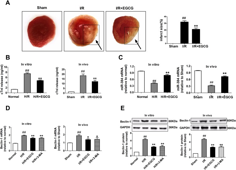
To gain insights into the EGCG's cardio-protective function, the TTC staining as golden standard for texting myocardial infract was evaluated. As shown in Figure 2A, in the sham group, there was little or zero percent of infract, however, it is critically increased in I/R group (p<0.05). Rats received EGCG pretreatment, which infarct size was significantly smaller than the I/R group (p <0.05). The levels of cTnI in cell culture supernatant and serum were significantly increased in H/R and I/R group compared with the normal or sham group (p<0.01 or p<0.05, Figure 2B), while EGCG treatment reversed the cTnI back toward normal levels. These findings suggested that EGCG protects myocardial cells from I/R- induced myocardial injury. In order to test whether the protective effect of EGCG in I/R-treated injury was related to the change of miR-384 and Beclin-1 expression, qRT-PCR and Western blotting assay were used to detect the relative miR-384 mRNA, Beclin-1 mRNA and protein levels. The results showed that I/R could decrease miR-384 levels and increase Beclin-1 levels as compared with the control group. However, EGCG gradually increased the expression of miR-384 (p<0.01 or p<0.05, Figure 2C) and decreased the expression of Beclin-1 both in vitro and in vivo (p<0.01 or p<0.05, Figure 2D and E), suggesting that the protective effect of EGCG on cardiomyocytes was associated with modulation of miR-384 and Beclin-1 expression. 3-MA played an opposite role in cell autophagy and Beclin-1 is the signature protein of autophagy. No significant difference between in EGCG and 3-MA pretreated group to respond to Beclin-1 level in vitro and in vivo (p > 0.05, Figure 2D and E). The results of the experiment proved again that EGCG could exert the protective effect on myocardial cells by inhibiting autophagy.
Figure 2.
EGCG increased the miR-384 and attenuated Beclin-1 levels in I/R- induced myocardial injury.
Notes: (A) Representative images of TTC-stained sections. (B) ELISA was performed to determine the expression levels of cTnI myocardial injury markers in cell culture supernatant and serum. (C) The expression levels of miR-384 in H9c2 cells and cardiac tissue of rats were determined with qRT-PCR. (D, E) The mRNA and protein levels of Beclin-1 were detected by qRT-PCR and Western blotting. Data among groups were compared using one-way analysis of variance. Values are expressed as mean±SD (n=6). ANOVA testing was performed; ## p<0.01 vs. normal (sham) group; * P<0.05, ** P<0.01 vs. H/R (I/R) group.
Abbreviations: ANOVA, Analysis of Variance; cTn-I, cardiac troponin-I; EGCG, Epigallocatechin gallate; GAPDH, glyceraldehyde-3-phosphate dehydrogenase; H/R, hypoxia/reoxygenation; I/R, ischemia/reperfusion; 3-MA, 3-methyladenine; miR-384, microRNA-384-5p; mRNA, Messenger RNA; qRT-RCR, quantitative real-time PCR; SD, Sprague-Dawley; TTC, 2,3,5-triphenyltetrazolium chloride.
MiR-384 Attenuated I/R-Stimulated Autophagy Both In Vitro And In Vivo
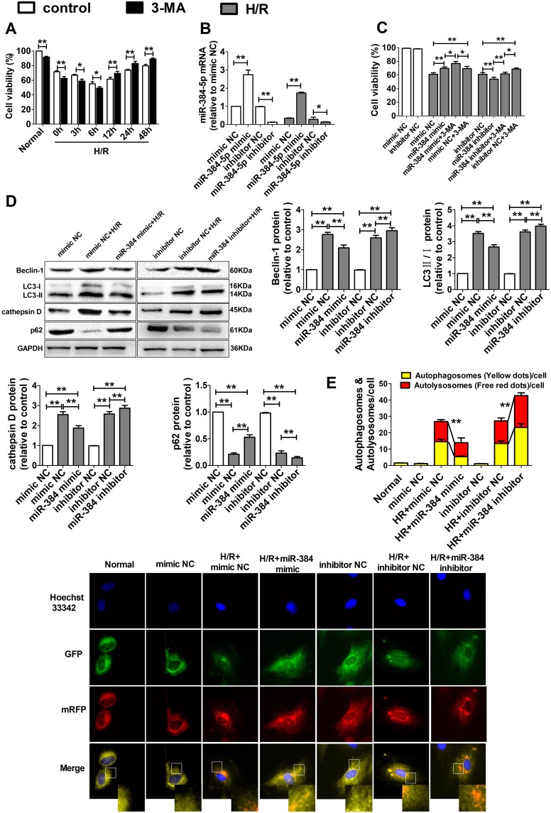
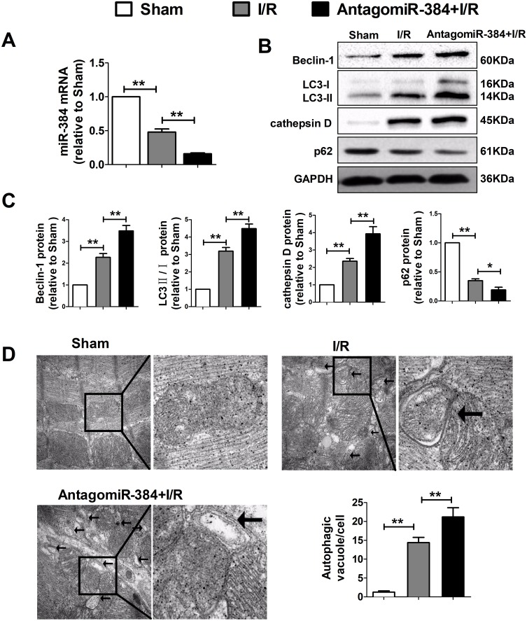
We subsequently pretreated H9c2 cells with 3-MA before hypoxia treatment. We found that after 6 hrs of hypoxia within the 12 h of reperfusion exposure, cell viability was decreased in the 3-MA group compared with that in the control group. However, we noted that cell viability was higher in the 3-MA group than in the control group after the cells had been exposed to reperfusion for 12 h. These results suggested that the effects of autophagy on H/R-induced cell injury can change over time and under the above conditions, autophagy is beneficial within the first 12 h of H/R exposure but harmful after 12 hrs of H/R exposure. We chose 12 hrs as the duration of H/R exposure in subsequent experiments. Thus, autophagy was harmful with respect to cell survival in those experiments (p<0.01 or p<0.05, Figure 3A). To further examine the effects of miR-384 on H/R-treated cardiomyocytes, we induced H9c2 cells by transfecting with miR-384 mimics or inhibitors in vitro and miR-384 knockdown in vivo by stereotactic injection of antagomiR-384. The efficiencies of these transfections were presented in Figures 3B and 4A and showed that miR-384 levels were significantly increased in the miR-384 mimic group and significantly decreased in the miR-384 inhibitor group compared with the control group (p<0.01 or p<0.05). As shown in Figure 3C, under H/R conditions, H9c2 cells pretransfected with miR-384 mimics had a higher percentage of viable cells than those with negative control (NC) miRNA, whereas cells pretransfected with miR-384 inhibitors exhibited significantly decreased viability compared with the corresponding control cells, thus, miR-384 has beneficial effects on cell survival (p<0.01 or p<0.05). We subsequently explored whether alterations in miR-384 expression could impact autophagy. Through experiment, we found that being transfected with miR-384 mimics exhibited a low autophagic flux compared with cells transfected with corresponding NC miRNAs, however, downregulating miR-384 and cardiac stereotactic injection with antagomiR-384 yielded contrasting results (p<0.01 or p<0.05, Figures 3D and 4B, C). MiR-384 silencing contributed to prevalent red dots in H9c2 cells, while miR-384 overexpression resulted in reduced red dots (p<0.01 or p<0.05, Figure 3E). Additionally, we observed that characteristic autophagosomes appeared in I/R group, whereas they were increased in the antagomiR-384 rat (p<0.01, Figure 4D). Therefore, miR-384 overexpression exhibited blocking effects on the autophagosome-lysosome fusion and impaired the autolysosome formation, but miR-384 inhibitor had the opposite effect.
Figure 3.
miR-384 attenuated H/R-stimulated H9c2 cells autophagy in vitro.
Notes: H9c2 cells were transfected with miR-384 mimic and miR-384 inhibitor. (A) H9c2 cells were incubated in the presence or absence of 3-MA (10 mM) for 4 hrs and subjected to hypoxia 6 hrs then reoxygenation for different periods of time. (B) The mRNA levels of miR-384 were detected by qRT-PCR. (C) H9c2 cells were pretreated with 3-MA for 4 hrs followed by hypoxia 6 hrs then reoxygenation 12 hrs (H/R) treatment and CCK-8 analysis. (D) Representative Western blotting results and quantitative data on autophagy-related protein expression in H9c2 cells following H/R in the presence or absence of miR-384 overexpression or inhibition. (E) Tandem mRFP-GFP-LC3B probe analysis of the autophagosome-lysosome fusion. Yellow dots represent the unfused autophagosome, whereas red dots denote the fusion, wherein autolysosomes are formed. H9c2 cells were transfected with mRFP-GFP-LC3B adenovirus for 6 h and then subjected to various treatments, after which the dots were observed under a confocal fluorescence microscope. Magnification: 630×. Scale bar: 5μm. Bar graph showing the number of LC3 puncta per cell. At least 10 cells per group were counted randomly in three independent experiments. Data among groups were compared using one-way analysis of variance. Values are expressed as mean±SD (n=6). ANOVA testing was performed; * P<0.05, ** P< 0.01.
Abbreviations: ANOVA, analysis of variance; CCK-8, cell counting kit-8; EGCG, epigallocatechin gallate; GAPDH, glyceraldehyde-3-phosphate dehydrogenase; H/R, hypoxia/reoxygenation; LC3, light chain 3; 3-MA, 3-methyladenine; miR-384, microRNA-384-5p; mRNA, messenger RNA; NC, negative control; qRT-RCR, quantitative real-time PCR; SD, Sprague-Dawley.
Figure 4.
MiR-384 attenuated I/R-stimulated cells autophagy in vivo.
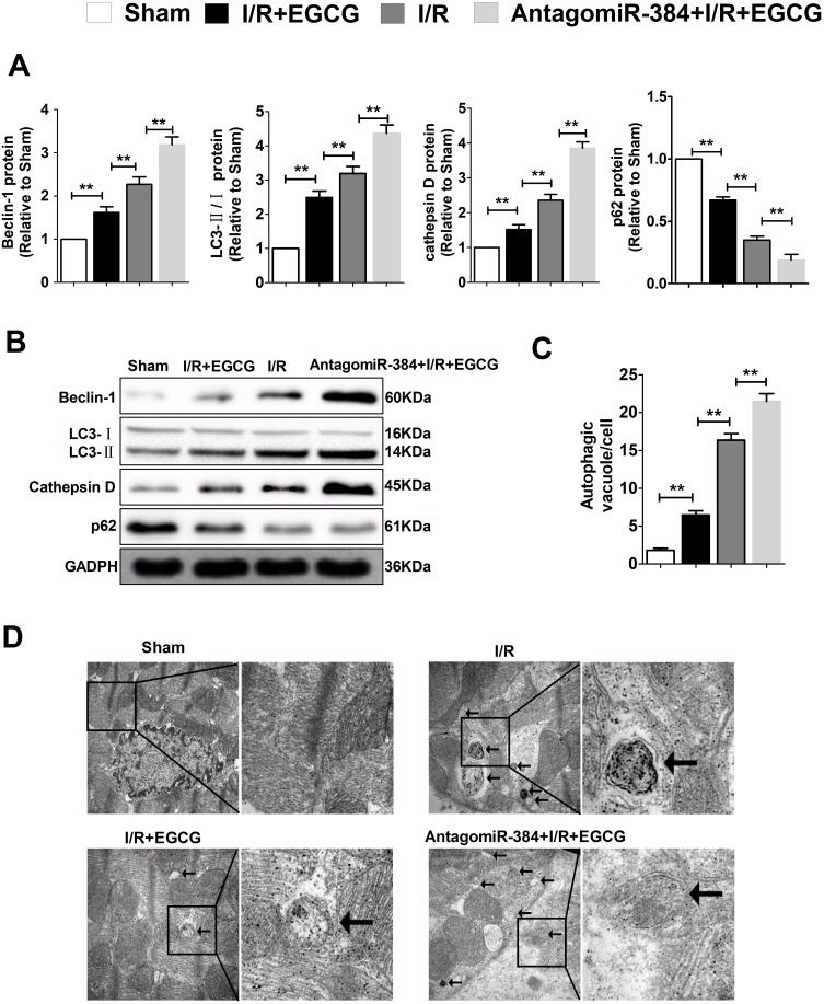
Notes: (A) The mRNA levels of miR-384 were detected by qRT-PCR. (B, C) Representative Western blotting results and quantitative data on autophagy-related protein expression in rats following I/R in the presence or absence of miR-384 inhibition. (D) Representative EM images were shown. Bar=2µM. The arrows depict autophagosome. Quantification of autophagic vacuoles was shown in the bar graph. Data among groups were compared using one-way analysis of variance. Values are expressed as mean±SD (n=6). ANOVA testing was performed; * P<0.05, ** P< 0.01.
Abbreviations: ANOVA, analysis of variance; antagomiR-384, miR-384 antagomir; EGCG, epigallocatechin gallate; GAPDH, glyceraldehyde-3-phosphate dehydrogenase; I/R, ischemia/reperfusion; LC3, light chain 3; miR-384, microRNA-384-5p; mRNA, Messenger RNA; qRT-RCR, quantitative real-time PCR; SD, Sprague-Dawley.
EGCG Protected Cardiomyocytes Against I/R-Induced Myocardial Injury Through Adjusting miR-384 To Inactivate Autophagy
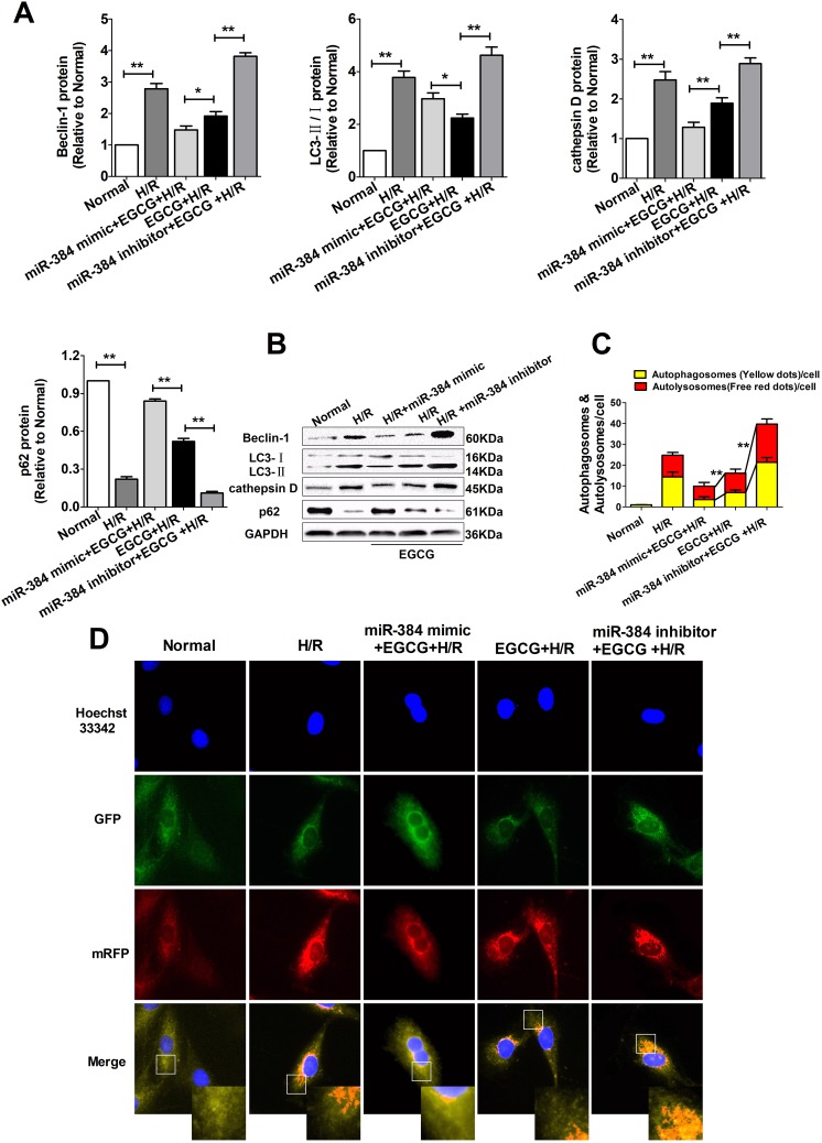
MiR-384 has been reported as an important autophagy regulator. Now we aim to further confirm the role of EGCG on modulation the miR-384 levels. As expected, I/R (H/R) remarkably increased the levels of autophagic flux, and pretreatment with EGCG reduced the levels of autophagic flux in vitro and in vivo (p<0.01 or p<0.05, Figures 5A, B and 6A, B). Furthermore, EGCG reduced the red dots fluorescence and autophagosomes, which illustrated that EGCG blocked the development of autophagy (p<0.01, Figures 5C, D and 6C, D). What is more, the effect of EGCG was enhanced by transfecting with miR-384 mimic, however, was significantly eliminated by miR-384 inhibitor. It suggested that EGCG might regulate miR-384 to protect myocardial cells from I/R-Induced injury.
Figure 5.
EGCG protected cardiomyocytes against H/R-Stimulated H9c2 cells injury through adjusting miRNA-384-5p to inactivate autophagy.
Notes: H9c2 cells were transfected with miR-384 mimic, miR-384 inhibitor. (A, B) Representative Western blotting results and quantitative data on autophagy-related protein expression in H9c2 cells following H/R in the presence or absence of miR-384. (C, D) Tandem mRFP-GFP-LC3B probe analysis of the autophagosome-lysosome fusion. Yellow dots represent the unfused autophagosome, whereas red dots denote the fusion, wherein autolysosomes are formed. H9c2 cells were transfected with a mRFP-GFP-LC3B adenovirus for 6 h and then subjected to various treatments, after which the dots were observed under a confocal fluorescence microscope. Magnification: 630×. Scale bar: 5μm. Bar graph showing the number of LC3 puncta per cell. At least 10 cells per group were counted randomly in three independent experiments. Data among groups were compared using one-way analysis of variance. Values are expressed as mean±SD (n=6). ANOVA testing was performed; * P<0.05, ** P<0.01.
Abbreviations: ANOVA, analysis of variance; EGCG, epigallocatechin gallate; GAPDH, glyceraldehyde-3-phosphate dehydrogenase; H/R, hypoxia/reoxygenation; LC3, light chain 3; miR-384, microRNA-384-5p; SD, Sprague-Dawley.
Figure 6.
EGCG protected cardiomyocytes against I/R-induced myocardial injury through adjusting miRNA-384-5p to inactivate autophagy.
Notes: (A, B) Representative Western blotting results and quantitative data on autophagy-related protein expression in rats following I/R in the presence or absence of miR-384 inhibition. (C-D) Representative EM images were shown. Bar=2µM. The arrows depict autophagosome. Quantification of autophagic vacuoles was shown in the bar graph. Data among groups were compared using one-way analysis of variance. Values are expressed as mean±SD (n=6). ANOVA testing was performed; ** P<0.01.
Abbreviations: ANOVA, analysis of variance; antagomiR-384, miR-384 antagomir; EGCG, Epigallocatechin gallate; GAPDH, glyceraldehyde-3-phosphate dehydrogenase; I/R, ischemia/reperfusion; LC3, light chain 3, miR-384, microRNA-384-5p; SD, Sprague-Dawley.
MiR-384 Prevented The Upregulation Of Beclin-1 In H/R-Stimulated H9c2 Cells
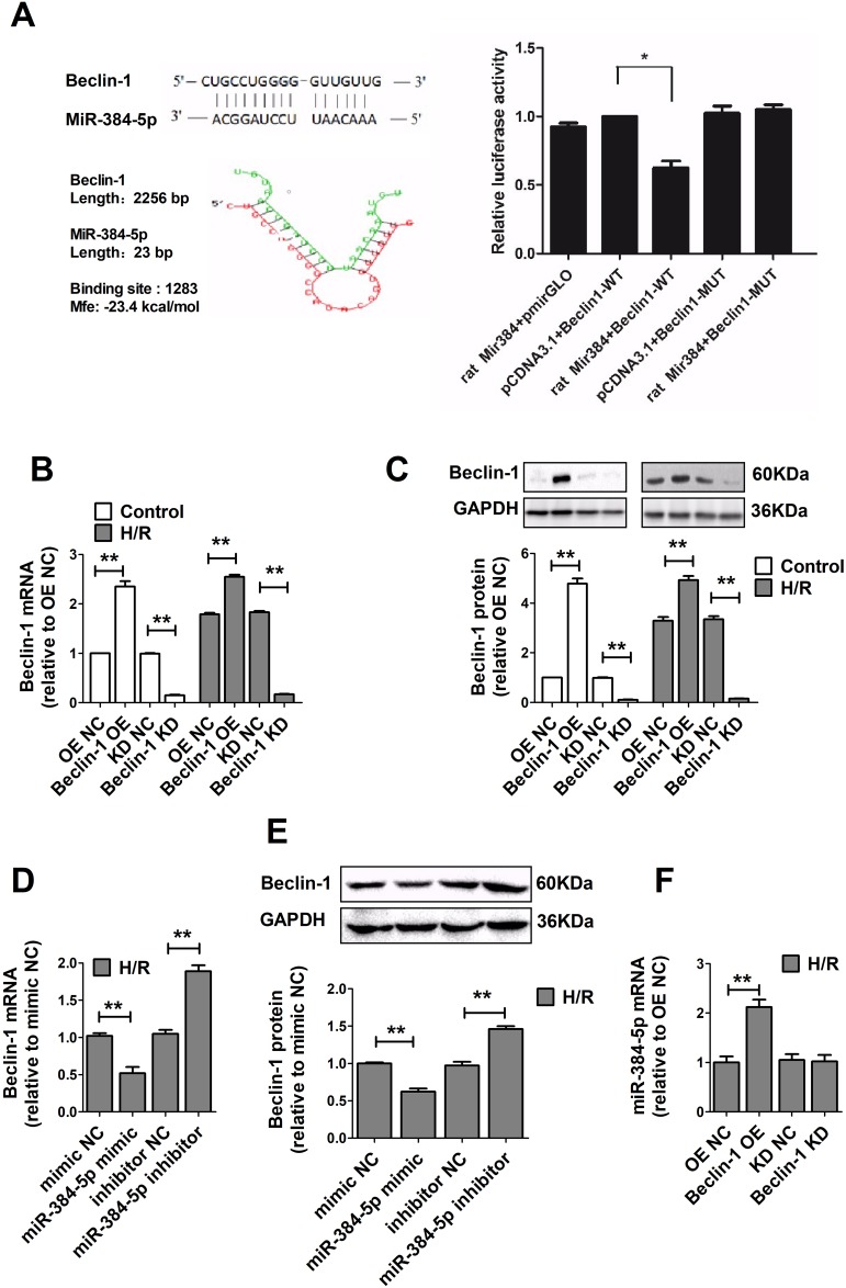
In order to determine biological role underlying the interaction between miR-384 and Beclin-1, the dual-luciferase reporter gene experiment was conducted. The outcome showed that miR-384 mimic acting on Beclin-1 mRNA WT 3ʹ-UTR plasmid, obviously reduced the firefly luciferase activity, whereas cotransfection with mutant type 3ʹ-UTR plasmid elicited significantly higher luciferase activity than that in the WT group (p<0.05, Figure 7A). In addition, to ulteriorly explain the relationship between miR-384 and Beclin-1 in H/R treated H9c2 cardiomyocytes, the H9c2 cells were also transfected with Beclin-1 OE which markedly enhanced Beclin-1 levels, whereas Beclin-1 KD significantly suppressed Beclin-1 expression (p<0.01, Figure 7B and C). Compared with the NC group, overexpression of miR-384 strikingly reversed the expression of Beclin-1 at both mRNA and protein in H/R treated H9c2 cardiomyocytes. Consistently, the effect of miR-384 inhibitor on enhancing Beclin-1 expression in H/R-induced H9c2 cardiomyocytes was significant (p<0.01, Figure 7D and E). In contrast, overexpression of Beclin-1 significantly increased the expression of miR-384 through negative feedback regulation, while silencing the expression of Beclin-1 had no effect on miR-384 expression (p<0.01, Figure 7F). Therefore, these findings suggested that Beclin-1 3ʹ-UTR was directly regulated by miR-384 at the post-transcriptional level in cardiomyocytes.
Figure 7.
miR-384 prevented the upregulation of Beclin-1 in H/R-stimulated H9c2 cells.
Notes: H9c2 cells were transfected with miR-384 mimic, miR-384 inhibitor, Beclin-1 OE and Beclin-1 KD. (A) Dual-luciferase gene reports experiments used to verify Beclin-1 was target gene of miR-384 in H9c2 cells. (B–E) The mRNA and protein levels of Beclin-1 were detected by qRT-PCR and Western blotting. (F) The mRNA levels of miR-384 were detected by qRT-PCR. Data among groups were compared using one-way analysis of variance. Values are expressed as mean±SD (n=6). ANOVA testing was performed; * P<0.05, ** P<0.01.
Abbreviations: ANOVA, analysis of variance; EGCG, epigallocatechin gallate; GAPDH, glyceraldehyde-3-phosphate dehydrogenase; H/R, hypoxia/reoxygenation; KD, knock-down; LC3, light chain 3; miR-384, microRNA-384-5p; mRNA, Messenger RNA; NC, negative control; OE, overexpression; SD, Sprague-Dawley.
EGCG Inhibited H/R-Induced H9c2 Autophagy By Downregulating Beclin-1
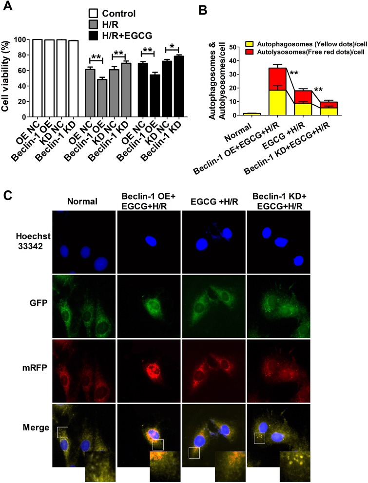
As shown in Figure 8A, under H/R conditions, H9c2 cells pretransfected with Beclin-1 KD had a higher percentage of viable cells, whereas cells pretransfected with Beclin-1 OE exhibited significantly decreased viability compared with the corresponding control cells, thus, Beclin-1 had harm effects on cell survival and EGCG failed to reduce the damage (p<0.01 or p<0.05). As expected, fluorescence imaging analysis showed that Beclin-1 OE increased the red dots fluorescence and Beclin-1 KD decreased them compared with the EGCG+H/R group (p<0.01 Figure 8B and C). This step confirmed that Beclin-1 expression could impact autophagy and it may be a target of EGCG.
Figure 8.
EGCG attenuated H/R-induced H9c2 autophagy by down-regulating Beclin-1.
Notes: (A) H9c2 cells viability rates were examined by the CCK-8 assay. (B-C) Tandem mRFP-GFP-LC3B probe analysis of the autophagosome-lysosome fusion. Yellow dots represent the unfused autophagosome, whereas red dots denote the fusion, wherein autolysosomes are formed. H9c2 cells were transfected with mRFP-GFP-LC3B adenovirus for 6 hrs and then subjected to various treatments, after which the dots were observed under a confocal fluorescence microscope. Magnification: 630×. Scale bar: 5μm. Bar graph showing the number of LC3 puncta per cell. At least 10 cells per group were counted randomly in three independent experiments. Data among groups were compared using one-way analysis of variance. Values are expressed as mean±SD (n=6). ANOVA testing was performed; * P<0.05, ** P<0.01.
Abbreviations: ANOVA, Analysis of Variance; EGCG, Epigallocatechin gallate; GAPDH, glyceraldehyde-3-phosphate dehydrogenase; H/R, hypoxia/reoxygenation; KD, knock-down; OE, overexpression; SD, Sprague-Dawley.
EGCG Inhibited H/R-Induced H9c2 Autophagy By Regulating miR-384/beclin-1 To Activate PI3K/Akt Pathway
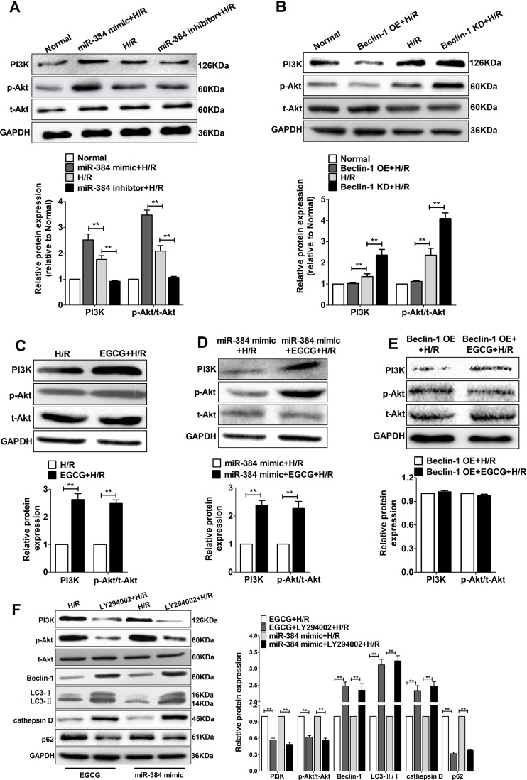
In order to examine the significance of miR-384/Beclin-1-mediated regulation of the PI3K/Akt signalling pathway during H/R injury, the expression levels of PI3K and Akt were detected by Western blotting assay. Compared to the H/R group, a significant upregulation of PI3K and p-Akt/t-Akt level was induced by transfection with miR-384 mimic and restrained by miR-384 inhibitor (p< 0.01, Figure 9A). Besides, Beclin-1 OE stimulation induced a conspicuous decrease in the expression of PI3K and p-Akt/t-Akt, whereas, Beclin-1 KD increased the expression of PI3K and p-Akt/t-Akt (p <0.01, Figure 9B). Still, we can speculate that PI3K/Akt-mediated signal pathway has been shown to be impaired in transfection with Beclin-1 OE, while transfection with miR-384 mimic in H9c2 cells may induce PI3K/Akt activation, to compensate for the impairment. Moreover, compared to the H/R group, pretreatment with EGCG activated PI3K and p-Akt/t-Akt (Figure 9C), enhanced the function of miR-384 mimic (p<0.01, Figure 9D). However, the inhibitory effect of Beclin-1 OE on PI3K/Akt pathway could not be reversed by pretreatment with EGCG (p<0.01, Figure 9E), namely, EGCG did not work if Beclin-1 was overexpressed. So, the results suggested that increasing miR-384 and lowering Beclin-1 expression may be an important mechanism in the protective effect of EGCG. Above all, EGCG could upregulate the expression of miR-384 and downregulate the expression of Beclin-1 to activate PI3K/Akt pathway.
Figure 9.
EGCG attenuated H/R-induced H9c2 autophagy by regulating miR-384-5p/Beclin-1 to activate the PI3K/Akt pathway.
Notes: H9c2 cells were transfected with miR-384-5p mimic and inhibitor, Beclin-1 OE and Beclin-1 KD, after EGCG pretreatment with 25 µM and LY294002 pretreatment with 10 µM for 4 h and H/R-stimulated H9c2 cells for another 24 hrs. (A–E) Western blotting results and quantitative data showing the expression of PI3K and Akt protein in H9c2 cells. (F) Representative Western blotting results and quantitative data of PI3K, Akt and autophagy-related protein expression in H9c2 cells. Data among groups were compared using one-way analysis of variance. Values are expressed as mean±SD (n=6). ANOVA testing was performed; ** P<0.01.
Abbreviations: ANOVA, analysis of variance; EGCG, epigallocatechin gallate; GAPDH, glyceraldehyde-3-phosphate dehydrogenase; H/R, hypoxia/reoxygenation; KD, knock-down; LC3, light chain 3; miR-384, microRNA-384-5p; OE, overexpression; PI3K, phosphoinositide 3 kinase; SD, Sprague-Dawley.
Furthermore, we inhibited PI3K/Akt signalling pathway using LY294002, a PI3K inhibitor, to determine whether there was a direct link between EGCG and miR-384 induced activation of the PI3K/Akt signalling pathway and autophagy. The upregulating PI3K and p-Akt/t-Akt effect of EGCG and miR-384 mimics were blocked by coadministration of LY294002. The effect of EGCG or miR-384 mimics on regulating autophagic protein expression was also blocked by LY294002 when H9c2 cells were exposured to H/R (p<0.01, Figure 9F). Altogether, these results suggested that EGCG and miR-384 could inhibit autophagy induced by H/R stimulation partially through activating PI3K/Akt signalling pathway.
Discussion
To date, this is the first study to show that EGCG could up-regulate miR-384 expression to protect cardiomyocytes from I/R injury by inhibiting excessive autophagy. Mechanistic studies have shown that miR-384 directly targets Beclin-1 and the protective effects of EGCG on myocardial ischemic injury are mediated by PI3K/Akt signaling pathway, which is downstream of miR-384/Beclin-1. Thus, we concluded that EGCG protected cardiomyocytes against I/R injury through adjusting miRNA-384 targets Beclin-1 to inhibit autophagy via PI3K/Akt pathway.
Autophagy plays an important role in the pathological mechanism of myocardial ischemia reperfusion injury. Enhanced levels of autophagy have been observed in the heart during both ischemia and reperfusion.23 Autophagy has a dual, opposite role in the heart. Whereas severe stress, such as prolonged hypoxia or subsequent reperfusion, results in excessive autophagy, which may lead to cell death and cardiac dysfunction by triggering excessive self-digestion of essential proteins and organelles.9 In our experiment, autophagy inhibitor (3-MA) reduced cell viability and aggravated cardiomyocytes injury in the early stage of reperfusion, while with the extension of reperfusion time, more than 12 hrs, 3-MA played a protective role in myocardial cells. The result was in accordance with other reported values that I/R injury could induce autophagy flux, inhibit cells survival, which indicating that I/R injury may cause excessive autophagy in cardiomyocytes.24 As is well known, EGCG, whose chemical formula is C22H18O11, has very strong antioxidant activity and plays an important role in myocardial ischemia reperfusion injury and autophagy.5 Our research also showed that EGCG pretreatment exhibited a significant decrease in cTnI level and inhibited Beclin-1, a autophagy associated protein, compared with the I/R group (Figure 2). This was consistent with the effect of autophagy inhibitor, 3-MA. So, it is clear that EGCG attenuated myocardial injury by inhibiting autophagy. But what is the specific mechanism of EGCG on adjusting autophagy?
A number of miRNAs were proven to be able to modulate autophagy by targeting their mRNAs or other signalling pathways.25 The high expression of miR-384 reduced the formation of autophagosomes and downregulated autophagy flux levels.26 Regrettably, few studies specifically focused on the role of miR-384 in I/R-induced cardiovascular disease-related cell autophagy, as well as its downstream pathways action. In this article, we found that upregulating miR-384 increased cell viability and decreased I/R-induced cell autophagy, whereas downregulating miR-384 accomplished the opposite effect (Figures 3 and 4). If drugs can regulate miR-384 expression and inhibit autophagy to anti-I/R injury, it will play an important guiding role in clinical treatment. So, we hypothesized that treatment with EGCG will upregulate miR-384 expression, which in turn contributes to restore the excessive autophagy and there has no report on the direct effect of EGCG on miR-384 expression in I/R injury. Our results showed that EGCG significantly increased the expression of miR-384 in cardiomyocytes in response to I/R injury (Figure 2). Beyond that, EGCG enhanced the protect effect of miR-384 mimic. For the same reason, miR-384 inhibitor significantly increased autophagy flux expression, thus attenuated the protective effects of EGCG (Figures 5 and 6). The above results also indicated that protective effect of EGCG was related to modulation of miR-384 levels.
MiRNAs act through downregulating multiple target genes, while Beclin-1 is known as a key initiator of the autophagic process.27 The authors reported that miR-384 could modulate autophagy to anti-atherosclerosis by inhibiting Beclin-1 expression.28 So, whether miR-384 act directly on Beclin-1 and what is their relationship to autophagy in the myocardial I/R injury are questions we need to explore. First of all, the miRNA target gene prediction software was used to indicate that miR-384 expression was significantly altered on the myocardial I/R injury. Secondly, upregulating miR-384 decreased Beclin-1 expression whereas downregulating miR-384 increased Beclin-1 expression at the mRNA and protein levels. The dual-luciferase reporter gene assay results confirmed that miR-384 interacts with Beclin-1. In addition, we found that inhibiting Beclin-1 alleviated the excessive autophagy induced by I/R treatment and has favorable effects on H9c2 cardiomyocyte survival. Interestingly, as shown in Figure 7, upregulating Beclin-1 increased miR-384 expression. We suspected that Beclin-1 has a negative feedback effect on miR-384, while Beclin-1 may be a downstream effector of miR-384 and potentially mediates the effects of miR-384. Besides, EGCG significantly decreased the expression of Beclin-1. Beyond that, Beclin-1 KD enhanced the protect effect of EGCG and Beclin-1 OE significantly increased autophagy, thus attenuated the protective effects of EGCG (Figure 8). Very coincidentally, the protective effect of EGCG was related to modulation of miR-384 and Beclin-1 levels. In other words, among those above, there are certain inherent connections that may interact and restrict each other. Thus, we can speculate that EGCG modulates the miR-384/Beclin-1 feedback loop.
Previous studies have demonstrated that PI3K/Akt pathway provides cardioprotection by inhibiting excessive autophagy and enhancing recovery in myocardial I/R injury.29 Bai et al noted that miR-384 could inhibit high glucose-induced lipogenesis in hepatocytes through the PI3K/Akt/mTORC1 pathway.30 So, we suspect that the protective effects of miR-384 in I/R injury may be associated with the inhibition of autophagy via the activation of the PI3K/Akt signaling pathway. As we expected, shown in Figure 9, miR-384 mimic and Beclin-1 KD increased PI3K and Akt phosphorylation under H/R conditions, whereas miR-384 inhibitor and Beclin-1 OE had opposite effects. Therefore, we concluded that miR-384/Beclin-1 plays an important regulatory role in myocardial ischemia and it may be the upstream target of PI3K/Akt pathway. Moreover, pretreating with EGCG reversed the suppressive of PI3K and Akt phosphorylation, which indicating that EGCG had positive effects on I/R injury. But more importantly, miR-384 mimic accumulated this positive effect, while Beclin-1 OE eliminated it. Furthermore, LY294002, an inhibitor of PI3K/Akt signaling pathway, abrogated the protective effects of EGCG and miR-384 against H/R-induced autophagy. So, the results suggested that the mechanism of miR-384/Beclin-1 regulates autophagy may be targetted PI3K/Akt, while EGCG inhibits autophagy by affecting the cross-talk of miR-384/Beclin-1 via PI3K/Akt pathway.
Conclusion
In summary, to our knowledge, this is the first study to show that EGCG could downregulate the autophagy in I/R-induced cardiomyocytes injury by increasing miR-384 levels and reducing the expression of Beclin-1 protein via PI3K/Akt pathway. This study provides a new and powerful evidence for the protective effect of EGCG in alleviating myocardial I/R injury. Hence, further investigation is warranted for the therapeutic potential of EGCG in I/R injury treatment.
Acknowledgments
We sincerely thank all the teachers from the Central Laboratory and the Pharmacological Laboratory of Guilin Medical University for their assistance with these experiments. This work was supported by the Natural Science Foundation of China (No. 81560665, No. 81760726) and Project of Natural Science Foundation of Guangxi, China (No. 2017 GXNSFAA 198244).
Disclosure
The authors report no conflicts of interest in this work.
References
- 1.Jennings RB. Historical perspective on the pathology of myocardial ischemia/reperfusion injury. Circ Res. 2013;113(4):428–438. [DOI] [PubMed] [Google Scholar]
- 2.Mak JC. Potential role of green tea catechins in various disease therapies: progress and promise. Clin Exp Pharmacol Physiol. 2012;39(3):265–273. doi: 10.1111/j.1440-1681.2012.05673.x [DOI] [PubMed] [Google Scholar]
- 3.Khurana S, Venkataraman K, Hollingsworth A, Piche M, Tai TC. Polyphenols: benefits to the cardiovascular system in health and in aging. Nutrients. 2013;5(10):3779–3827. doi: 10.3390/nu5103779 [DOI] [PMC free article] [PubMed] [Google Scholar]
- 4.Wang W, Huang X, Shen D, Ming Z, Zheng M, Zhang J. Polyphenol epigallocatechin-3-gallate inhibits hypoxia/reoxygenation-induced H9C2 cell apoptosis. Minerva Med. 2018;109(2):95–102. doi: 10.23736/S0026-4806.17.05349-6 [DOI] [PubMed] [Google Scholar]
- 5.Xuan F, Jian J. Epigallocatechin gallate exerts protective effects against myocardial ischemia/reperfusion injury through the PI3K/Akt pathway-mediated inhibition of apoptosis and the restoration of the autophagic flux. Int J Mol Med. 2016;38(1):328–336. doi: 10.3892/ijmm.2016.2615 [DOI] [PubMed] [Google Scholar]
- 6.Li ZL, Lerman LO. Impaired myocardial autophagy linked to energy metabolism disorders. Autophagy. 2012;8(6):992–994. doi: 10.4161/auto.20285 [DOI] [PMC free article] [PubMed] [Google Scholar]
- 7.Mintern JD, Villadangos JA. Autophagy and mechanisms of effective immunity. Front Immunol. 2012;3:60. doi: 10.3389/fimmu.2012.00198 [DOI] [PMC free article] [PubMed] [Google Scholar]
- 8.Zou J, Yue F, Jiang X, Li W, Yi J, Liu L. Mitochondrion-associated protein LRPPRC suppresses the initiation of basal levels of autophagy via enhancing Bcl-2 stability. Biochem J. 2013;454(3):447–457. doi: 10.1042/BJ20130306 [DOI] [PMC free article] [PubMed] [Google Scholar]
- 9.Jian J, Xuan F, Qin F, Huang R. Bauhinia championii flavone inhibits apoptosis and autophagy via the PI3K/Akt pathway in myocardial ischemia/reperfusion injury in rats. Drug Des Devel Ther. 2015;9:5933–5945. doi: 10.2147/DDDT.S92549 [DOI] [PMC free article] [PubMed] [Google Scholar]
- 10.Zhao YG, Zhang H. Autophagosome maturation: an epic journey from the ER to lysosomes. J Cell Biol. 2019;218(3):757–770. doi: 10.1083/jcb.201810099 [DOI] [PMC free article] [PubMed] [Google Scholar]
- 11.Ha SW, Weitzmann MN, Beck GR Jr. Bioactive silica nanoparticles promote osteoblast differentiation through stimulation of autophagy and direct association with LC3 and p62. ACS Nano. 2014;8(6):5898–5910. doi: 10.1021/nn5009879 [DOI] [PMC free article] [PubMed] [Google Scholar]
- 12.Lampert MA, Orogo AM, Najor RH, et al. BNIP3L/NIX and FUNDC1-mediated mitophagy is required for mitochondrial network remodeling during cardiac progenitor cell differentiation. Autophagy. 2019;15:1–17. [DOI] [PMC free article] [PubMed] [Google Scholar]
- 13.Liu L, Wu Y, Huang X. Orientin protects myocardial cells against hypoxia-reoxygenation injury through induction of autophagy. Eur J Pharmacol. 2016;776:90–98. doi: 10.1016/j.ejphar.2016.02.037 [DOI] [PubMed] [Google Scholar]
- 14.Liu MW, Su MX, Tang DY, Hao L, Xun XH, Huang YQ. Ligustrazin increases lung cell autophagy and ameliorates paraquat-induced pulmonary fibrosis by inhibiting PI3K/Akt/mTOR and hedgehog signalling via increasing miR-193a expression. BMC Pulm Med. 2019;19(1):35. doi: 10.1186/s12890-019-0850-6 [DOI] [PMC free article] [PubMed] [Google Scholar]
- 15.Alberti C, Cochella L. A framework for understanding the roles of miRNAs in animal development. Development. 2017;144(14):2548–2559. doi: 10.1242/dev.146613 [DOI] [PubMed] [Google Scholar]
- 16.Sun T, Dong YH, Du W, et al. The role of MicroRNAs in myocardial infarction: from molecular mechanism to clinical application. Int J Mol Sci. 2017;18:4. doi: 10.3390/ijms18040745 [DOI] [PMC free article] [PubMed] [Google Scholar]
- 17.Bao Y, Lin C, Ren J, Liu J. MicroRNA-384-5p regulates ischemia-induced cardioprotection by targeting phosphatidylinositol-4,5-bisphosphate 3-kinase, catalytic subunit delta (PI3K p110δ). Apoptosis. 2013;18(3):260–270. doi: 10.1007/s10495-013-0802-1 [DOI] [PubMed] [Google Scholar]
- 18.Zheng J, Liu X, Wang P, et al. CRNDE promotes malignant progression of glioma by attenuating miR-384/PIWIL4/STAT3 axis. Mol Ther. 2016;24(7):1199–1215. doi: 10.1038/mt.2016.71 [DOI] [PMC free article] [PubMed] [Google Scholar] [Retracted]
- 19.Zhang W, Cheng P, Hu W, et al. Inhibition of microRNA-384-5p alleviates osteoarthritis through its effects on inhibiting apoptosis of cartilage cells via the NF-κB signaling pathway by targeting SOX9. Cancer Gene Ther. 2018;25(11–12):326–338. doi: 10.1038/s41417-018-0029-y [DOI] [PubMed] [Google Scholar]
- 20.Bøtker HE, Hausenloy D, Andreadou I, et al. Practical guidelines for rigor and reproducibility in preclinical and clinical studies on cardioprotection. Basic Res Cardiol. 2018;113(5):39. [DOI] [PMC free article] [PubMed] [Google Scholar]
- 21.Prasad KM, Xu Y, Yang Z, Acton ST, French BA. Robust cardiomyocyte-specific gene expression following systemic injection of AAV: in vivo gene delivery follows a Poisson distribution. Gene Ther. 2011;18(1):43–52. doi: 10.1038/gt.2010.105 [DOI] [PMC free article] [PubMed] [Google Scholar]
- 22.Liu Z, Li X, Zhang JT, et al. Autism-like behaviours and germline transmission in transgenic monkeys overexpressing MeCP2. Nature. 2016;530(7588):98–102. doi: 10.1038/nature16533 [DOI] [PubMed] [Google Scholar]
- 23.Tao L, Bei Y, Lin S, et al. Exercise training protects against acute myocardial infarction via improving myocardial energy metabolism and mitochondrial biogenesis. Cell Physiol Biochem. 2015;37(1):162–175. doi: 10.1159/000430342 [DOI] [PubMed] [Google Scholar]
- 24.Wu S, Chang G, Gao L, et al. Trimetazidine protects against myocardial ischemia/reperfusion injury by inhibiting excessive autophagy. J Mol Med (Berl). 2018;96(8):791–806. doi: 10.1007/s00109-018-1664-3 [DOI] [PubMed] [Google Scholar]
- 25.Füllgrabe J, Klionsky DJ, Joseph B. The return of the nucleus: transcriptional and epigenetic control of autophagy. Nat Rev Mol Cell Biol. 2014;15(1):65–74. doi: 10.1038/nrm3716 [DOI] [PubMed] [Google Scholar]
- 26.Yan L, Wu K, Du F, Yin X, Guan H. miR-384 suppressed renal cell carcinoma cell proliferation and migration through targeting RAB23. J Cell Biochem. 2018. [DOI] [PubMed] [Google Scholar]
- 27.Ranjan K, Pathak C. Expression of cFLIPL determines the basal interaction of Bcl-2 with beclin-1 and regulates p53 dependent ubiquitination of beclin-1 during autophagic stress. J Cell Biochem. 2016;117(8):1757–1768. doi: 10.1002/jcb.25474 [DOI] [PubMed] [Google Scholar]
- 28.Wang B, Zhong Y, Huang D, Li J. Macrophage autophagy regulated by miR-384-5p-mediated control of Beclin-1 plays a role in the development of atherosclerosis. Am J Transl Res. 2016;8(2):606–614. [PMC free article] [PubMed] [Google Scholar]
- 29.Ye G, Fu Q, Jiang L, Li Z. Vascular smooth muscle cells activate PI3K/Akt pathway to attenuate myocardial ischemia/reperfusion-induced apoptosis and autophagy by secreting bFGF. Biomed Pharmacother. 2018;107:1779–1785. doi: 10.1016/j.biopha.2018.05.113 [DOI] [PubMed] [Google Scholar]
- 30.Bai PS, Xia N, Sun H, Kong Y. Pleiotrophin, a target of miR-384, promotes proliferation, metastasis and lipogenesis in HBV-related hepatocellular carcinoma. J Cell Mol Med. 2017;21(11):3023–3043. doi: 10.1111/jcmm.13213 [DOI] [PMC free article] [PubMed] [Google Scholar]


