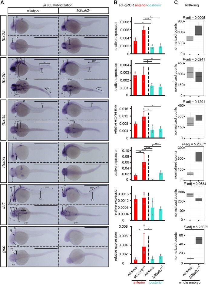
Fig. 5.
Transcription factor expression is spatially dysregulated in MZezh2 mutant (MZezh2−/−) embryos. (A-C) Spatial expression analysis by (A) in situ hybridization, (B) RT-qPCR on anterior half and posterior half, and (C) RNA-seq results of transcription factors tbx2a, tbx2b, tbx3a, tbx5a, isl1 and gsc in 24 hpf embryos. Scale bars: 1 mm. Experiments were performed in biological duplicates of a least 15 pooled embryos for in situ hybridization and in triplicates or quadruplicates of 20 pooled larval halves for RT-qPCR. Relative expression was calculated based on expression of the housekeeping gene actb1. Data are mean±s.e.m. in B with dots representing individual RT-qPCR samples. Relative expression was compared between anterior (red) or posterior (turquoise) parts in MZezh2−/− and wild-type embryos (one-way ANOVA with post-hoc tests, ***P<0.001, **P<0.01, *P<0.05). (C) Box plots represent normalized counts from RNA-seq experiments in whole MZezh2−/− and wild type after differential expression analysis with DEseq2. all, anterior lateral lane ganglion; ba, branchial arch; cmn, cranial motor neurons; de, diencephalon; drp, distal region of the pronephros; dscn, dorsal spinal cord neurons; e, eye; ep, epiphysis; fn, forebrain nuclei; h, heart; hmn, hindbrain motor neurons; llg, lateral lane ganglion; mot, primary motor neurons; og, olfactory ganglion; ov, otic vesicle; pa, pharyngeal arches; pan, pancreas; pf, pectoral fin; pro, pronephros; sc, spinal cord; tdn, telencephalon and diencephalon nuclei; te, telencephalon; vg, ventral ganglion. The box represents the first quartile, median and third quartile. The whiskers below and above the box represent the minimum and maximum values.

