Abstract
Idiopathic basal ganglia calcification-1 (IBGC1) is an autosomal dominant disorder characterized by calcification in the basal ganglia, which can manifest a range of neuropsychiatric symptoms, including parkinsonism. We herein describe a 64-year-old Japanese IBGC1 patient with bilateral basal ganglia calcification carrying a novel SLC20A2 variant (p.Val322Glufs*92). The patient also presented with dopa-responsive parkinsonism with decreased dopamine transporter (DAT) density in the bilateral striatum and decreased cardiac 123I-meta-iodobenzylguanidine uptake.
Subject terms: Neurological disorders, Medical genetics
Idiopathic basal ganglia calcification (IBGC), also known as Fahr disease or primary familial brain calcification (PFBC), is a disorder characterized by bilateral calcifications in the basal ganglia and other brain regions. Clinical manifestations of IBGC range from asymptomatic to neuropsychiatric symptoms, including dystonia, parkinsonism, ataxia, and cognitive impairment1. Typically, the inheritance mode of familial IBGC is an autosomal dominant one and to date, four dominant causal genes of familial IBGC have been identified, including SLC20A2 (IBGC1, MIM: #213600), PDGFRB (IBGC4, MIM: #615007), PDGFB (IBGC5, MIM: #615483), and XPR1 (IBGC6, MIM: #616413)2–5. Recently, MYORG was reported as an autosomal recessive causal gene for IBGC (IBGC7, MIM: #618317)6,7. Variants in SLC20A2, encoding the type III sodium-dependent phosphate transporter 2 (PiT-2), are a major cause of IBGC8,9. Herein, we report an IBGC1 patient with a novel variant in SLC20A2 associated with dopa-responsive parkinsonism.
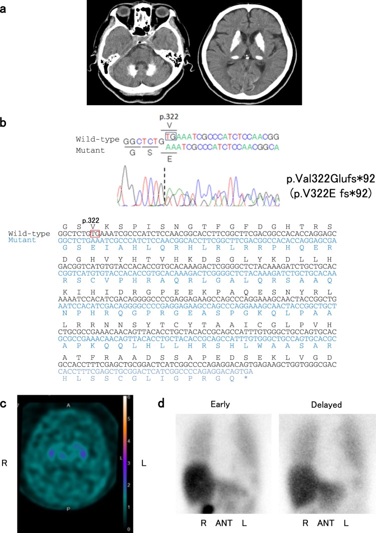
The patient was a 63-year-old Japanese woman who presented to our hospital with a one-month history of lumbago and unsteady gait. Neurological examination revealed gait disturbance with stooped posture and short steps, but rigidity, tremor, weakness, and cerebellar symptoms were not observed. Computed tomography (CT) images of her brain revealed marked calcifications in the bilateral basal ganglia, thalami, and dentate nuclei (Fig. 1a). Laboratory tests showed that serum calcium, phosphate, and intact parathyroid hormone levels were all within the normal ranges. There was no family history of IBGC or parkinsonism. After written informed consent was obtained, we analyzed all the coding regions of the IBGC causative genes, SLC20A2, PDGFRB, and PDGFB, by Sanger sequencing as previously reported10. We diagnosed her as IBGC1 based on the identification of a novel heterozygous frameshift variant, p.Val322Glufs*92 (NM_006749.4:c.965_966delTG, exon 8), in SLC20A2 (Fig. 1b). The variant was absent in the following genome databases: dbSNP 151 (https://www.ncbi.nlm.nih.gov/projects/SNP/), Integrative Japanese Genome Variation Database (http://ijgvd.megabank.tohoku.ac.jp/), Exome Aggregation Consortium database version 0.3.1 (http://exac.broadinstitute.org/), and Human Gene Mutation Database (HGMD® Professional 2019.1).
Fig. 1. Imaging and sequencing findings of the patient.
a Computed tomographic (CT) images of the patient show calcifications in the bilateral basal ganglia, thalami, and dentate nuclei. b Electropherogram and sequence of SLC20A2 (NM_006749.4) from the patient’s DNA shows the c.965_966delTG variant. DNA and corresponding amino acid sequences of wild-type and mutant SLC20A2 alleles are also shown. The c.965_966delTG variant causes a frameshift variant (p.Val322Glufs*92). c Dopamine transporter (DAT) single photon emission CT shows diffusely decreased DAT density in the bilateral striatum. The specific binding ratios (SBRs) of both striatum were 0.51 (right) and 0.14 (left). d 123I-meta-iodobenzylguanidine (123I-MIBG) myocardial scintigraphy shows decreased cardiac 123I-MIBG uptake with early and delayed heart to mediastinum (H/M) rates of 1.995 and 1.585, respectively
Ten months after her first visit, she was hospitalized because of difficulties in standing up without assistance at the age of 64. She showed severe bradykinesia, postural instability, and mild symmetric rigidity without tremor. Her Unified Parkinson Disease Rating Scale part III (UPDRS-III) score was 43 of 108 on the ninth hospital day. Her Mini-Mental State Examination score was 24 of 30, and her Hasegawa dementia scale revised was 22 of 30. Dopamine transporter (DAT) single photon emission CT using 123I-ioflupane showed diffusely decreased DAT density in the bilateral striatum (Fig. 1c). The specific binding ratios (SBRs) of both striatum were 0.51 (right) and 0.14 (left). Her 123I-meta-iodobenzylguanidine (123I-MIBG) myocardial scintigraphy revealed reduced cardiac 123I-MIBG uptake with early and delayed heart to mediastinum (H/M) rates of 1.995 and 1.585, respectively (Fig. 1d). Levodopa therapy (200 mg/day) was started on the 14th hospital day and was effective against bradykinesia and postural instability. She was able to walk without assistance in her room. On the 122nd hospital day, she received 600 mg/day of levodopa, and her UPDRS-III score markedly improved from 43 to 11.
The variants associated with IBGC are located widely in SLC20A2 among the patients with IBGC, and the correlation of genotype and phenotype remains unclear1,9,11.
Parkinsonism is one of the common clinical symptoms of IBGC. Tadic et al. showed that 13% of patients with SLC20A2 or PDGFRB variants presented with parkinsonism1. Another review reported motor improvement with dopatherapy in five patients with genetically confirmed IBGC12. Genetically confirmed Japanese IBGC1 patients presenting with parkinsonism have also been reported (Table 1)10,13,14. Among the five variants summarized in Table 1, two variants (c.516+1G>A and c.965_966delTG) are frameshift variants, presumably resulting in loss of function of SLC20A2. In addition, a decreased level of SLC20A2 protein was described in the case with the missense variant (c.1909A>C, S637R), raising the possibility of unstable mutant protein13. Although the functional investigations were not reported for the two missense variants (R71H and G90V), loss-of-function variants are considered for the three variants shown in Table 1. Consistent with previous reports, the majority of variants associated with IBGC are loss-of-function variants8,9, and the present study also suggests that loss-of-function mechanisms are likely involved in at least of the three variants. The present case demonstrated decreased DAT density in the bilateral striatum and decreased cardiac 123I-MIBG uptake (Fig. 1c, d). The decreased DAT density in the bilateral striatum suggested presynaptic dopaminergic dysfunction, which was reported in patients with IBGC14–17. Saito et al. also showed that postsynaptic dopaminergic dysfunction in the bilateral striatum matched calcified regions16. These findings suggested that basal ganglia calcification might result in dopaminergic dysfunction in IBGC patients. The three cases with reduced DAT density in the striatum (cases 2, 5, and 7. Table 1) also presented with decreased cardiac 123I-MIBG uptake, which was indistinguishable from that observed in patients with Lewy body diseases, including idiopathic Parkinson disease (PD)18. Since PD is a relatively common disease in Japan (prevalence of ~150 per 100,000 persons in Japan)19, the coincidental presence of idiopathic PD and IBGC remains a possibility concerning dopa-responsive parkinsonism of patients with IBGC1. However, it is important to pay attention to patients with IBGC who show dopa-responsive parkinsonism to provide appropriate treatment. To clarify the etiologies of dopa-responsive parkinsonism occasionally observed in patients with IBGC, further functional analyses including DAT SPECT and 123I-MIBG myocardial scintigraphy will be required in a larger number of patients with genetically confirmed IBGC.
Table 1.
Variants of SLC20A2 and clinical features of genetically confirmed IBGC1 Japanese patients with parkinsonism
| Case | 1 | 2 | 3 | 4 | 5 | 6 | 7 | 8 | 9 |
|---|---|---|---|---|---|---|---|---|---|
| Variant | c.212G>A R71H Exon 2 | c.269G>T G90V Exon 2 | c.516+1G>A V144Gfs*85 IVS 4 | c.965_966delTG V322Efs*92 Exon 8 | c.1909A>C S637R Exon 11 | ||||
| Patient | Proband | Proband | Son | Mother | Proband | Son | Proband | Brother | Proband |
| Age/sex | 73/F | 79/M | 52/M | 89/F | 62/M | 27/M | 64/F | NA/M | 62/M |
| Age at onset (years) | 71 | 74 | 50 | NA | 60 | 63 | NA | 62 | |
| Onset symptom | Clumsiness of hands and unsteady gait | Dementia | Depression | NA | Slowness and gait disturabance | Asymptomatic | Unsteady gait | NA | Difficulty in driving a car |
| Parkinsonism | (+) | (+) | None | (+) | (+) | None | (+) | (+) | (+) |
| Levodopa responsiveness | (+) | NA | NA | (+) | (+) | NA | NA | ||
| Cognitive impairment | (+) | (+) | None | NA | None | None | Mild | NA | (+) |
| MMSE | 16/30 | 13/30 | 30/30 | NA | 30/30 | NA | 24/30 | NA | NA |
| HDS-R | NA | NA | NA | NA | NA | NA | 22/30 | NA | 14/30 |
| FAB | NA | 3/18 | NA | NA | NA | NA | Not examined | NA | NA |
| DAT SPECT | NA | Decreased | Normal | NA | Decreased | NA | Decreased | NA | NA |
| MIBG scintigraphy | NA | Decreased | Normal | NA | Decreased | NA | Decreased | NA | NA |
| Early H/M | NA | 1.62 | 3.24 | NA | 1.43 | NA | 1.995 | NA | NA |
| Delayed H/M | NA | NA | NA | NA | NA | NA | 1.585 | NA | NA |
| Autopsy | (+) | NA | NA | NA | NA | NA | (−) | NA | (+) |
| Lewy bodies | (+) | (+) | |||||||
| Reference | Yamada et al.10 | Koyama et al.14 | Koyama et al.14 | This report | Kimura et al.13 | ||||
F female, M male, NA not applicable, MMSE Mini-Mental State Examination, HDS-R Hasegawa dementia scale revised, FAB frontal assessment battery, DAT SPECT dopamine transporter single photon emission CT, MIBG scintigraphy 123I-meta-iodobenzylguanidine myocardial scintigraphy
Acknowledgements
This work was supported by Grant-in-Aid (No. H26-Jitsuyoka [Nanbyo]-Ippan-080) from the Ministry of Health, Labour and Welfare, Japan (S.T.).
HGV Database
The relevant data from this Data Report are hosted at the Human Genome Variation Database at 10.6084/m9.figshare.hgv.2603
Conflict of interest
The authors declare that they have no conflict of interest.
Footnotes
Publisher’s note: Springer Nature remains neutral with regard to jurisdictional claims in published maps and institutional affiliations.
References
- 1.Tadic V, et al. Primary familial brain calcification with known gene mutations: a systematic review and challenges of phenotypic characterization. JAMA Neurol. 2015;72:460–467. doi: 10.1001/jamaneurol.2014.3889. [DOI] [PubMed] [Google Scholar]
- 2.Wang C, et al. Mutations in SLC20A2 link familial idiopathic basal ganglia calcification with phosphate homeostasis. Nat. Genet. 2012;44:254–256. doi: 10.1038/ng.1077. [DOI] [PubMed] [Google Scholar]
- 3.Keller A, et al. Mutations in the gene encoding PDGF-B cause brain calcifications in humans and mice. Nat. Genet. 2013;45:1077–1082. doi: 10.1038/ng.2723. [DOI] [PubMed] [Google Scholar]
- 4.Nicolas G, et al. Mutation of the PDGFRB gene as a cause of idiopathic basal ganglia calcification. Neurology. 2013;80:181–187. doi: 10.1212/WNL.0b013e31827ccf34. [DOI] [PubMed] [Google Scholar]
- 5.Legati A, et al. Mutations in XPR1 cause primary familial brain calcification associated with altered phosphate export. Nat. Genet. 2015;47:579–581. doi: 10.1038/ng.3289. [DOI] [PMC free article] [PubMed] [Google Scholar]
- 6.Yao XP, et al. Biallelic mutations in MYORG cause autosomal recessive primary familial brain calcification. Neuron. 2018;98:1116–1123. doi: 10.1016/j.neuron.2018.05.037. [DOI] [PubMed] [Google Scholar]
- 7.Arkadir D, et al. MYORG is associated with recessive primary familial brain calcification. Ann. Clin. Transl. Neurol. 2019;6:106–113. doi: 10.1002/acn3.684. [DOI] [PMC free article] [PubMed] [Google Scholar]
- 8.Hsu SC, et al. Mutations in SLC20A2 are a major cause of familial idiopathic basal ganglia calcification. Neurogenetics. 2013;14:11–22. doi: 10.1007/s10048-012-0349-2. [DOI] [PMC free article] [PubMed] [Google Scholar]
- 9.Lemos RR, et al. Update and mutational analysis of SLC20A2: a major cause of primary familial brain calcification. Hum. Mutat. 2015;36:489–495. doi: 10.1002/humu.22778. [DOI] [PubMed] [Google Scholar]
- 10.Yamada M, et al. Evaluation of SLC20A2 mutations that cause idiopathic basal ganglia calcification in Japan. Neurology. 2014;82:705–712. doi: 10.1212/WNL.0000000000000143. [DOI] [PubMed] [Google Scholar]
- 11.Ding Y, Dong HQ. A novel SLC20A2 mutation associated with familial idiopathic basal ganglia calcification and analysis of the genotype-phenotype association in Chinese patients. Chin. Med J. (Engl.) 2018;131:799–803. doi: 10.4103/0366-6999.228245. [DOI] [PMC free article] [PubMed] [Google Scholar]
- 12.Nicolas G, et al. Phenotypic spectrum of probable and genetically-confirmed idiopathic basal ganglia calcification. Brain. 2013;136:3395–3407. doi: 10.1093/brain/awt255. [DOI] [PubMed] [Google Scholar]
- 13.Kimura T, et al. Familial idiopathic basal ganglia calcification: Histopathologic features of an autopsied patient with an SLC20A2 mutation. Neuropathology. 2016;36:365–371. doi: 10.1111/neup.12280. [DOI] [PubMed] [Google Scholar]
- 14.Koyama S, et al. Clinical and radiological diversity in genetically confirmed primary familial brain calcification. Sci. Rep. 2017;7:12046. doi: 10.1038/s41598-017-11595-1. [DOI] [PMC free article] [PubMed] [Google Scholar]
- 15.Paschali A, et al. Dopamine transporter SPECT/CT and perfusion brain SPECT imaging in idiopathic basal ganglia calcinosis. Clin. Nucl. Med. 2009;34:421–423. doi: 10.1097/RLU.0b013e3181a7d195. [DOI] [PubMed] [Google Scholar]
- 16.Saito T, et al. Neuroradiologic evidence of pre-synaptic and post-synaptic nigrostriatal dopaminergic dysfunction in idiopathic Basal Ganglia calcification: a case report. J. Neuroimaging. 2010;20:189–191. doi: 10.1111/j.1552-6569.2008.00314.x. [DOI] [PubMed] [Google Scholar]
- 17.Paghera B, Caobelli F, Giubbini R. 123I-ioflupane SPECT in Fahr disease. J. Neuroimaging. 2013;23:157–158. doi: 10.1111/j.1552-6569.2011.00581.x. [DOI] [PubMed] [Google Scholar]
- 18.Orimo S, Suzuki M, Inaba A, Mizusawa H. 123I-MIBG myocardial scintigraphy for differentiating Parkinson’s disease from other neurodegenerative parkinsonism: a systematic review and meta-analysis. Park. Relat. Disord. 2012;18:494–500. doi: 10.1016/j.parkreldis.2012.01.009. [DOI] [PubMed] [Google Scholar]
- 19.Yamawaki M, Kusumi M, Kowa H, Nakashima K. Changes in prevalence and incidence of Parkinson’s disease in Japan during a quarter of a century. Neuroepidemiology. 2009;32:263–269. doi: 10.1159/000201565. [DOI] [PubMed] [Google Scholar]
Associated Data
This section collects any data citations, data availability statements, or supplementary materials included in this article.
Data Availability Statement
The relevant data from this Data Report are hosted at the Human Genome Variation Database at 10.6084/m9.figshare.hgv.2603


