Abstract
Background
Bovine tuberculosis (bTB) is prevalent in dairy cattle in Ethiopia. Currently used diagnostic tools such as the single intradermal comparative tuberculin test (SICTT) are time consuming and labor intensive. A rapid, easy-to-use and cost-effective diagnostic test would greatly contribute to the control of bTB in developing countries like Ethiopia. In the present study, two point-of-care diagnostic tests were evaluated for the detection of bTB: LIONEX® Animal TB Rapid test, a membrane-based test for the detection of antibodies to Mycobacterium bovis in blood and ALERE® Determine TB Lipoarabinomannan (LAM) Ag, an immunoassay for the detection of lipoarabinomannan (LAM) antigen (Ag) of mycobacteria in urine. A combination of the SICTT and gamma interferon (IFN-γ) test was used as the gold standard for the validation of these point-of-care tests, as it was not feasible to slaughter the study animals to carry out the historical gold standard of mycobacterial culture. A total of 175 heads of cattle having three different bTB infection categories (positive SICTT, negative SICTT, and unknown SICTT status) were used for this study.
Result
The sensitivity and specificity of TB LAM Ag were 72.2% (95% CI = 62.2, 80.4) and 98.8% (95% CI = 93.6, 99.7), respectively, while the sensitivity and specificity of the LIONEX Animal TB rapid test assay were 54% (95% CI = 44.1 64.3) and 98.8% (95% CI = 93.6, 99.7) respectively. The agreement between TB LAM Ag and SICTT was higher (κ = 0.85; 95% CI = 0.65–0.94) than between TB LAM Ag and IFN-γ (κ = 0.67; 95% CI = 0.52–0.81). The agreement between LIONEX Animals TB Rapid blood test and SICTT was substantial, (κ = 0.63; 95% CI = 0.49–0.77) while the agreement between LIONEX Animal TB rapid blood test and IFN-γ test was moderate (κ = 0.53; 95% CI = 0.40–0.67). Analysis of receiver operating curve (ROC) indicated that the area under the ROC curve (AUC) for TB LAM Ag was 0.85 (95% CI = 0.79–0.91) while it was 0.76 (95% CI; =0.69–0.83) for LIONEX Animal TB rapid test assay.
Conclusion
This study showed that TB LAM Ag had a better diagnostic performance and could potentially be used as ancillary either to SICTT or IFN-γ test for diagnosis of bTB.
Keywords: TB LAM antigen, LIONEX animal TB rapid, Bovine tuberculosis, Diagnostic performance, Ethiopia
Background
Bovine tuberculosis (bTB) is a chronic progressive disease of mammals caused primarily by Mycobacterium bovis, a member of the M. tuberculosis complex (TBC), and is characterized by the development of granulomatous lesions (tubercles) in the lymph nodes, lungs and other tissues. M. bovis can be transmitted from animal to human and is estimated to cause approximately 10–15% of human TB cases in low- and middle-income countries (LMIC) [1]. The economic impact of bovine TB is significant and accounts for over $3 billion in annual expenditures worldwide [2]. Although high-income countries have been implementing the test-and-slaughter control policy, LMIC are unable to support the cost of a test-and-slaughter policy. Consequently, in Africa, where 85% of cattle and 82% of the human population reside, there is absent or only a partial bTB control policy [3].
In Ethiopia, studies have shown that there is a widespread but variable occurrence of bTB throughout the country based on cattle breed and dairy farm conditions [4, 5]. A review and meta-analysis of the prevalence of bTB over 16 years (2000–2016) showed a pooled prevalence of 5.8% [5]. Higher prevalence (21.6%) was observed in the Holstein-Friesian breed compared to a low prevalence (4.1%) recorded in the zebu breed. Furthermore, the same review showed higher prevalence (16.6%) in intensive farms as compared to low prevalence (4.6%) in extensive farms.
Because of its chronic nature, bTB is difficult to detect clinically until its late stage [6]. Furthermore, the presently used diagnostic tests have constraints that compromise their diagnostic performance. The two most widely used methods are 1) the single intradermal comparative tuberculin test (SICTT), based on cutaneous measurement of a delayed-type hypersensitivity response and 2) the interferon-gamma (IFN-γ) release assay, an enzyme-linked immunosorbent assay that measures the production of IFN-γ from activated whole blood incubated with M. bovis-specific antigens. Attempts to evaluate bTB diagnostic tests in naturally infected cattle are rare and complicated by the absence of the status of the truly diseased animals [7–9]. The constraints associated with SICTT include poor specificity, repeat visits needed for skin test placement and reading, desensitization after tuberculin inoculation, false negativity in late pregnancy and early parturition, and cross-reactivity with other nontuberculous mycobacteria antigens [10]. The IFN-γ assay also has constraints that limit its diagnostic potential, including low specificity particularly in young cattle due to underdeveloped immune system [10], the need to incubate unfrozen blood samples within 18 h of collection, and high cost of the test for LMIC [10]. Hence, it is paramount to search for diagnostic tests that are rapid, cost-effective and can easy to use in developing countries such as Ethiopia.
Currently, two point-of-care diagnostic tests are available for detecting TBC, the ALERE® Determine LAM urine test and the LIONEX® Animal TB Rapid blood test [11–13]. Mannose capped liporabinomannan (LAM) is the major surface antigen of the M. bovis cell wall and may account for up to 15% of the total bacterial weight. In active TBC disease, LAM is released from both metabolically active and degrading bacteria. LAM is subsequently cleared through the kidneys and can be detected in urine. Detection of LAM in the urine can be used for the diagnosis of bTB using the LAM kit that is currently used for the diagnosis of human TBC disease in individuals with HIV/AIDS (ALERE® Determine LAM, United States) [11, 12]. LIONEX® Animal TB Rapid test has developed a number of highly purified mycobacterial antigens, which can be used for sero-diagnosis of TB in whole blood, serum, or plasma samples from cattle or other mammals (LIONEX® Animal TB Rapid test, Germany) [13].
In this study the LIONEX Animal-TB Rapid test and the ALERE Determine TB LAM Ag test were evaluated for sensitivity, specificity and agreement (Kappa statistic) in comparison to SICTT and IFN-γ. Cattle obtained from herds with known bTB infection were enrolled in the study and used for the validation of the two tests.
Result
Antibody detection test
In the present study, a total of 175 animals from the three different herd categories were enrolled: Group one consisted of 51 cattle with known positive SICTT and IFN-γ; Group two consisted of 64 cattle with negative SICTT and/ or IFN-γ; and Group three with 60 cattle with unknown SICTT status. Each animal underwent testing by SICTT, IFN-γ, TB LAM Ag test and LIONEX Animal TB rapid test. On the basis of SICTT, 45% (79/175) of the animals were positive for bTB. These 79 animals consisted of all 51 animals from a known TB infected herd (group one), 3 from the previously known TB negative herd (group two) and 25 animals from the unknown bTB status herd (group three) (Table 1).
Table 1.
Agreement of TB LAM Ag or LIONEX animal TB tests with SICTT in detecting bovine TB
| Type of test | SICTT | Kappa statistics 95% Confidence Interval | ||
|---|---|---|---|---|
| Positive | Negative | Total | ||
| LAM TB Ag | ||||
| Positive | 64 | 2 | 66 | 0.85 (0.65–0.94) |
| Negative | 15 | 94 | 109 | |
| Total | 79 | 96 | 175 | Strong agreement |
| LIONEX Animal TB | ||||
| Positive | 49 | 1 | 50 | 0.63(0.49–0.76) |
| Negative | 30 | 95 | 125 | |
| Total | 79 | 96 | 175 | Moderate Agreement |
TB Tuberculosis, SICTT Single intradermal comparative tuberculin test, LAM Lipoarabinomannan, IFN-γ Gamma interferon, Ag Antigen
Agreement of TB LAM Ag or LIONEX animal TB rapid test with the SICTT
The results of both the ALERE Determine TB LAM Antigen (Ag) urine test and LIONEX Animal TB rapid blood test were read visually. From the total of 79 SICTT positive animals, 66 animals were positive by the TB LAM Antigen test, comprised of 37 from TBC-infected cattle, 26 from herds with unknown TB status and 3 from the “infection-free” herd. Fifty cattle were found to be positive by the LIONEX Animal TB rapid blood test: 28 animals from the herd known to be TBC-infected herd and 22 from herds with unknown TB status. Forty-nine of the 50 animals with positive LIONEX Animal TB rapid blood test animals also tested positive by SICTT (Table 1). The discordant test result between positive LIONEX Animal TB rapid blood test and negative SICTT was from an animal with unknown TB status. All of the 96 SICTT negative cattle had negative TB LAM Antigen as well as negative LIONEX Animal TB rapid blood test. As seen in Table 1, the test agreement between SICTT and TB LAM Ag test was strong (κ = 0.85 95% CI = 0.65–0.94). There was also good agreement between the LIONEX animal TB rapid test and SICTT (κ = 0.63; 95%CI = 0.49–0.76).
Agreement of TB LAM Ag or LIONEX animal TB rapid test with the IFN-γ test
A total of 86 animals that were negative for the IFN-γ test were also negative by the LIONEX Animal TB rapid blood test and the TB LAM Antigen test. However, of the 64 cattle from the known negative herd (group two), one animal was positive by the LIONEX Animal TB rapid blood test and the TB LAM Antigen tests. From 89 IFN-γ assay positive animals, 63 were positive by the TB LAM antigen: 36/63 from the herd with known TB infection (group one), 3/63 from the herd known to be negative (group two), and 26/63 from the herd with unknown status (group three) (Table 2). The agreement between TB LAM Ag and IFN-γ test was substantial (κ = 0.67; 95% CI = 0.52–0.81), while the agreement between LIONEX Animal Ag test and IFN-γ test was moderate (κ = 0.53; 95%CI = 0.40–0.66).
Table 2.
Agreement of tuberculosis (TB) LAM Ag or LIONEX animal TB tests with IFN-γ test in detecting bovine TB
| Type of test | IFN-γ test PPD (B-A) | Kappa statistics 95% Confidence Interval | ||
|---|---|---|---|---|
| Positive | Negative | Total | ||
| TB LAM Ag | ||||
| Positive | 63 | 3 | 66 | 0.67 (0.52–0.81) |
| Negative | 26 | 83 | 109 | |
| Total | 89 | 86 | 175 | Substantial agreement |
| LIONEX Animal TB | ||||
| Positive | 49 | 1 | 50 | 0.53 (0.40–0.66) |
| Negative | 40 | 85 | 125 | |
| Total | 89 | 86 | 175 | Moderate agreement |
TB Tuberculosis, LAM Lipoarabinomannan, IFN-γ, SICTT Single intradermal comparative tuberculin test
Sensitivity and specificity of TB LAM Ag and LIONEX animal TB rapid tests
The sensitivity and specificity of the TB LAM Ag and LIONEX Animal TB rapid tests were estimated by considering the combination of SICTT and IFN-γ as a gold standard. Out of 175 tested animals, 90 were positive for both SICTT and the IFN-γ test. Of these 90 animals, 65 animals tested positive, 25 negative, and one falsely negative. Accordingly, the sensitivity of TB LAM Ag test was 72%, while its specificity was 98.8% (Table 3). The sensitivity and specificity of the LIONEX Animal TB rapid test were 54.4 and 98.8%, respectively (Table 3).
Table 3.
Diagnostic performance (sensitivity and specificity) of TB LAM Ag or LIONEX animal TB rapid test
| Type of test | Combined of SICTT and IFN-γ tests | Sensitivity Specificity |
||
|---|---|---|---|---|
| Positive | Negative | Total | ||
| LAM TB Ag | ||||
| Positive | 65 | 1 | 66 |
72.2%(62.2,80.42) 98.8% (93.6, 99.79) |
| Negative | 25 | 84 | 109 | |
| Total | 90 | 85 | 175 | |
| LIONEX Animal TB | ||||
| Positive | 49 | 1 | 50 |
54%(44.1, 64.3) 98.9%(93.6, 99.79) |
| Negative | 41 | 84 | 125 | |
| Total | 90 | 85 | 175 | |
TB Tuberculosis, LAM Lipoarabinomannan, IFN-γ, SICTT Single intradermal comparative tuberculin test
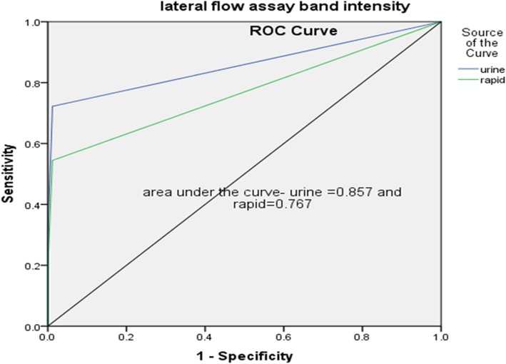
Receiver Operating Characteristics (ROC) analysis was performed for assessing the performances of the two tests. Accordingly, the area under ROC curve (AUC) of TB for TB LAM Ag test was 0.85 (95%CI = 0.79–0.91) while it was 0.76 (95%CI; =0.69–0.83) for the LIONEX Animal TB rapid blood test (Fig. 1).
Fig. 1.
Receiver Operating Characteristics (ROC) curve for TB LAM Ag and LIONEX Animal TB rapid blood test. The area under ROC curve (AUC) of TB for TB LAM Ag test was 0.85 (95%CI = 0.79–0.91) and for the LIONEX Animal TB rapid blood test was 0.76 (95%CI; =0.69–0.83)
Discussion
In this preliminary study, diagnostic performance of the TB LAM Ag urine test and LIONEX Animal TB Rapid blood test was evaluated using the combined result of SICTT and IFN-γ test as a gold standard.
The test results of each assay were compared with those of the standard tests i.e. with those of the SICTT or with those of the IFN-γ test [14]. There was high concordance between the TB LAM Ag test and that of SICTT. Furthermore, the agreement between TB LAM Ag and IFN-γ test was substantial. The sensitivity and specificity of the TB LAM Ag were 72 and 98.8%, respectively. A similar finding was reported by Lamont et al. a study with a similar design that was conducted in California [15]. The result of a meta-analysis published by Minion et al. on human subjects showed that the sensitivity of the TB LAM Ag test ranged from 13 to 93% while its specificity ranged from 87 to 99%. Thus, the sensitivity and specificity of TB LAM Ag recorded by the present study were within the ranges reported by Minion et al. Our findings are encouraging and could suggest the potential use of TB LAM Ag test for the diagnosis of bTB. Technically, a test such as the TB LAM Ag that uses urine samples is preferable to one that relies on blood or serum. Bacterial metabolism and degraded cell wall shed into body fluids could be detected by TB LAM Ag, which implies presence of active TB disease [16].
Diagnostic performance of TB LAM Ag from urine of bTB-infected animals has been studied by different groups [15, 17]. In experimentally infected animals, evaluation of LAM excretion [17] indicated that LAM is more frequently detected in the first 7 to 14 days post-infection and then decreases onward. Cross-reactivity of the TB LAM Ag was also reported with no significant changes. The ability of the test to differentiate bTB infected and not infected animals was as good as observed in experimentally and naturally infected bovine serum [15].
Additionally, test agreement between the SICTT and LIONEX Animal TB rapid blood test was substantial, while the test agreement between IFN-γ test and LIONEX Animal TB rapid blood test was moderate. Furthermore, the sensitivity and specificity of LIONEX Animal TB rapid blood test were 54 and 98.8%, respectively. Thus, according to the results of this preliminary study, both the sensitivity and agreement of LIONEX Animal TB rapid blood test with either SICTT or IFN-γ test was inferior to those of the TB LAM Ag. LIONEX Animal TB rapid is new and its field evaluation is still ongoing in different geographic areas like Gonder, Ethiopia and with milk and urine specimens from Michigan, USA (unpublished data). LIONEX TB animal rapid kit failed to identify many SICTT-positive animals and had relatively lower sensitivity compared to the other serological tests [18, 19].
Because of a number of limitations of SICTT, including cross reactivity with environmental mycobacteria, animal desensitization after severe infection, long test performance time, and interference with vaccination [10], there remains a need to identify tests with better performance characteristics.
Serological assays could be additional tools to detect animals that do not react to cell mediated immune based tests [20]. Such methods could aim for detecting either mycobacterial specific antigens that are an immunologically active component or possibly mycobacterial specific antibodies. In general, serological tests perform better during the latter stages of infection [21, 22] as the immune response to bTB shifts from cellular to humoral [22]. It has been indicated that specificity of serological tests is higher and minimizes the proportion of false positive reactions in non-infected animals [23].
Conclusion
In a low resource setting where infrastructure is limited, a rapid, field-deployable test which can improve diagnosis of bTB would allow for a decrease in zoonotic transmission and thereby improve public health. The TB LAM Ag had a better diagnostic performance than the LIONEX animal TB rapid blood test and could potentially be used as ancillary either to SICTT or IFN-γ test for the diagnosis of bTB. Additional studies in a real-time field setting to determine its utility and implementation capacity are needed. .
Methods
Study animals and settings
Animals
The study was conducted in central Ethiopia and 175 study animals were enrolled from different farms and divided into distinct groups: 1) group one, with 51 SICTTand IFN-γ positive animals recruited from the National Animal Health Diagnostic and Investigation Center (NAHDIC) in Sebeta; 2) group two, with 64 SICTT and/or IFN-γ-negative animals recruited from a farm located at Muke-Turi, in the North Shewa Zone of the Oromia Region; and 3) Group three: 60 animals with unknown status of bTB were recruited from Chancho and Sululta, North Shewa of the Oromia Region. Since there is no prevention and control strategy in Ethiopia for BTB at this time, the animals that tested positive were not removed from the farms. Farmers were provided with the results of tests as well as educational awareness in order to minimize the risk of transmission to other animals and human.
Single intradermal comparative tuberculin test (SICTT)
Two sites on one side of the mid-neck 12 cm apart were shaved and the skin thickness was measured with a caliper. One site was injected intradermally with an aliquot of 0.1 ml 3000 IU/ml bovine purified protein derivative (PPD) (Prionics Leslystad BV, Lelystad Netherland). The second site was injected with 0.1 ml 3000Ul/ml avian PPD (Prionics Leslystad BV, Lelystad Netherland). After 72 h the skin thickness at injection site was measured. An animal was defined as a reactor if the bovine site reaction exceeded the avian site reaction by > 4 mm at 72 h [24].
LIONEX animal TB rapid blood test
The animal TB rapid test is a lateral flow immunochromatography and membrane-based screening test for the rapid detection of antibodies to M. bovis in sample plasma or serum from animals, developed by LIONEX Diagnostic and Therapeutic GmbH Company, Germany [13]. Whole blood was collected into a heparinized tube and transported to the Aklilu Lemma Institute of Pathobiology immunology lab. Plasma was harvested after centrifugation at 1500 rpm for 10 min, and then the samples were stored at -200c until processed.
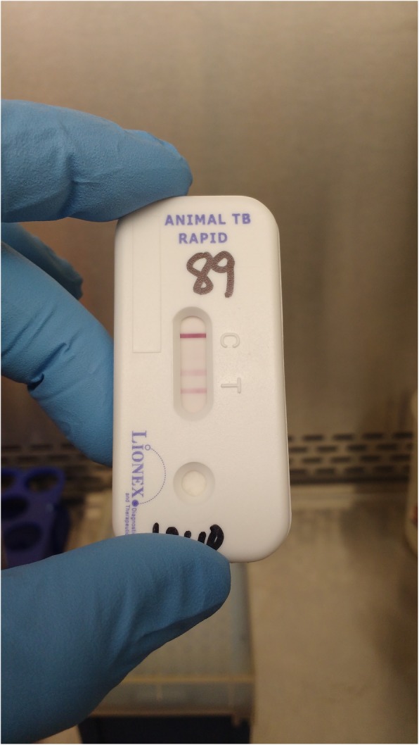
On the test day the kit and the plasma samples were thawed, after which one drop (about 20 μl) of plasma was added to the designated test kit pad area. Two drops of the diluent buffer were added to the pad, mixed, and then kept at room temperature for 5 min. Next, 1 drop of diluent buffer was added to the solution and kept at room temperature for an additional 20 min, at which point the result was read. Interpretation of the result was based on manufacturer instructions, i.e., the test was considered positive when bands were observed at control bar C and sample bar T and negative if a band was observed only at control bar C. Intensity of the test band was graded per grading intensity scales supplied by the manufacturer: Zero for no band, 1 for faint band intensity, 2 for moderate intensity and 3 for strong test band intensity (Fig. 2).
Fig. 2.

LIONEX Animal TB Rapid Blood Test. Positive LIONEX Animal TB rapid blood test is shown in the figure: One strong line appear in the control zone “C” and one or more test lines appear in the test zone “T”
ALERE determine LAM TB urine antigen test
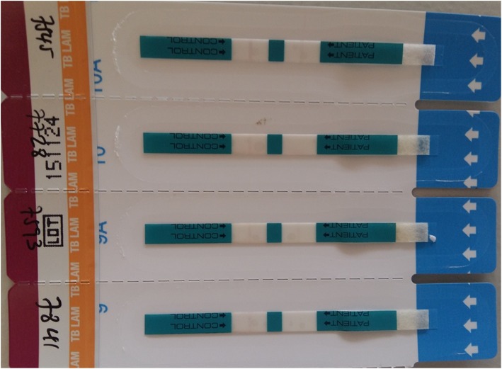
TB LAM Ag is an immunochromatographic test for the qualitative detection of LAM antigen of mycobacteria in urine, based on the use of highly purified antibodies specific to LAM (Fig. 3) [11]. The capture antibodies are adsorbed onto the nitrocellulose membrane of the test strip. This strip was originally designed for human TB detection, and we evaluated the assay for bTB infection diagnosis.
Fig. 3.
ALERE Determine LAM TB Urine Antigen Test. The TB LAM antigen urine test has Control Zone and Patient Zone. If a band is visualized in the Control zone and the patient zone, then the test is positive. If a band is visualized in the Control zone but no band is seen in the patient zone, then the test is negative
Urine samples were collected after manual stimulation of the vulva area and transported within 8 h of collection to the Aklilu Lemma Institute of Pathobiology (immunology laboratory). After removal of the protective foil cover from each TB LAM Ag test strip, 60 μl urine was added onto the sample pad on the test strip and kept at room temperature for at least 25 min to read the result. All test results were interpreted according to manufacturer instructions. Band intensity was graded per the manufacturer-supplied reference card: Zero for no band, 1 for faint band intensity, 2 for moderate band intensity, and 3 for strong test band intensity.
INF-γ test and whole blood culture
A 3 ml blood sample from each animal was collected into a heparinized tube after shaving and disinfecting the groove of the animal’s neck. All collected blood was transported to the Aklilu Lemma Institute of Pathobiology (ALIPB, immunology laboratory) within 8 h of collection. About 250 μl of whole blood was dispensed in duplicate into 96-well flat-bottom culture plates. Whole blood was stimulated using 25 μl of Avian-PPD and Bovine-PPD, giving a final assay concentration of 10 μg/ml in each well. Lectin from Phytolacca americana (pokeweed, Sigma) (5 μg/ml) was used as a positive control, and saline was used as a negative control, each dispensed at 25 μl into the corresponding wells. The culture was incubated at 37 °C in 5% CO2 atmosphere for 48 h and then the supernatant was harvested and frozen. IFN-γ in the supernatants were measured by an enzyme-linked immunosorbent assay using the Bovigam® test kit as per product insert (Prionics AG, Schlieren, Switzerland) [25]. The result was interpreted as per the manufacturer recommendation as positive, negative with the cut-off value of IFN- γ ≥0.1.
Data analysis
Test sensitivity (proportion of positive results found among infected animals), specificity (proportion of negative results among non- infected animals), positive predictive value (PPV) and accuracy (proportion of correct result among all tested animals) and their 95% confidence intervals (CI) were calculated using open Agreement between tests were evaluated using Kappa-statistic (k) using SPSS statistic version 24.
Acknowledgements
The work was done by the support of Aklilu Lemma Institute of Pathobiology, Addis Ababa University and The Ohio State University.
Abbreviations
- Ag
Antigen
- bTB
Bovine tuberculosis
- HIV/AIDS
Human immunodeficiency virus; acquired immunodeficiency syndrome
- IFN-ɤ
Gamma interferon
- LAM
Lipoarabinomannan
- LMIC
Low- and middle- income countries
- PPD
Purified protein derivative
- SICTT
Single intradermal comparative tuberculin test
- TBC
Mycobacterium tuberculosis complex
Authors’ contributions
GA, SHW, and AZ conceived the current study protocol. WGH, XP, JMB, WG, JBT, SHW developed the initial rapid diagnostic testing proposal. AZ, TM, LT collected the specimen and performed all of the tests. AZ analyzed the initial data and drafted the initial manuscript. AZ, TM, LT, WGH, XP, JMB, WG, JBT, SHW, GA have all reviewed and have provided critical input for the manuscript. All authors read and approved the final manuscript.
Funding
The project described was supported in part by Award Number UL1TR001070 from the National Center for Advancing Translational Sciences awarded to The Ohio State University (SW, GW, JBT, XP, GH, JB). Funding obtained was used for the purchase of LIONEX Animal TB Rapid Blood Test and ALERE Determine LAM TB Urine Antigen Test used in this study. The content is solely the responsibility of the authors and does not necessarily represent the official views of the National Center for Advancing Translational Sciences or the National Institutes of Health. The funding body did not participate in the design of the study, the specimen collection, analysis, and interpretation of data or any involvement in the writing of the manuscript.
Availability of data and materials
The data used for analysis to support this study are mostly included in the published article. Additional data can be available from corresponding author on reasonable request.
Ethics approval and consent to participate
Ethical clearance: The study was reviewed and approved by the Institutional review Board of the Aklilu Lemma Institute of Pathobiology by its Minutes Ref. No. ALIPB/002/2017/2018. The aim of the study was explained to farm owners and conducted in farms where owners consented verbally and permitted access to their farms and animals. The Addis Ababa Institutional Ethics Committee approved verbal consent for animal testing for this study protocol.
Consent for publication
Not applicable.
Competing interests
The authors declare that they have no competing interests.
Footnotes
Publisher’s Note
Springer Nature remains neutral with regard to jurisdictional claims in published maps and institutional affiliations.
Shu-Hua Wang and Gobena Ameni are contributed as senior authors.
Contributor Information
Aboma Zewude, Email: akoyeroon@yahoo.com.
Temesgen Mohammed, Email: lematd63@gmail.com.
Lemma Terfassa, Email: temsgenmoh@yahoo.com.
W. Garrett Hunt, Email: W.Garrett.Hunt@nationwidechildrens.org.
Xueliang Pan, Email: Jeff.Pan@osumc.edu.
Joan Miquel Balada-Llasat, Email: Joan-Miquel.Balada-Llasat@osumc.edu.
Wondwossen Gebreyes, Email: gebreyes.1@osu.edu.
Jordi B. Torrelles, Email: jtorrelles@txbiomed.org
Shu-Hua Wang, Email: Shu-Hua.Wang@osumc.edu.
Gobena Ameni, Email: gobena.ameni@aau.edu.et.
References
- 1.Adem J. Review on zoonotic importance of bovine tuberculosis and its control. Open Access Libr J. 2016;3:e2504. [Google Scholar]
- 2.Schiller I, Oesch B, Vordermeier HM, Palmer MV, Harris BN, Orloski KA, et al. Bovine tuberculosis: a review of current and emerging diagnostic techniques in view of their relevance for disease control and eradication. Transbound Emerg Dis. 2010;57(4):205–220. doi: 10.1111/j.1865-1682.2010.01148.x. [DOI] [PubMed] [Google Scholar]
- 3.Cosivi O, Grange JM, Daborn CJ, Raviglione MC, Fujikura T, Cousins D, et al. Zoonotic tuberculosis due to Mycobacterium bovis in developing countries. Emerg Infect Dis. 1998;4(1):59–70. doi: 10.3201/eid0401.980108. [DOI] [PMC free article] [PubMed] [Google Scholar]
- 4.Berg S, Firdessa R, Habtamu M, Gadisa E, Mengistu A, Yamuah L, et al. The burden of mycobacterial disease in ethiopian cattle: implications for public health. PLoS One. 2009;4(4):e5068. doi: 10.1371/journal.pone.0005068. [DOI] [PMC free article] [PubMed] [Google Scholar]
- 5.Sibhat B, Asmare K, Demissie K, Ayelet G, Mamo G, Ameni G. Bovine tuberculosis in Ethiopia: a systematic review and meta-analysis. Prev Vet Med. 2017;147:149–157. doi: 10.1016/j.prevetmed.2017.09.006. [DOI] [PMC free article] [PubMed] [Google Scholar]
- 6.Alito A, McNair J, Girvin RM, Zumarraga M, Bigi F, Pollock JM, et al. Identification of Mycobacterium bovis antigens by analysis of bovine T-cell responses after infection with a virulent strain. Braz J Med Biol Res. 2003;36(11):1523–1531. doi: 10.1590/S0100-879X2003001100011. [DOI] [PubMed] [Google Scholar]
- 7.Buddle BM, Wilson T, Denis M, Greenwald R, Esfandiari J, Lyashchenko KP, et al. Sensitivity, specificity, and confounding factors of novel serological tests used for the rapid diagnosis of bovine tuberculosis in farmed red deer (Cervus elaphus) Clin Vaccine Immunol. 2010;17(4):626–630. doi: 10.1128/CVI.00010-10. [DOI] [PMC free article] [PubMed] [Google Scholar]
- 8.Coad M, Downs SH, Durr PA, Clifton-Hadley RS, Hewinson RG, Vordermeier HM, et al. Blood-based assays to detect Mycobacterium bovis-infected cattle missed by tuberculin skin testing. Vet Rec. 2008;162(12):382–384. doi: 10.1136/vr.162.12.382. [DOI] [PubMed] [Google Scholar]
- 9.Muller B, Vounatsou P, Ngandolo BN, Diguimbaye-Djaibe C, Schiller I, Marg-Haufe B, et al. Bayesian receiver operating characteristic estimation of multiple tests for diagnosis of bovine tuberculosis in Chadian cattle. PLoS One. 2009;4(12):e8215. doi: 10.1371/journal.pone.0008215. [DOI] [PMC free article] [PubMed] [Google Scholar]
- 10.Rua-Domenech R, Goodchild AT, Vordermeier HM, Hewinson RG, Christiansen KH, Clifton-Hadley RS. Ante mortem diagnosis of tuberculosis in cattle: a review of the tuberculin tests, gamma-interferon assay and other ancillary diagnostic techniques. Res Vet Sci. 2006;81(2):190–210. doi: 10.1016/j.rvsc.2005.11.005. [DOI] [PubMed] [Google Scholar]
- 11.Alere Determine Product Information: https://www.alere.com/en/home/product-details/determine-tb-lam.html REF Type: Online source. Accessed 14 Oct 2019.
- 12.Nicol MP, Allen V, Workman L, Isaacs W, Munro J, Pienaar S, et al. Urine lipoarabinomannan testing for diagnosis of pulmonary tuberculosis in children: a prospective study. Lancet Glob Health. 2014;2(5):e278–e284. doi: 10.1016/S2214-109X(14)70195-0. [DOI] [PMC free article] [PubMed] [Google Scholar]
- 13.Lionex, Lionex Diagnosis and Therapeutics: Lionex LIO-Detect-TB test. www.lionex.de. REF Type: Online source. Accessed 14 Oct 2019.
- 14.Engel, Backer J, Buist W. Evaluation of the accuracy of diagnostic tests from repeated measurements without a gold standard. J Agric Biol Environ Stat. 2010;15(1):83–100. doi: 10.1007/s13253-009-0013-y. [DOI] [Google Scholar]
- 15.Lamont EA, Ribeiro-Lima J, Waters WR, Thacker T, Sreevatsan S. Mannosylated lipoarabinomannan in serum as a biomarker candidate for subclinical bovine tuberculosis. BMC Res Notes. 2014;7:559. doi: 10.1186/1756-0500-7-559. [DOI] [PMC free article] [PubMed] [Google Scholar]
- 16.Fukuda T, Matsumura T, Ato M, Hamasaki M, Nishiuchi Y, Murakami Y, et al. Critical roles for lipomannan and lipoarabinomannan in cell wall integrity of mycobacteria and pathogenesis of tuberculosis. MBio. 2013;4(1):e00472–e00412. doi: 10.1128/mBio.00472-12. [DOI] [PMC free article] [PubMed] [Google Scholar]
- 17.Vu DM, Sakamuri RM, Waters WR, Swanson BI, Mukundan H. Detection of lipomannan in cattle infected with bovine tuberculosis. Anal Sci. 2017;33(4):457–460. doi: 10.2116/analsci.33.457. [DOI] [PubMed] [Google Scholar]
- 18.Green LR, Jones CC, Sherwood AL, Garkavi IV, Cangelosi GA, Thacker TC, et al. Single-antigen serological testing for bovine tuberculosis. Clin Vaccine Immunol. 2009;16(9):1309–1313. doi: 10.1128/CVI.00028-09. [DOI] [PMC free article] [PubMed] [Google Scholar]
- 19.O'Brien A, Whelan C, Clarke JB, Hayton A, Watt NJ, Harkiss GD. Serological analysis of tuberculosis in goats by use of the enferplex caprine TB multiplex test. Clin Vaccine Immunol. 2017;24(2). 10.1128/CVI.00518-16. [DOI] [PMC free article] [PubMed]
- 20.Ramos DF, Silva PE, Dellagostin OA. Diagnosis of bovine tuberculosis: review of main techniques. Braz J Biol. 2015;75(4):830–837. doi: 10.1590/1519-6984.23613. [DOI] [PubMed] [Google Scholar]
- 21.Sada E, Brennan PJ, Herrera T, Torres M. Evaluation of lipoarabinomannan for the serological diagnosis of tuberculosis. J Clin Microbiol. 1990;28(12):2587–2590. doi: 10.1128/jcm.28.12.2587-2590.1990. [DOI] [PMC free article] [PubMed] [Google Scholar]
- 22.Welsh MD, Cunningham RT, Corbett DM, Girvin RM, McNair J, Skuce RA, et al. Influence of pathological progression on the balance between cellular and humoral immune responses in bovine tuberculosis. Immunology. 2005;114(1):101–111. doi: 10.1111/j.1365-2567.2004.02003.x. [DOI] [PMC free article] [PubMed] [Google Scholar]
- 23.Casal C, Infantes JA, Risalde MA, Diez-Guerrier A, Dominguez M, Moreno I, et al. Antibody detection tests improve the sensitivity of tuberculosis diagnosis in cattle. Res Vet Sci. 2017;112:214–221. doi: 10.1016/j.rvsc.2017.05.012. [DOI] [PubMed] [Google Scholar]
- 24.OIE . Manual of diagnostic tests and vaccines for terrestrial animals 2009. Paris: World Organiation for Animal Health 2009; 2008. [Google Scholar]
- 25.A. Biosystems, BOVIGAM TB KIT-https://assets.thermofisher.com/TFS-Assets/GSD/Flyers/bovigam-tb-flyer.pdf Applied Biosystems, 2019. Online source: Accessed 14 Oct 2019.
Associated Data
This section collects any data citations, data availability statements, or supplementary materials included in this article.
Data Availability Statement
The data used for analysis to support this study are mostly included in the published article. Additional data can be available from corresponding author on reasonable request.


