

Abstract
Analyses in the clinical area need quick and reliable analytical methods and devices. For this purpose, biosensors can be a suitable option, whereas they are constructed to be simple for use, specific for the target analyte, capable of continuous monitoring and giving quick results, potentially low‐costing and portable. In this article, we describe electrochemical biosensors developed for clinical diagnosis, namely for glucose, lactate, cholesterol, urea, creatinine, DNA, antigens, antibodies, and cancer markers assays. Chosen biosensors showed desirable sensitivity, selectivity, and potential for application on real samples. They are often designed to avoid interference with undesired components present in the monitored systems.
Keywords: amperometric, analysis, biosensor, clinical diagnosis, enzyme
INTRODUCTION
Today's situation in clinical diagnostics and monitoring requires rapid and accurate analyses. Very important factors complicating these procedures are prices of analytical devices and expenses for whole measurements. In addition, if we consider that a qualified personnel is needed for clinical analyses, the natural necessity for alternative analytical technologies is present. For this purpose, biosensors can serve as an option for solving problems mentioned before, or become a helpful tool at least 1, 2.
Kissinger has defined a biosensor as a device where a biological recognition element is built in (physically attached or confined) and is the primary selectivity element (Fig. 1). Many sensors used for biological purposes are therefore not biosensors, including those for temperature, pressure, electrocardiograms, pH, Ca2+, catecholamines and the like 3. As a biorecognition system, an enzyme, antibody, DNA, microorganism, etc., can be used. These are capable of recognizing their specific analytes and also regulate the specificity and sensitivity of the device. Numerous techniques of immobilization, such as covalent linkage, physical adsorption, cross‐linking, encapsulation, and entrapment, have been known for stabilization of enzymes or other components for the development of biosensing devices 4. Biosensors existing today use several types of transducers for converting a biochemical event resulting from the interaction between the bioreceptor molecule and analyte into measurable signal. These are mainly electrochemical including amperometric, conductometric, and potentiometric. The basic principle for this class of biosensors is that chemical reactions between immobilized biomolecule and target analyte produce or consume ions or electrons which cause some change in the measurable electrical properties of the solution, such an electric current, conductance, potential, and ionic strength. The second class is optical transducers utilizing optical fibers based on fluorescence or optical diffraction, when measured signal is light 5. Other class, thermometric biosensors, is constructed by combining immobilized biomolecules with temperature sensors measuring change in heat reaction 6. The mass‐sensitive one is based on the coupling of the biological recognition element with a piezoelectric component, usually a quartz‐crystal coated with gold electrodes. These vibrate at a specific frequency with the application of an electrical signal of a specific frequency. The frequency of oscillation is therefore dependent on the electrical frequency applied to the crystal as well as the crystal's mass. Therefore, when the mass increases due to binding of chemicals, the oscillation frequency of the crystal changes and this can be measured electrically to determine the additional mass of the crystal 7, 8, 9. Other type of a mass‐sensitive biosensor is a microcantilever. This device can be used as a physical, chemical, or biological sensor for detection of changes in cantilever bending or vibrational frequency. The main principle of detection is the transduction of molecular adsorption and specific molecular interactions on a cantilever surface into the mechanical response change of a cantilever. Viscosity, density, and flow rate can be measured by detecting changes in the vibrational frequency. The most commonly used class is electrochemical biosensors 10, 11. The main advantages of well‐constructed biosensors are their selectivity, rapid response, low time‐consumption during analysis, easy handling, and lower price of analysis. During developing process of biosensors for commercial use, many obstacles have to be overcomed, such as unwilling interferences, instability of biological components, insufficient reproducibility, or an accuracy of results. Last but not least it is very important to make it the simplest for users. In this review, we describe biosensors for medical and diagnostic application for notable parameters.
Figure 1.

Working principle of an amperometric biosensor. Analyte present in a sample reacts with a bioelement immobilized on the working electrode. Biochemical event results in the current change, which is proportional to the analyte concentration.
BIOSENSORS FOR IMPORTANT MARKERS ANALYSIS
Glucose
Glucose concentration is a crucial indicator in many diseases, such as diabetes and other endocrine metabolic disorders. Also, blood glucose is the most common analyte measured after electrolytes and blood gases 4. Glucose fluctuations within the normal physiological range of 110 ± 25 mg/dL (around 6 μM) are considered to be acceptable while diabetics may reach values of 360 mg/dL (20 μM) or higher 12. Since diabetic patients need to control their blood glucose levels carefully, the importance of self‐monitoring of blood glucose represent an effective method of measuring blood sugar not only in clinics but also at home and in the working place 13.
Glucose biosensor is a first developed biosensor ever. Clark, known as the father of the biosensor concept, described in 1962 an experiment in which glucose oxidase (GOX) was entrapped at a Clark oxygen electrode using dialysis membrane 14. The majority of glucose biosensors are amperometric based on a reaction in which the oxidized form of GOX reacts with ‐glucose and produces ‐gluconic acid and reduced GOX, involving two electrons and two protons. This reaction also consumes oxygen, thus forms hydrogen peroxide and oxidized GOX, so depletion of oxygen or produced hydrogen peroxide can be measured 15.
| (1) |
In other system, a natural substrate oxygen is replaced by a small redox molecule, which serve as the redox mediator and exchange electrons between electrodes and enzymes. Various soluble redox molecules, such as ferricyanide, ferrocene, thionine, methylene blue, methyl viologen, or methazolium sulfate, were used to improve the sensor performance. In case of commercial glucose biosensors, most of them are based on the following reaction:
| (2) |
However, the response of these biosensors is susceptible to oxygen concentration in the measuring media and in case of utilizing artificial mediator increasing in the O2 concentration can leads to a decrease in the signal and underestimation of the glucose levels 16. Moreover, if the detection is based on measuring the H2O2 level, the oxidation of H2O2 often requires higher working potential (usually over +600 mV vs. standard electrode) and thus many other electroactive compounds commonly present in biological fluids can also be oxidized and cause false current response. Many articles describe biosensors utilizing GOX with novel methods, for example Li et al. immobilized GOX on TiO2/SiO2 nanocomposite. The detection of glucose may be accomplished by monitoring the formation of hydrogen peroxide. Usage of a 96‐hole polyporous plate accessory of fluorescence spectrophotometer led to linear response to glucose concentrations ranging from 1 nM to 100 mM with a detection limit of 0.12 nM. The TiO2/SiO2 nanocomposite can be used as both a supporting material and a signal transducer. The biosensor has been successfully applied to the determination of glucose in human blood serum 17. Biosensor with a lifetime at least 520 days has been constructed by immobilization of GOX within poly(dimethylamino) ethyl methacrylate microparticles. It was found that the best biosensor response corresponds to microparticles synthesized with 1.19 M monomer and 0.37% cross‐linking reagent content. The biosensor was used to analyze glucose in human serum samples with satisfactory results 18. Wu et al. prepared the bionanocomposite film consisting of GOX/Pt/functional graphene sheets/chitosan (GOX/Pt/FGS/chitosan) for glucose sensing. With the electrocatalytic synergy of FGS and Pt nanoparticles to hydrogen peroxide, a sensitive biosensor with a detection limit of 0.6 μM glucose was achieved. This system represents new opportunity for clinical diagnosis and point‐of‐care applications 19.
Hsu et al. evaluated the long‐term stability of glucose strips made of barrel plating gold electrodes. This biosensor was fabricated by inserting two barrel plating gold electrodes onto an injection‐molding plastic base followed by immobilizing with a bio‐reagent layer and membrane. This concept showed superior stability up to 2.5 years at 25°C. Results indicated that the GOX/barrel plating gold biosensing platform is suitable for long‐term accurate glycemic control 20. Other article reports the advantages of incorporating an iron in a carbon paste electrode containing GOX. The catalytic activity of iron nanoparticles toward the reduction of hydrogen peroxide has made possible the quantification of glucose at a very low potential of −100 mV (usually it requires a potential of +600 mV) and thus avoiding, in this way, the interference of easily oxidizable compounds like ascorbic acid and uric acid without the need of redox mediators or permselective membranes. Good correlation with the spectrophotometric methods when determining glucose in human blood serum was achieved 21. Other interesting concepts for glucose biosensors used for clinical diagnostics were reviewed by Malhotra and Chaubey 4 and Yoo and Lee 22. The detailed review article summarizing available commercial glucose biosensors was written by Newman and Turner 23.
Amperometric biosensors based on nicotinamide‐adenine‐dinucleotide‐dependent dehydrogenases can be employed to overcome the problem present in case of GOX‐based biosensors 24, but the necessity for a free diffusing coenzyme NAD+ handicap this concept. Suitable candidates for the construction of biosensors, which do not require O2 as an electron acceptor or NAD+ as a coenzyme, are pyrroloquinoline quinone‐(PQQ‐) 25 or FAD‐dependent glucose dehydrogenase (GDH‐FAD) 26.
| (3) |
Utilizing GDH‐PQQ in case of biosensors designed for commercial use is complicated by the fact that GDH‐PQQ isolated from outer surface of the cytoplas‐mic membrane requires suitable detergents for solubili‐zation 27. On the other hand, water‐soluble GDH‐PQQ present in cell interior has low specificity and it can oxidize various saccharides, such as mannose, maltose, lactose etc., which can cause interferences 26. Hence, the most suitable concept seems to be a biosensor utilizing GDH‐FAD based on the reaction 3. Glucose biosensors found massive use in the real diagnostics and they represent about 90% of the biosensor market. Examples of various commercial biosensors are given in Table 1.
Table 1.
Examples of commercial biosensor based systems
| Analyte | Company | Models |
|---|---|---|
| Glucose | Roche | Series Accu‐Chek (Active, Advantage, Aviva, Compact Plus) |
| Bayer | Contour, Didget, Breeze2, Ascensia ENTRUST | |
| LifeScan/Johnson&Johnson | Series OneTouch (Basic, FastTake, SureStep, Ultra, UltraMini, UltraSmart) | |
| Abbott | Series FreeStyle (Lite and Freedom Lite) | |
| BST Bio Sensor Technology | GLUKOMETERPRO | |
| A.Menarini Diagnostics | GlucoCard, GlucoMen, GlucoFix | |
| Arkray | Series GLUCOCARD (01, 01‐mini, X‐METER, Vital) | |
| ACON Diabetes Care | On‐Call Plus | |
| Infopia Co. Ltd. | Finetest, EasyGluco, GlucoLab 5 sec | |
| Hypoguard | Hypoguard quick tek glucose meter | |
| NIPRO diagnostics | series TRUE (2go,result, track, balance, read) | |
| National Diagnostic Products | BETACHECK G5 | |
| Nova Biomedical | Nova Max, Nova Max Link | |
| Diabetic Supply of Suncoast | Advocate, Advocate Duo, Advocat Redicode | |
| Prodigy Diabetes Care | Series Prodigy (Autocode meter, Pocket, Voice meter) | |
| ReliOn | Confirm, Micro, Ultima | |
| Entra Health Systems | MyGlucoHealth Meter | |
| U.S. Diagnostics | Maxima, EasyGluco, Control AST, Infinity, Acura | |
| AgaMatrix | KeyNote, Presto, Jazz | |
| Glucose/Ketone | Abbott | Precision Xtra |
| Nova Biomedical | StatStrip Glucose/Ketone, Statstrip Xpress Glucose/Ketone | |
| YSI Life Sciences | YSI 2300 STAT Plus Glucose & Lactate Analyzer | |
| Glycated hemoglobin | Bayer | A1CNOW SELFCHECK |
| Lactate | BST Bio Sensor Technology | LACPRO |
| Arkray | Lactate Pro LT‐1710 | |
| YSI Life Sciences | YSI 1500 SPORT Lactate Analyzer | |
| nova biomedical | StatStrip Lactate, StatStrip Lactate Xpress | |
| Roche | Accutrend Lactate | |
| Cholesterol, Glucose, Triglyceride, Lactate | Roche | Accutrend Plus System |
| Total Cholesterol, HDL‐Cholesterol, Triglyceride | Infopia Co. Ltd. | LipidPro |
| HealthCheckSystems | CardioChek | |
| Creatinine | Nova Biomedical | StatSensor Creatinine, StatSensor Xpress Creatinine |
Lactate
Its determination is helpful in the monitoring of respiratory insufficiency, shocks, heart failure, and metabolic disorders 28. Any change or increase in blood lactate is an indication of surviving capability 29. Lactic acidosis is known to accompany decreased tissue oxygenation, left ventricular failure, and drug toxicity 30. Most lactate amperometric biosensors reported in literature are based on immobilized lactate dehydrogenase (LDH) 31, 32 or lactate oxidase (LOX) 33, 34.
| (4) |
| (5) |
The multiwalled carbon nanotubes (MWCNTs) and sol–gel film was used to immobilize LOX on the surface of glassy carbon electrode (GCE). Analytical characteristics and dynamic parameters of the biosensors with and without MWCNTs in the hybrid film showed that performance of the biosensor could be improved greatly after introduction of the MWCNTs. The biosensor without MWCNTs and with MWCNTs showed a linear range of 0.3–1.5 mM and 0.2–2.0 mM, respectively. This biosensor has been used to determine the ‐lactate concentration in real human blood samples 35. Dispersed platinum nanoparticles (Ptnano) on the surface of the GCE were used in combination with MWCNTs for fabricating electrochemical lactate biosensor. The resulting LOX/MWCNTs/Ptnano electrode was covered by a thin layer of silica sol–gel to avoid the loss of LOX in determination and to improve the anti‐interferent ability. The biosensor showed a large determination range (0.2–2.0 mM) and a short response time (within 5 sec). The prepared biosensor had practically good selectivity against interferences. The results for blood samples showed a good agreement with those measured by spectrophotometric method 36. Seeing that normal lactate value in a human body ranges from 0.5 to 2.2 mM/L 37, the higher level indicating lactic acidosis would not be determined without dilution of sample using these biosensors considering their linear range. However, dilution of whole blood is not an option due to unpredictable hematocrit effects. Dilution of serum does not pose this problem. The high end of the linear range is more important than the lower limit of detection and the mentioned biosensors are not optimal for clinical application. However, the upper limit of the linear range can be increased by an introduction of a diffusion barrier, such as outer membrane or a polymer film 38.
MWCNTs were also used for the construction of amperometric biosensor based on LDH and Meldola's blue (MB). The amperometric response was based on the electrocatalytical properties of MB to oxidize NADH2, which was generated in the enzymatic reaction of lactate with NAD+ under catalysis of LDH. The biosensor showed linear response range from 0.1 to 10 mM. These characteristics allowed its application for direct measurements of lactate in blood samples. Results were in good agreement with other analytical method 39. Cui et al. nanoengineered chitosan/polyvinylimidazole‐Osmium (PVI‐Os)/carbon nanotube/LOX network on gold electrode for detection of lactate. Positively charged chitosan and PVI‐Os were used as the matrix and the mediator to immobilize the negatively charged LOX. This network can be extended to other enzyme biosensors, and to have potential applications in diagnostics, life science and food analysis 40, but it is questionable whether using carbon nanotubes is suitable for mass production considering their cost and complicated biosensor construction.
Other lactate biosensor was developed through the immobilization of LOX in an albumin and mucin composed hydrogel. The performance of the biosensor was evaluated in matrixes with different amounts of albumin, mucin, and glutaraldehyde as a crosslinking reagent. The detection limit calculated from the signal‐to‐noise ratio was 0.7 μM. Only 0.1 units of enzyme was used for this biosensor, which markedly reduces fabrication expenses. High reproducibility in the response was obtained, but biosensor has to be tested for interferences in real samples such as serum and blood 33. Suman et al. immobilized LOX through glutar‐aldehyde coupling onto polyaniline‐co‐fluoroaniline film deposited on an Indium tin oxide (ITO) coated glass plate. The biosensor showed a linear range from 0.1 to 5.5 mM with the minimum detection limit of 0.1 mM. This electrode was used for the determination of lactate in serum. Among the various serum substances tested, only 8‐hydroxyquinoline, urea, ammonium molybdate, and uric acid caused 64, 38, 34, and 31% inhibition, respectively 41. Liu et al. developed biosensor for the analysis of ‐lactate in athletes serum samples. The biosensor was fabricated with gold thin‐film two‐electrode system and was modified with platinum‐black nanoparticles and ferricyanide mediator. A wide linear range was achieved from 1 to 20 mM lactate with fast detection time of 50 sec. The biosensor was successfully applied for the determination of ‐lactate in serum samples without dilution 42. Romero et al. developed an amperometric lactate biosensor which required only 0.2U of LOX. Enzyme was immobilized in a mucin/ albumin hydrogel matrix. By protecting the platinum surface with a Nafion membrane, typical interferences have been minimized to practically undetectable levels. Electrochemical properties associated with the Nafion membrane are assessed as a function of Nafion concentration. The lactate biosensor presented remarkable operational stability and sensitivity 0.537±0.007 mA/M with a detection limit of 0.8 μM. The biosensor kept the same sensitivity for 5 months, while the linear range decreases until an upper value of 0.8 μM was reached. Assays performed with whole blood samples spiked with 100 μM lactate gave (89 ± 6)% of recovery 43. In other interesting concept all reagents, i.e., luminol, LOX, BSA, electrolyte and buffer immobilized by a Methocel superTM membrane were placed on the working electrode of the screen‐printed electrochemical cell. The measurement of the electrochemiluminescence was made via a photocounting head when sample was placed into the cell with a circular container containing the disposable sensing membrane. The disposable biosensor responds to lactate after 20 sec. The biosensor showed a dynamic range from 100 to 500 μM with a detection limit of 5 μM and a sensor‐to‐sensor repeatability, as relative standard deviation of 3.30% at the medium level of the range. Presented biosensor was applied to the analysis of lactate in human saliva. The procedure was validated for use in human saliva, comparing the results against an enzymatic reference procedure. In case of two samples, the correlation was above 99%, but in the third sample it was only 82%, so more extensive analysis of real samples would be valuable to confirm accuracy of the presented biosensor 44.
Cholesterol
The alarming increase in the rate of clinical disorders such as heart disease, hypertension, coronary artery disease, arteriosclerosis cerebral thrombosis, etc., due to high levels of cholesterol in blood has increased public concern about the determination of cholesterol level in the world 45. Keeping cholesterol level in optimal values (up to 5 mM) can reduce the risk of a heart attack or stroke.
In the construction of a cholesterol biosensor, cholesterol oxidase (ChOX) is the most commonly used enzyme.
| (6) |
Amperometric cholesterol biosensors utilizing ChOX immobilized in a Prussian blue (PB)/polypyrrole (PPy) composite film with the detection limit of 8 μM and linear range 0.025–0.3 mM were published and lifetime up to 25 days of use 46. Umar et al. fabricated cholesterol amperometric biosensor based on the immobilization of ChOX onto the ZnO nanoparticles. Achieved detection limit was 0.37 nM, response time less than 5 s, and linear range from 1.0 to 500.0 nM 47. The similar system was also used in other article from Umar et al. In this case, ChOX was immobilized on well‐crystallized flower‐shaped ZnO structures composed of hexagonal‐shaped ZnO nanorods grown by low‐temperature simple solution process. The fabricated cholesterol biosensors reported a very high and reproducible sensitivity of 61.7 μA/μM cm2 with a response time less than 5 sec and detection limit (based on S/N ratio) of 0.012 μM. The biosensor exhibited a linear dynamic range from 1.0 to 15.0 μM. However, experiments with real samples were not performed 48. The disadvantage of presented biosensors is their low linear range and thus they are not suitable for the analysis of real samples.
Israr et al. constructed a potentiometric biosensor based on ZnO nanorods. Hexagon‐shaped ZnO nanor‐ods were directly grown on a silver wire having a diameter of 250 μM using low temperature aqueous chemical approach that produced ZnO nanorods with a diameter of 125–250 nm and a length of ∼1 μM. ChOX was immobilized by a physical adsorption method onto ZnO nanorods. The electrochemical response of the ChOX/ZnO/Ag biosensor against a standard reference electrode (Ag/AgCl) was investigated as a logarithmic function of the cholesterol concentration (1 μM to 10 mM) showing good linearity with a sensitivity of 35.2 mV per decade and the stable output signal was obtained at around 10 sec 49.
In other work, cholesterol biosensor was constructed by co‐immobilizing cholesterol esterase (ChE) and ChOX in polyvinyl membrane and by mounting over the sensing part of oxygen electrode. The detection limit of the cholesterol was 50 mg/dL. A good correlation was found among cholesterol values obtained by colorimetric, enzymatic‐kit, and autoanalyzer method. The shelf life of the biosensor was more than three months at 4°C and enzyme membrane can be re‐used 20 times without any significant loss in enzyme activity 50. Shih et al. developed a prototype chronoamperometric biosensor consisting of a homemade potentiostat and disposable strips immobilized with Fe3O4, ChOX, and ChE. The co‐immobilization of ChE and ChOX allows the sensor to detect both concentrations of esterified and free cholesterol (reaction 7). The sensing device displays a linear response over the range of 100–400 mg/dL for cholesteryl oleate. No significant interferences were observed 51.
| (7) |
Aravind et al. reported a cholesterol biosensor using graphene nanoplatelets by gold nanoparticles. Thermally exfoliated graphene nanoplatelets acted as a suitable support for the deposition of Au nanoparticles. Cholesterol biosensor electrodes have been constructed with nafion solubilized functionalized graphene nanoplatelets (f‐G) as well as Au nanoparticles decorated f‐G, immobilized over GCE. f‐G and Au/f‐G thin film deposited GCEs were further functionalized with ChOX by physical adsorption. Au nanoparticles dispersed over f‐G demonstrate the ability to substantially raise the response current. The fabricated Au/f‐G based cholesterol biosensor exhibits sensitivity of 314 nA/μM cm2 for the detection of cholesterol with a linear response up to 135 μM. Furthermore, it has been observed that the biosensor exhibits a good anti‐interference ability and favorable stability over a month's period 52.
Tsai et al. electrochemically deposited platinum nanoparticles in MWCNT–chitosan matrix by a cyclic voltammetry method. The influence of enzyme loading within the MWCNT–chitosan–Pt–cholesterol oxidase nanobiocomposite was explored to optimize the electro‐analytical performance of the cholesterol biosensor. Additions of 100 μM glucose and 1 μM ascorbic acid to 100 μM cholesterol had no influence. The prepared cholesterol biosensor retained 60% of initial activity after 7 days when stored in 0.1 M phosphate buffer solution at 41°C 53. In case of biosensor developed by Fang et al., the integrated reagent layer was formed by coating a working ink containing cholesterol esterase, cholesterol dehydrogenase, coenzyme, redox mediator, surfactant, stabilizer, filler, and at least one aqueous thickening agent. The biosensor showed the linearity for 50–500 mg/dL cholesterol acetate. The minimum detection limit of the cholesterol was 50 mg/dL. A good correlation was found among cholesterol values obtained by commercial colorimetric test strip and clinical/laboratory methods 54.
Safavi and Farjami applied an electrodeposition method to form gold–platinum (AuPt) alloy nanopar‐ticles on the GCE modified with a mixture of an ionic liquid (IL) and chitosan (Ch) (AuPt–Ch–IL/GCE). AuPt–Ch–IL/GCE electrocatalyzed the reduction of H2O2 and thus could be used for the preparation of biosensors. ChOX was immobilized on the surface of the electrode by cross‐linking ChOX and chitosan through addition of glutaraldehyde (ChOX/AuPt–Ch–IL/GCE). The fabricated biosensor exhibited two wide linear ranges of responses to cholesterol in the concentration ranges of 0.05–6.2 mM and 6.2–11.2 mM. The sensitivity of the biosensor was 90.7 μA/mM cm2 and the limit of detection was 10 μM of cholesterol. The response time was less than 7 sec. To check the possible interferents, the effect of the addition of 1 mM ascorbic acid and glucose was tested on the amperometric response of 0.5 mM cholesterol and no response was observed 55.
Urea
Physicians can use urea levels to detect diseases and disorders that affect the kidneys, such as acute kidney failure or end‐stage renal disease. The blood urea nitrogen (BUN) and the urine urea nitrogen (UUN) tests, which measure urea nitrogen levels in the blood and urine, are often used to assess how well a patient's kidneys are functioning. Increased or decreased urea levels, however, do not always indicate kidney problems, but instead may reflect dehydration or increased protein intake. Urea biosensors utilize immobilized urease, which catalyzes the hydrolysis of urea to ammonium and bicarbonate ions. Many urea biosensors are based on the detection of or 56, 57, 58.
| (8) |
Alqasaimeh et al. fabricated an optical urea biosensor by stacking several layers of sol‐gel films, which allowed the immobilization of a Nile Blue chromoionophore (ETH 5294) and urease enzyme separately without the need of any chemical attachment procedure. The deprotonation of the chromoionophore was monitored as the absorbance response of the biosensor at 550 nm. Results were in good agreement with those obtained by a spectrophotometric method using the reagent p‐dimethylaminobenzaldehyde 59. Vostiar et al. used polytoluidine blue (PTOB) film for the construction of an amperometric urea biosensor. PTOB film increased stability and higher electrochemical activity compared to the adsorbed monomeric dye. This biosensor has been operating at a working potential of −200 mV vs. SCE and showed linearity in the concentration range up to 0.8 μM with the detection limit of 0.02 μM 60. Karakus immobilized urease with four different procedures on poly(vinylchloride) ammonium membrane electrode containing palmitic acid by using nonactine as an ammonium‐ionophore. The urea assay in serum was successfully carried out by using the standard addition method 61. A potentiometric urea biosensor based developed for biomedical applications by Lakard et al. had urease immobilized either onto an electro‐deposited polyaniline film (PANI), or on a layer‐by‐layer film (LbL) assembled over the PANI film, that was obtained by the alternate deposition of charged polysaccharides (carboxymethylpullulan) and chitosan. In the latter case, the urease enzyme was either physically adsorbed or covalently grafted to the LbL film using carbodiimide‐coupling reaction. Biosensor showed dynamic range from 10 μM to 0.1 M urea. A stability study showed a higher stability over time for the response of the sensor with the enzyme‐grafted LbL film, testifying for the protective nature of the polysaccharide coating and the interest of covalent grafting 62.
Srivastava et al. immobilized urease purified from pigeonpea seeds on gelatin beads via cross‐linking with glutaraldehyde. Beads stored in 50 mM Tris/acetate buffer (pH 7.3) at 4°C showed a half‐life of 240 days and can be reused more than 30 times (with 24‐hr intervals). At least 14 samples of urea can be measured with this biosensor within an hour. Applicability of the beads and the biosensor were tested on real clinical samples and the results were similar to those obtained with the commonly employed biochemical/autoanalyzer methods used. The easy availability of used enzyme and its simple immobilization lowered fabrication expenses and made it suitable for applications in therapy and diagnosis 63. Other urea biosensor was developed by using the urease entrapped in polyvinyl alcohol and polyacrylamide composite polymer membrane. The membrane was prepared on the cheesecloth support by δ‐irradiation induced free radical polymerization. The produced ammonia was monitored potentiometrically and sensor working range was 1–1,000 mM urea with a response time of 120 sec. The biosensor was tested for BUN estimation in clinical serum samples and results were in good correlation with commercial InfinityTM BUN reagent method using a clinical chemistry autoanalyzer 64. Pizzariello et al. used the natural dye haematein in water solution as a pH‐sensitive redoxactive mediator for amperometric pH‐sensing. Several types of urea biosensors were constructed with urease on the surface of platinum or in the bulk of the graphite composite electrodes. They were used for the amperometric urea determination at a working potential of 0 mV (vs. SCE) using 0.5 mM haematein. The sensitivity of the sensors did not change after 3 hr of discontinuous use and a repeated drying of the biosensors (at least three times) did not show any effect, either. Methods to extend the operational stability of the biosensor should be investigated. The presented biosensors displayed excellent storage characteristics when stored dry in dessicator at room temperature. The surface‐modified composite biosensors remained for stable 3 months. After 6 months, 71.3 ± 6.5% (n = 5) of the initial sensitivity was found. The bulk‐modified biosensors exhibited 94% sensitivity after 6 months when the surface was renewed by a mechanical polishing 65. A new matrix for immobilization of urease was obtained by incorporating rhodium nanoparticles (5% on activated charcoal) and chemical bonding of chitosan in previously chemically modified acrylonitrile copolymer membrane. A linear interval was detected along the calibration curve from 1.6 to 8.2 μM. The sensitivity of the constructed biosensor was calculated to be 3.1927 μA/ μM cm2 and the detection limit 0.5 μM at a signal‐to‐noise ratio of 3. The biosensor was employed for 10 days, while the maximum response to urea retained 86.8% 66.
Other interesting articles describing urea biosensors suitable for medical application were reviewed by Malhotra and Chaubey 4, Singh et al. 67, or Dhawan et al. 68.
Creatine and Creatinine
Creatine and creatinine are analytes used for the determination of renal, thyroid, and muscular dysfunction. Muscular young or middle‐aged adults may have more creatinine in their blood than the norm for the general population (0.6–1.2 mg/dL in adult males and 0.5–1.1 mg/dL in adult females). Elderly persons, on the other hand, may have less creatinine in their blood than the norm.
The detailed study and characterization of creatinine biosensor was described by Berberich et al. The biosensor has been constructed on a clinical blood analyzer platform utilizing creatinine amidohydrolase. Enzyme was significantly stabilized by immobilization in polyurethane polymers. The effect of silver ions leached from amperometric reference electrodes on enzyme and sensor performance was investigated. Also, the use of cellulose acetate cover membranes to prevent silver from reaching the enzyme was studied. Sensors prepared with cover membranes have half‐lives almost an order of magnitude greater than those prepared with no cover membrane over the silver electrode 69, 70, 71.
| (9) |
| (10) |
| (11) |
Radomska et al. immobilized creatinine deaminase on the surface of the polymeric ion‐sensitive membrane in the form of monomolecular layer using one‐step carbodiimide covalent attachment method. Ammonium ion selective membranes were prepared by dissolving the membrane components in tetrahydrofuran. The solution was spilled out on a polytetrafluoroethylene plate inside a glass ring and left overnight to dry. The composition of membranes after solvent evaporation was as follows: 3 wt% of nonactin, 32 wt% of carboxylated poly (vinyl chloride), 65 wt% of bis‐(2‐ethylhexyl)‐sebacate (DOS, plasticizer). The thickness of the formed membrane was around 0.5 mm. The discs of 5 mm diameter were cut out and attached to electrode bodies filled with inner solution (0.1M NH4Cl). The silver/silver chloride electrode played a role of internal electrode.
| (12) |
The resulting enzyme electrodes are useful for the measurement under flow injection analysis (FIA) and are able to retain over 70% of initial sensitivity after ten weeks of work. The simple biosensor/FIA system has been successfully used for the determination of creatinine in urine, serum, and posthemodialysate samples 72. Ramanavicius‐coated carbon rod electrode surface with sarcosine oxidase (SOX) and creatine amidinohydrolase by cross‐linking under glutaraldehyde vapor. The natural SOX electron acceptor, oxygen, was replaced by a redox mediating system, which allowed amperometric detection of an analytical signal at +400‐mV potential. However, this working potential is too high and its reduction should lead to an improvement in the selectivity. The response time of the biosensor was less than 1 min. The biosensor showed a linear range at physiological creatine concentration levels. The half‐life of the biosensor was 8 days in 0.1 M Tris‐HCl buffer (pH 8.0) at 201°C 73. Other concept utilizes carbon paste‐based biosensors integrated into a sequential injection system for the determination of creatine and creatinine. Applying the multi‐enzyme sequence of creatininase and/or creatinase with SOX, H2O2 has been detected amperometrically. The proposed SIA system can be utilized reliably for the online simultaneous detection of creatine and creatinine in pharmaceutical products, as well as in serum samples, with a rate of 34 samples per hour and RSD values better than 0.16% (n = 10) 74. Other creatinine potentiometric or amperometric biosensors are described in reviews written by Killard and Smyth 75 and Lad et al. 76.
Immunological Substances and Cancer Markers
Immunosensors are devices based on the sensitive and highly specific recognition of antigens with antibodies coupled to a signal transducer (Fig. 2) 77. Sometimes it can be difficult to distinguish between immunosensors and immunoassays employing similar electrochemical steps as the approaches and technologies used are very similar. Immunosensors have several advantages compared with conventional analysis like the low costs per sample, the high sensitivity, and high sample throughput.
Figure 2.

Scheme showing different biosensor immunoassay formats using amperometric detection. (A) Biosensor to detect an antigen (Ag) using a competitive immunoassay format, with a redox‐enzyme‐labeled antigen and the natural substrate of the enzyme. (B) Biosensor to detect a specific antibody (Ab) using an indirect immunoassay format 77.
On the other hand, their use is limited by the high development expenses and the fact that the result of the measurement is only a single value of a substance equivalent 78. There are two main types of immuno‐sensors. The first, an indirect immunosensors, use a separate labeled species that is detected after binding by, e.g., fluorescence or luminescence. Principle of the homogeneous assay is based on a change of the label signal that occurs when the analyte–label conjugate forms immuno‐complex with antibody. In the complex, the active site of enzyme becomes shielded and the access of substrate molecules is either partially or completely blocked. The reaction components are mixed with sample and the response is measured usually kinetically. Heterogeneous formats are studied more widely as lower limits of detection are generally achieved. Usually, the conventional enzyme‐linked solid phase immunoassay (ELISA) is performed in microplates, tubes, capillaries, or on glass strips, and some kind of electrochemical sensor is finally coupled to measure the label‐generated signal.
The second option is a direct detection by means of the binding by a change in potential difference, current, resistance, mass, heat, or optical properties (i.e., a homogeneous immunoassay). The direct immunosensor follows the actual binding event, i.e., antigen–antibody interaction, usually continuously and in real time 79. Heterogeneous formats are more sensitive but have the disadvantage of requiring multiple washing and separation steps and have disadvantage of nonspecific binding effects. Direct sensors are capable of real‐time monitoring of the antigen–antibody reaction 80.
Liang et al. develop a highly sensitive, label‐free amperometric sensor for immunoassays using functiona‐lized gold nanoparticles (SV‐GNPs) with covalently capping the surface of gold nanoparticles GNPs with 1,1′‐bis‐(2‐mercapto)‐4,4′‐bipyridinium dibromide. An immunosensor was fabricated in a multi‐step fashion, by first coating the SV‐GNPs onto a GCE surface. The resulting electrode core then adsorbs a suitable antibody in a second step to afford the desired immunosensor. α‐Fetoprotein (AFP) was used as a model analyte and the anti‐AFP/SV‐GNP‐modified electrode was sensitive to α‐fetoprotein with a linear range between 1.25 and 200 ng/mL. The detection limit was 0.23 ng/mL 81. Yang and Wang prepared a label‐free immunosensor for the detection of human immunoglobulin G based on gold nanoparticle–silver (GNP) enhancement detection with a simple charge‐coupled device (CCD) detector. With the addition of silver enhancement buffer, metallic silver will deposit onto GNP, causing darkness that can be optically measured by the CCD camera and quantified using ImageJ software 82.
The impedimetric immunosensor based on ‐carbox‐ymethylchitosan surface modified Fe3O4 nanoparticles (OCMCS‐Fe3O4 nanoparticles) was successfully demonstrated for the detection of Campylobacter jejuni in diarrhea patient's stool samples. Monoclonal antibodies 2D12 (2D12McAbs) were immobilized on OCMCS‐Fe3O4 nanoparticles and the detection was performed by measuring relative change in impedance before and after 2D12McAbs‐Campylobacter jejuni reaction with the technique of electrochemical impedance spectroscopy 83. The amperometric immunosensor for detecting human chorionic gonadotrophin (HCG) was constructed by using a novel multilayer film based on GNP and poly‐(2,6‐pyridinediamine)/multiwall carbon nanotubes composite (PPA/MWNTs). In this method, PPA/MWNTs composite was prepared by electropolymerizing PA onto MWNTs‐modified electrode, and then GNP were used as a linker to immobilize HCG antibody onto the PPA/MWNTs modified electrode. The obtained immunosensor exhibited a wide linear response to HCG in two range from 1.0 to 10.0 mIU/mL and 10.0 to 160.0 mIU/mL with a relatively low detection limit of 0.3 mIU/mL 84. Other interesting immuno sensors with described technologies, their merits, limitations, and applications were reviewed by Morgan et al. 80.
An immunosensor for the determination of carcinoem‐bryonic antigen was fabricated by positively charged dye toluidine blue (TB) coated onto negatively charged poly‐sulfanilic acid (PSAA) modified GCE. TB/PSAA film was formed and utilized for immobilization of carcinoembryonic antibody and HRP instead of bovine serum albumin to block sites against nonspecific binding. Achieved detection limit was 0.2 ng/mL 85. Other prepared immunosensor was able to capture breast cancer MCF7 and T47D cells, under laminar flow, onto antibody‐coated long alkylsilane self‐assembled monolayers in a parallel plate flow chamber. The surface floor of the laminar flow chamber was grafted with an amino‐terminated long alkyl chain spacer, 21‐aminohenicosyl trichlorosilane (AHTS). Spacer was followed by tethering a specific monoclonal antibody directed against the human epithelial cell adhesion molecule antigen, which is overexpressed in primary breast cancer. High cell capture was yielded on antibody‐tethered long alkyl AHTS surface 86.
Several review articles deeply describe biosensors for cancer markers, for example Chen et al. described protein chips and nanomaterials for application in tumor marker immunoassays 87. Rasooly and Jacobson summarized some of the basic elements of cancer biology and cancer biomarkers relevant for the development of biosensors for cancer clinical testing, along with the challenges in using this approach 88. Other review provides an overview of the current thinking on molecular profiling for diagnosis and prognosis of cancers and also, provides insight into the current state‐of‐the‐art in the biosensor field and new strategies that must be considered to bring this important technology into the cancer field 89. Tothill described areas of a biosensor technology which are currently being developed and researched for cancer markers diagnosis and a consideration of future prospects for the technology 90.
Nucleic Acids
Nucleic acid diagnosis has various applications, such a paternity tests, genetic research, forensic analyses, sex and ancestry determination, disease diagnostic, etc. In case of construction of electrochemical DNA biosensors nucleic acid layers immobilized on electrochemical transducers. These sensors can detect the presence of genes or mutant genes associated with inherited human diseases, also they can be employed to obtain diagnoses of infectious agents in various environments, or can be exploited for monitoring sequences for specific hybridization events directly or by DNA intercalators 91. Conventional methods for the analysis of specific gene sequences are based on either direct sequencing or DNA hybridization 92. Several articles describing electrochemical DNA biosensors utilizing these principles were reported 93, 94, 95, 96, 97.
A progressive way is to use carbon nanotubes (CNTs) for construction of DNA biosensors (Fig. 3) 98. CNTs enable immobilization of DNA molecules and amplify signal transduction of hybridization. CNTs also work as a novel indicator of hybridization and the application of arrayed CNTs into DNA chip requires small amount of sample 99. Cai et al. constructed biosensor based on multi‐walled carbon nanotubes functionalized with a carboxylic acid group for covalent DNA immobilization and enhanced hybridization detection. The hybridization reaction on the electrode was monitored by differential pulse voltammetry (DPV) analysis using an electroactive intercalator daunomycin as an indicator 100. Other article reports the effect of single‐walled carbon nanotube (SWCNT) DNA binding on DNA functionality. The protocol for label‐free detection of DNA hybridization was demonstrated with random sequence 15mer and 30mer oligonucleotides. DNA hybridization on gold electrodes, instead of on SWCNT sidewalls, was mainly responsible for the acute electrical conductance change due to the modulation of energy level alignment between SWCNT and gold contact 101. Liao et al. used other system for developing species‐specific sensor array for the detection of bacterial pathogens (Escherichia coli, Proteus mirabilis, Pseudomonas aeruginosa, Enterocococcus spp., and the Klebsiella‐Enterobacter group) for rapid diagnosis of urinary. A bacterial 16S rRNA target was hybridized both to the biotin‐modified capture probe on the sensor surface and to a second, fluorescein‐modified detector probe. Detection of the target‐probe hybrids was achieved through binding of an HRP‐conjugated anti‐fluorescein antibody to the detector probe. The sensor array had 100% sensitivity for direct detection of gram‐negative bacteria without nucleic acid purification or amplification. Identification was demonstrated for 98% of Gram‐negative bacteria for which species‐specific probes were available 102.
Figure 3.
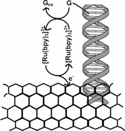
Ultrasensitive DNA sensing by Ru(bpy)3 2+ mediator amplified guanine (G) oxidation on a electrode covered with MWCNT 98. MWCNT, multiwalled carbon nanotube.
Meric et al. developed a biosensor for the detection of DNA sequences related to the Hepatitis B virus (HBV) and TT virus (TTV). The biosensor relies on the immobilization of the 21‐ or 24‐mer single stranded oligonucleotides (probe) related to the HBV and TTV sequences and hybridization of these oligonucleotides at carbon paste electrode (CPE). The extent of hybridization between the probe and target sequences was determined by using square wave voltammetry (SWV) with moving average baseline correction and methylene blue (MB) as the hybridization indicator. The difference between the MB signals, obtained from the hybrid modified and the probe‐modified CPE, is used to detect the DNA sequences of the infectious diseases from PCR‐amplified real samples 103. A new platform for universal DNA biosensing and its implications for the future of molecular diagnosis were presented in the review written by Teles and Fonseca 104.
CONCLUSION
Biosensor development made a huge progress in recent years, but their application in clinical diagnosis is not very common, except for glucose biosensors representing about 90% of the global biosensor market. The biggest problems are interferences with undesired molecules during measurements with real samples. High selectivity and accuracy is required because treatment is often dependent on concrete levels of clinical markers. Today's progress in biosensor constructions and real importance of small portable analytical devices for rapid clinical tests gives a perspective for better implementation of biosensors to the commercial sphere. In this review some chosen important analytes were described in the text containing basic principles, characteristics, and utilization. This study highlights the importance of biosensors in clinical diagnosis and summarizes the most recent biosensoric principles and techniques. The most of the described biosensors are based on amperometric techniques what indicates the trends in biosensors development. The important task for developers is to reach appropriate stability, to eliminate undesirable interferences, and make them applicable for commercial use.
Grant sponsor: Slovak Research and Development Agency; Grant number: VMSP‐P‐0073‐09; Grant sponsor: Agency of the Ministry of Education, Science, Research and Sport of the Slovak Republic for the Structural Funds; Grant number: ITMS 26240220040.
REFERENCES
- 1. Rechnitz GA. Biosensors: An overview. J Clin Lab Anal 1987;1:308–312. [Google Scholar]
- 2. Rechnitz GA. Future of biosensors in the clinical laboratory. J Clin Lab Anal 1988;2:131–133. [Google Scholar]
- 3. Kissinger PT. Biosensors‐a perspective. Biosens Bioelectron 2005;20:2512–2516. [DOI] [PubMed] [Google Scholar]
- 4. Malhotra BD, Chaubey A. Biosensors for clinical diagnostics industry. Sensor Actuator BChem 2003;91:117–127. [Google Scholar]
- 5. Velasco‐Garcia MN. Optical biosensors for probing at the cellular level: A review of recent progress and future prospects. Semin Cell Dev Biol 2009;20:27–33. [DOI] [PubMed] [Google Scholar]
- 6. Serna Cock L, Zetty Arenas AM, Ayala Aponte A. Use of enzymatic biosensors as quality indices: A synopsis of present and future trends in the food industry. Chil J Agric Res 2009;69:270–280. [Google Scholar]
- 7. Vo‐Dinh T, Cullum B. Biosensors and biochips: Advances in biological and medical diagnostics. Fresenius J Anal Chem 2000;366:540–551. [DOI] [PubMed] [Google Scholar]
- 8. Cooper MA. Label‐free screening of bio‐molecular interactions. Anal Bioanal Chem 2003;377:834–842. [DOI] [PubMed] [Google Scholar]
- 9. Janshoff A, Galla HJ, Steinem C. Piezoelectric mass‐sensing devices as biosensors‐An alternative to optical biosensors? Angew Chem Int Ed 2000;39:4004–4032. [DOI] [PubMed] [Google Scholar]
- 10. Chaubey A, Malhotra BD. Mediated biosensors. Biosens Bioelectron 2002;17:441–456. [DOI] [PubMed] [Google Scholar]
- 11. Monosik R, Stredansky M, Tkac J, Sturdik E. Application of enzyme biosensors in analysis of food and beverages. Food Anal Method 2011;doi 10.1007/s12161-011-9222-s12161-011-9224. [DOI] [Google Scholar]
- 12. Ye J‐S, Wen Y, De Zhang W, Ming Gan L, Xu GQ, Sheu F‐S. Nonenzymatic glucose detection using multi‐walled carbon nanotube electrodes. Electrochem Commun 2004;6:66–70. [Google Scholar]
- 13. Hu J. The evolution of commercialized glucose sensors in China. Biosens Bioelectron 2009;24:1083–1089. [DOI] [PubMed] [Google Scholar]
- 14. Clark LC, Lyons C. Electrode systems for continuous monitoring in cardiovascular surgery. Ann N Y Acad Sci 1962;102:29–45. [DOI] [PubMed] [Google Scholar]
- 15. Song S, Xu H, Fan C. Potential diagnostic applications of biosensors: Current and future directions. Int J Nanomedicine 2006;1:433–440. [DOI] [PMC free article] [PubMed] [Google Scholar]
- 16. Zuping T, Louie RF, Lee JH, Lee DM, Miller EE, Kost GJ. Oxygen effects on glucose meter measurements with glucose dehydrogenase‐ and oxidase‐based test strips for point‐of‐care testing. Crit Care Med 2001;29:1062–1070. [DOI] [PubMed] [Google Scholar]
- 17. Li Y, Liu X, Yuan H, Xiao D. Glucose biosensor based on the room‐temperature phosphorescence of TiO2/SiO2 nanocompo‐site. Biosens Bioelectron 2009;24:3706–3710. [DOI] [PubMed] [Google Scholar]
- 18. Pérez JPH, López‐Cabarcos E, López‐Ruiz B. Amperometric glucose biosensor based on biocompatible poly(dimethylami‐noethyl) methacrylate microparticles. Talanta 2010;81:1197–1202. [DOI] [PubMed] [Google Scholar]
- 19. Wu H, Wang J, Kang X, Wang C, Wang D, Liu J, Aksay IA, Lin Y. Glucose biosensor based on immobilization of glucose oxidase in platinum nanoparticles/graphene/chitosan nanocomposite film. Talanta 2009;80:403–406. [DOI] [PubMed] [Google Scholar]
- 20. Hsu C‐T, Hsiao H‐C, Fang M‐Y, Zen J‐M. Superior long‐term stability of a glucose biosensor based on inserted barrel plating gold electrodes. Biosens Bioelectron 2009;25:383–387. [DOI] [PubMed] [Google Scholar]
- 21. Comba FN, Rubianes MD, Herrasti P, Rivas GA. Glucose biosensing at carbon paste electrodes containing iron nanopar‐ticles. Sensor Actuator B Chem 2010;149:306–309. [Google Scholar]
- 22. Yoo E‐H, Lee S‐Y. Glucose biosensors: An overview of use in clinical practice. Sensors 2010;10:4558–4576. [DOI] [PMC free article] [PubMed] [Google Scholar]
- 23. Newman JD, Turner APF. Home blood glucose biosensors: A commercial perspective. Biosens Bioelectron 2005;20:2435–2453. [DOI] [PubMed] [Google Scholar]
- 24. Schelter‐Graf A, Schmidt HL, Huck H. Determination of the substrates of dehydrogenases in biological material in flow‐injection systems with electrocatalytic NADH oxidation. Anal Chim Acta 1984;163(C):299–303. [Google Scholar]
- 25. Li G, Xu H, Huang W, Wang Y, Wu Y, Parajuli R. A pyrrole quinoline quinone glucose dehydrogenase biosensor based on screen‐printed carbon paste electrodes modified by carbon nanotubes. Meas Sci Technol 2008;19:065203. [Google Scholar]
- 26. Tsujimura S, Kojima S, Kano K, Ikeda T, Sato M, Sanada H, Omura H. Novel FAD‐dependent glucose dehydrogenase for a dioxygen‐insensitive glucose biosensor. Biosci Biotechnol Bio‐chem 2006;70:654–659. [DOI] [PubMed] [Google Scholar]
- 27. Buchert J. A xylose‐oxidizing membrane‐bound aldose dehydro‐genase of Gluconobacter oxydans ATCC 621. J Biotechnol 1991;18:103–113. [Google Scholar]
- 28. Gomes SP, Odlozilikova M, Gabriela Almeida M, Araujo AN, Couto CMCM, Montenegro MCBSM. Application of lactate amperometric sol‐gel biosensor to sequential injection determination of l‐lactate. J Pharm Biomed Anal 2007;43:1376–1381. [DOI] [PubMed] [Google Scholar]
- 29. Cowan BN, Burns HJG, Boyle P, Ledingham IM. The relative prognostic value of lactate and haemodynamic measurements in early shock. Anaesthesia 1984;39:750–755. [DOI] [PubMed] [Google Scholar]
- 30. Bakker J, Gris P, Coffernils M, Kahn RJ, Vincent J‐L. Serial blood lactate levels can predict the development of multiple organ failure following septic shock. Am J Surg 1996;171:221–226. [DOI] [PubMed] [Google Scholar]
- 31. Rahman MM, Shiddiky MJA, Rahmana A, Shim YB. A lactate biosensor based on lactate dehydrogenase/nictotinamide adenine dinucleotide (oxidized form) immobilized on a conducting polymer/multiwall carbon nanotube composite film. Anal Bio‐chem 2009;384:159–165. [DOI] [PubMed] [Google Scholar]
- 32. Cai X, Yan J, Chu H, Wu M, Tu Y. An exercise degree monitoring biosensor based on electrochemiluminescent detection of lactate in sweat. Sensor Actuator B Chem 2010;143:655–659. [Google Scholar]
- 33. Romero MR, Garay F, Baruzzi AM. Design and optimization of a lactate amperometric biosensor based on lactate oxidase cross‐linked with polymeric matrixes. Sensor Actuator B Chem 2008;131:590–595. [Google Scholar]
- 34. Gamero M, Pariente F, Lorenzo E, Alonso C. Nanostructured rough gold electrodes for the development of lactate oxidase‐based biosensors. Biosens Bioelectron 2010;25:2038–2044. [DOI] [PubMed] [Google Scholar]
- 35. Huang J, Song Z, Li J, Yang Y, Shi H, Wu B, Anzai J‐i, Osa T, Chen Q. A highly‐sensitive l‐lactate biosensor based on sol‐gel film combined with multi‐walled carbon nanotubes (MWCNTs) modified electrode. Mat Sci Eng C Bio 2007;27:29–34. [Google Scholar]
- 36. Huang J, Li J, Yang Y, Wang X, Wu B, Anzai J‐i, Osa T, Chen Q. Development of an amperometric l‐lactate biosensor based on l‐lactate oxidase immobilized through silica sol‐gel film on multi‐walled carbon nanotubes/platinum nanoparticle modified glassy carbon electrode. Mat Sci Eng C Bio 2008;28:1070–1075. [Google Scholar]
- 37. A service of the U.S. National Library of Medicine, web page: http://www.nlm.nih.gov/medlineplus/ency/article/003507.htm
- 38. Maines A, Ashworth D, Vadgama P. Diffusion restricting outer membranes for greatly extended linearity measurements with glucose oxidase enzyme electrodes. Anal Chim Acta 1996;333:223–231. [Google Scholar]
- 39. Pereira AC, Aguiar MR, Kisner A, Macedo DV, Kubota LT. Amperometric biosensor for lactate based on lactate dehydro‐genase and Meldola Blue coimmobilized on multi‐wall carbon‐nanotube. Sensor Actuator B Chem 2007;124:269–276. [Google Scholar]
- 40. Cui X, Li CM, Zang J, Yu S. Highly sensitive lactate biosensor by engineering chitosan/PVI‐Os/CNT/LOD network nanocompo‐site. Biosens Bioelectron 2007;22:3288–3292. [DOI] [PubMed] [Google Scholar]
- 41. Suman S, Singhal R, Sharma AL, Malthotra BD, Pundir CS. Development of a lactate biosensor based on conducting copolymer bound lactate oxidase. Sensor Actuator B Chem 2005;107:768–772. [Google Scholar]
- 42. Liu C‐X, Liu H‐M, Yang Q‐D, Tian Q, Cai X‐X. Development of amperometric lactate biosensor modified with Pt‐black nanopar‐ticles for rapid assay. Chinese J Anal Chem 2009;37:624–628. [Google Scholar]
- 43. Romero, MR , Ahumada F, Garay F, Baruzzi AM. Ampero‐metric biosensor for direct blood lactate detection. Anal Chem 2010;82:5568–5572. [DOI] [PubMed] [Google Scholar]
- 44. Claver JB, Miron MCV, Capitan‐Vallvey LF. Disposable electrochemiluminescent biosensor for lactate determination in saliva. Analyst 2009;134:1423–1432. [DOI] [PubMed] [Google Scholar]
- 45. Arya SK, Datta M, Malhotra BD. Recent advances in cholesterol biosensor. Biosens Bioelectron 2008;23:1083–1100. [DOI] [PubMed] [Google Scholar]
- 46. Vidal J‐C, Espuelas J, Garcia‐Ruiz E, Castillo J‐R. Ampero‐metric cholesterol biosensors based on the electropolymerization of pyrrole and the electrocatalytic effect of Prussian‐Blue layers helped with self‐assembled monolayers. Talanta 2004;64:655–664. [DOI] [PubMed] [Google Scholar]
- 47. Umar A, Rahman MM, Vaseem M, Hahn Y‐B. Ultra‐sensitive cholesterol biosensor based on low‐temperature grown ZnO nanoparticles. Electrochem Commun 2009;11:118–121. [Google Scholar]
- 48. Umar A, Rahman MM, Al‐Hajry A, Hahn YB. Highly‐sensitive cholesterol biosensor based on well‐crystallized flower‐shaped ZnO nanostructures. Talanta 2009;78:284–289. [DOI] [PubMed] [Google Scholar]
- 49. Israra MQ, Sadafa JR, Asifa MH, Nura O, Willandera M, Danielsson B. Potentiometric cholesterol biosensor based on ZnO nanorods chemically grown on Ag wire. Thin Solid Films 2010;519:1106–1109. [Google Scholar]
- 50. Kumar A, Suman. Cholesterol biosensor based on polyvinyl formal membrane bound cholesterol esterase and oxidase. Sens Trans 2007;83:1555–1563. [Google Scholar]
- 51. Shih W‐C, Yang M‐C, Lin MS. Development of disposable lipid biosensor for the determination of total cholesterol. Biosens Bioelectron 2009;24:1679–1684. [DOI] [PubMed] [Google Scholar]
- 52.Aravinda SSJ, Babya TT, Arockiadossa T, Rakhia RB, Ramaprabhu S. A cholesterol biosensor based on gold nano‐particles decorated functionalized graphene nanoplatelets. Thin Solid Films 2011;doi: 10.1016/j.tsf.2011.03.032. [DOI]
- 53. Tsai Y‐C, Chen S‐Y, Lee C‐A. Amperometric cholesterol biosensors based on carbon nanotube‐chitosan‐platinum‐choles‐terol oxidase nanobiocomposite. Sensor Actuator B Chem 2008;135:96–101. [Google Scholar]
- 54. Fang Ch, He J, Chen Z. A disposable amperometric biosensor for determining total cholesterol in whole blood. Sensor Actuator B Chem 2011;doi: 10.1016/j.snb.2011.01.005. [DOI]
- 55. Safavi A, Farjami F. Electrodeposition of gold–platinum alloy nanoparticles on ionic liquid–chitosan composite film and its application in fabricating an amperometric cholesterol biosensor. Biosens Bioelectron 2011;26:2547–2552. [DOI] [PubMed] [Google Scholar]
- 56. Senda M, Yamamoto Y. Urea biosensor based on amperometric ammonium ion electrode. Electroanalysis 1993;5:775–779. [Google Scholar]
- 57. Gambhir A, Gerard M, Mulchandani A, Malhotra B. Coimmo‐bilization of urease and glutamate dehydrogenase in electro‐chemically prepared polypyrrole‐polyvinyl sulfonate films. Appl Biochem Biotech 2001;96:249–257. [DOI] [PubMed] [Google Scholar]
- 58. Singhal R, Gambhir A, Pandey MK, Annapoorni S, Malhotra BD. Immobilization of urease on poly(N‐vinyl carbazole)/stearic acid Langmuir‐Blodgett films for application to urea biosensor. Biosens Bioelectron 2002;17:697–703. [DOI] [PubMed] [Google Scholar]
- 59. Alqasaimeh M, Heng L, Ahmad M. A urea biosensor from stacked sol‐gel films with immobilized nile blue chromoionophore and urease enzyme. Sensors 2007;7:2251–2262. [DOI] [PubMed] [Google Scholar]
- 60. Vostiar I, Tkac J, Sturdik E, Gemeiner P. Amperometric urea biosensor based on urease and electropolymerized toluidine blue dye as a pH‐sensitive redox probe. Bioelectrochemistry 2002;56:113–115. [DOI] [PubMed] [Google Scholar]
- 61. Karakus E, Pekyardimci S, Esma K. Urea biosensors based on PVC membrane containing palmitic acid. Artif Cell Blood Substit Biotechnol 2005;33:329–341. [DOI] [PubMed] [Google Scholar]
- 62. Lakarda B, Magnin D, Deschaume O, Vanlancker G, Glinel, K , Demoustier‐Champagne S, Nysten B, Jonas AM, Bertrand P, Yunus S. Urea potentiometric enzymatic biosensor based on charged biopolymers and electrodeposited polyaniline. Biosens Bioelectron 2011;26:4139–4145. [DOI] [PubMed] [Google Scholar]
- 63. Srivastava PK, Kayastha AM, Srinivasan. Characterization of gelatin‐immobilized pigeonpea urease and preparation of a new urea biosensor. Biotech Appl Biochem 2001;34:55–62. [DOI] [PubMed] [Google Scholar]
- 64. Jha SK, Topkar A, D'Souza SF. Development of potentiometric urea biosensor based on urease immobilized in PVA‐PAA composite matrix for estimation of blood urea nitrogen (BUN). J Biochem Biophys Methods 2008;70:1145–1150. [DOI] [PubMed] [Google Scholar]
- 65. Pizzariello A, Stredansky M, Stredanska S, Miertus S. Urea biosensor based on amperometric pH‐sensing with haematein as a pH‐sensitive redox mediator. Talanta 2001;54:763–772. [DOI] [PubMed] [Google Scholar]
- 66. Gabrovska K, Ivanov J, Vasileva I, Dimova N, Godjevargova T. Immobilization of urease on nanostructured polymer membrane and preparation of urea amperometric biosensor. Int J Biol Macromol 2011;48:620–626. [DOI] [PubMed] [Google Scholar]
- 67. Singh M, Verma N, Garg AK, Redhu N. Urea biosensors. Sensor Actuat B‐Chem 2008;134:345–351. [Google Scholar]
- 68. Dhawan G, Sumana G, Malhotra BD. Recent developments in urea biosensors. Biochem Engl J 2009;44:42–52. [Google Scholar]
- 69. Berberich JA, Yang LW, Madura J, Bahar I, Russell AJ. A stable three‐enzyme creatinine biosensor. 1. Impact of structure, function and environment on PEGylated and immobilized sarcosine oxidase. Acta Biomater 2005;1:173–181. [DOI] [PubMed] [Google Scholar]
- 70. Berberich JA, Yang LW, Bahar I, Russell AJ. A stable three enzyme creatinine biosensor. 2. Analysis of the impact of silver ions on creatine amidinohydrolase. Acta Biomater 2005;1:183–191. [DOI] [PubMed] [Google Scholar]
- 71. Berberich JA, Chan A, Boden M, Russell AJ. A stable three‐enzyme creatinine biosensor. 3. Immobilization of creatinine amidohydrolase and sensor development. Acta Biomater 2005;1:193–199. [DOI] [PubMed] [Google Scholar]
- 72. Radomska A, Bodenszac E, Glab S, Koncki R. Creatinine biosensor based on ammonium ion selective electrode and its application in flow‐injection analysis. Talanta 2004;64:603–608. [DOI] [PubMed] [Google Scholar]
- 73. Ramanavicius A. Amperometric biosensor for the determination of creatine. Anal Bioanal Chem 2007;387:1899–1906. [DOI] [PubMed] [Google Scholar]
- 74.Stefan‐van Staden RI, Bokretsion RG, van Staden JF, Aboul‐Enein HY. Simultaneous detection of creatine and creatinine using a sequential injection analysis/biosensor system. Prep Biochem Biotechnol 2006;36:287–296. [DOI] [PubMed] [Google Scholar]
- 75. Killard AJ, Smyth MR. Creatinine biosensors: Principles and designs. Trends Biotechnol 2000;18:433–437. [DOI] [PubMed] [Google Scholar]
- 76. Lad U, Khokhar S, Kale GM. Electrochemical creatinine biosensors. Anal Chem 2008;80:7910–7917. [DOI] [PubMed] [Google Scholar]
- 77. Belluzo M, Ribone M, Lagier C. Assembling amperometric biosensors for clinical diagnostics. Sensors 2008;8:1366–1399. [DOI] [PMC free article] [PubMed] [Google Scholar]
- 78. Schuetz AJ, Winklmair M, Weller MG, Niessner R. Selection of hapten structures for indirect immunosensor arrays. Fresenius J Anal Chem 1999;363:625–631. [Google Scholar]
- 79. Skladal P. Advances in electrochemical immunosensors. Electro‐analysis 1997;9:737–745. [Google Scholar]
- 80. Morgan C, Newman D, Price C. Immunosensors: Technology and opportunities in laboratory medicine. Clin Chem 1996;42:193–209. [PubMed] [Google Scholar]
- 81. Liang W, Yi W, Li S, Yuan R, Chen A, Chen S, Xiang G, Hu C. A novel, label‐free immunosensor for the detection of [alpha]‐fetoprotein using functionalised gold nanoparticles. Clin Biochem 2009;42:1524–1530. [DOI] [PubMed] [Google Scholar]
- 82. Yang M, Wang C. Label‐free immunosensor based on gold nanoparticle silver enhancement. Anal Biochem 2009;385:128–131. [DOI] [PubMed] [Google Scholar]
- 83. Huang J, Yang G, Meng W, Wu L, Zhu A, Jiao Xa. An electrochemical impedimetric immunosensor for label‐free detection of Campylobacter jejuni in diarrhea patients’ stool based on O‐carboxymethylchitosan surface modified Fe3O4 nanoparticles. Biosens Bioelectron 2010;25:1204–1211. [DOI] [PubMed] [Google Scholar]
- 84. Wang J, Yuan R, Chai Y, Cao S, Guan S, Fu P, Min L. A novel immunosensor based on gold nanoparticles and poly‐(2,6‐pyridinediamine)/multiwall carbon nanotubes composite for immunoassay of human chorionic gonadotrophin. Biochem Engl J 2010;51:95–101. [Google Scholar]
- 85. Li X, Yuan R, Chai Y, Zhang L, Zhuo Y, Zhang Y. Amperometric immunosensor based on toluidine blue/nano‐Au through electrostatic interaction for determination of carcinoembryonic antigen. J Biotechnol 2006;123:356–366. [DOI] [PubMed] [Google Scholar]
- 86. Ehrhart J‐C, Bennetau B, Renaud L, Madrange J‐P, Thomas L, Morisot J, Brosseau A, Allano S, Tauc P, Tran P‐L. A new immunosensor for breast cancer cell detection using antibody‐coated long alkylsilane self‐assembled monolayers in a parallel plate flow chamber. Biosens Bioelectron 2008;24:467–474. [DOI] [PubMed] [Google Scholar]
- 87. Chen H, Jiang C, Yu C, Zhang S, Liu B, Kong J. Protein chips and nanomaterials for application in tumor marker immunoas‐says. Biosens Bioelectron 2009;24:3399–3411. [DOI] [PubMed] [Google Scholar]
- 88. Rasooly A, Jacobson J. Development of biosensors for cancer clinical testing. Biosens Bioelectron 2006;21:1851–1858. [DOI] [PubMed] [Google Scholar]
- 89. Soper SA, Brown K, Ellington A, Frazier B, Garcia‐Manero G, Gau V, Gutman SI, Hayes DF, Korte B, Landers JL, Larson D, Ligler F, Majumdar A, Mascini M, Nolte D, Rosenzweig Z, Wang J, Wilson D. Point‐of‐care biosensor systems for cancer diagnostics/prognostics. Biosens Bioelectron 2006;21:1932–1942. [DOI] [PubMed] [Google Scholar]
- 90. Tothill IE. Biosensors for cancer markers diagnosis. Semin Cell Dev Biol 2009;20:55–62. [DOI] [PubMed] [Google Scholar]
- 91. Mascini M, Palchetti I, Marrazza G. DNA electrochemical biosensors. Fresen J Anal Chem 2001;369:15–22. [DOI] [PubMed] [Google Scholar]
- 92. Marrazza G, Chianella I, Mascini M. Disposable DNA electrochemical sensor for hybridization detection. Biosens Bioelectron 1999;14:43–51. [DOI] [PubMed] [Google Scholar]
- 93. Drummond TG, Hill MG, Barton JK. Electrochemical DNA sensors. Nat Biotechnol 2003;21:1192–1199. [DOI] [PubMed] [Google Scholar]
- 94. Kerman K, Kobayashi M, Tamiya E. Recent trends in electrochemical DNA biosensor technology. Meas Sci Technol 2004;15:R1–R11. [Google Scholar]
- 95. Cagnin S, Caraballo M, Guiducci C, Martini P, Ross M, SantaAna M, Danley D, West T, Lanfranchi G. Overview of electrochemical DNA biosensors: New approaches to detect the expression of life. Sensors 2009;9:3122–3148. [DOI] [PMC free article] [PubMed] [Google Scholar]
- 96. Odenthal KJ, Gooding JJ. An introduction to electrochemical DNA biosensors. Analyst 2007;132:603–610. [DOI] [PubMed] [Google Scholar]
- 97. Erdem A, Ozsoz M. Electrochemical DNA biosensors based on DNA‐brug interactions. Electroanalysis 2002;14:965–974. [Google Scholar]
- 98. Koehne J, Li J, Cassell AM, Chen H, Ye Q, Ng HT, Han J, Meyyappan M. The fabrication and electrochemical characterization of carbon nanotube nanoelectrode arrays. J Mater Chem 2004;14:676–684. [Google Scholar]
- 99. He P, Xu Y, Fang Y. Applications of carbon nanotubes in electrochemical DNA biosensors. Microchim Acta 2006;152:175–186. [Google Scholar]
- 100. Cai H, Cao X, Jiang Y, He P, Fang Y. Carbon nanotube‐enhanced electrochemical DNA biosensor for DNA hybridization detection. Anal Bioanal Chem 2003;375:287–293. [DOI] [PubMed] [Google Scholar]
- 101. Tang X, Bansaruntip S, Nakayama N, Yenilmez E, Chang Y‐l, Wang Q. Carbon nanotube DNA sensor and sensing mechanism. Nano Lett 2006;6:1632–1636. [DOI] [PubMed] [Google Scholar]
- 102. Liao JC, Mastali M, Gau V, Suchard MA, Moller AK, Bruckner DA, Babbitt JT, Li Y, Gornbein J, Landaw EM, McCabe ERB, Churchill BM, Haake DA. Use of electrochemical DNA biosensors for rapid molecular identification of uropathogens in clinical urine specimens. J Clin Microbiol 2006;44:561–570. [DOI] [PMC free article] [PubMed] [Google Scholar]
- 103. Meric B, Kerman K, Ozkan D, Kara P, Erensoy S, Akarca US, Mascini M, Ozsoz M. Electrochemical DNA biosensor for the detection of TT and Hepatitis B virus from PCR amplified real samples by using methylene blue. Talanta 2002;56:837–846. [DOI] [PubMed] [Google Scholar]
- 104. Telesa FRR, Fonseca LP. Trends in DNA biosensors. Talanta 2008;77:606–623. [Google Scholar]