Abstract
Cognitive brain networks such as the default-mode network (DMN), frontoparietal network, and salience network, are key functional networks of the human brain. Here we show that the rapid evolutionary cortical expansion of cognitive networks in the human brain, and most pronounced the DMN, runs parallel with high expression of human-accelerated genes (HAR genes). Using comparative transcriptomics analysis, we present that HAR genes are differentially more expressed in higher-order cognitive networks in humans compared to chimpanzees and macaques and that genes with high expression in the DMN are involved in synapse and dendrite formation. Moreover, HAR and DMN genes show significant associations with individual variations in DMN functional activity, intelligence, sociability, and mental conditions such as schizophrenia and autism. Our results suggest that the expansion of higher-order functional networks subserving increasing cognitive properties has been an important locus of genetic changes in recent human brain evolution.
Subject terms: Evolutionary genetics, Cognitive neuroscience, Genetics of the nervous system, Neural circuits
Several cortical association areas have rapidly expanded in size during human evolution, including elements of the central cognitive default mode network (DMN). Here, the authors show that genes highly divergent between humans and other primates (HAR genes) are particularly expressed in these brain regions.
Introduction
The human brain is capable of supporting a wide range of complex cognitive abilities, more so than other highly developed and intelligent great apes, such as the chimpanzee, one of our closest living evolutionary relatives with which we share the majority of our genetic material1. This distinction in cognitive abilities is commonly believed to be associated with the rapid expansion of multimodal association areas and their structural and functional connections in the human brain2–4, with cognitive functional networks, such as the frontoparietal network, salience network, and default-mode network (DMN), playing an essential role in higher-order brain functions5–7. These cognitive functional networks are highly heritable8,9 and relate to genetic effects associated with neuron growth and metabolism10. Uncovering the evolutionary genetic underpinnings of cognitive functional networks, and in particular, to what extent cognitive functional networks have developed in recent human evolution, is crucial for our understanding of the high cognitive complexity of human brain function.
The DMN in particular has been identified as a central network in human cognition, consisting of densely connected areas such as the posterior cingulate, precuneus, inferior parietal, middle temporal, and medial prefrontal cortices5,6. Comparative neuroimaging studies have shown default-mode activity in chimpanzees11 and macaques12, but with potential subtle differences in both the spatial and topological layout of this central network13–changes that may relate to enhancement of cognitive functions in humans compared to other primate species. The DMN is central to social cognition, including aspects of mental self-projection14, mental rehearsal of future actions15, and understanding of another person’s mental perspective. These advanced social abilities are likely to have been highly adaptive during recent human evolution16, potentially enabling humans to make more complex social inferences14.
Here, we studied the expansion and evolutionary genetics of higher-order cognitive networks in recent human brain evolution, with a particular focus on the evolutionarily genetic drive of the DMN. Recent genome-wide studies comparing the human genome with that of the chimpanzee have identified a unique set of loci that displayed accelerated divergence in the human lineage17,18. Genes associated with these so-called human accelerated regions (HAR) have been linked to neuron development, but also the development of brain disorders such as autism spectrum disorder (ASD)19. We integrate genetic data with comparative neuroimaging and present that regions of higher-order cognitive networks are highly expanded in recent human brain evolution. We show that HAR genes likely have played a crucial role in this, being highly expressed in expanded cognitive networks (and in particular the DMN) and being differentially expressed in the human brain compared to chimpanzees and macaques. We provide further evidence of HAR and DMN genes to be important in human cognitive functioning, social behavior, and mental disorders, such as autism and schizophrenia.
Results
Human cortical expansion
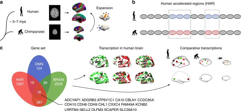
We started by mapping the expansion of the human cortex (Homo sapiens) compared to the cortex of the chimpanzee (Pan troglodytes), one of our closest living evolutionary relatives along with the bonobo (Pan paniscus). Cortical morphometry of the chimpanzee and human cortex was assessed using a surface-to-surface mapping of 3D reconstructions of the cortical mantle across both species, based on in vivo T1-weighted MRI (29 chimpanzees, 30 humans; Fig. 1a). The largest expansion of the human cortex was found in areas of bilateral orbital inferior frontal gyrus (×4.0 expansion), rostral middle frontal lobe (×3.8 expansion), inferior/middle temporal lobe (×3.0/2.9 expansion), lingual gyrus (×2.9 expansion), right inferior parietal lobe (×3.7 expansion), and left precuneus (×2.7 expansion; two-sample t-test on the normalized expansion, q < 0.001, false discovery rate [FDR] corrected; Cohens’d > 0.989; Fig. 2a). The lowest expansion was found in primary areas, including bilateral precentral gyrus (×1.3 expansion), postcentral gyrus (×1.4 expansion), and paracentral lobe (×1.2 expansion; q < 0.001, FDR corrected; Cohens’d < −1.047; Fig. 2a and Supplementary Data 1).
Fig. 1.
Methods overview. a The human and chimpanzee cortex were constructed using MRI data, with chimpanzee-to-human cortical expansion computed based on the reconstructed cortical maps. b Genes associated with human accelerated regions (HAR), which represent genomic loci with accelerated divergence in humans, were examined. c Cortical gene expression of HAR genes and HAR-BRAIN genes were examined using human transcription data from the Allen Human Brain Atlas (AHBA) and comparative transcription data of the human, chimpanzee, and macaque from the PsychENCODE database
Fig. 2.
Cortical expansion. a Cortical expansion from chimpanzees to humans. Blue: below-median human expansion (i.e., < × 2 expansion compared to chimpanzee); red: above-median expansion (i.e., > × 2). b Brain maps of the seven resting-state functional networks according to the DK-114 atlas, describing the visual (VN), somatomotor (SMN), dorsal-attention (DAN), limbic (LN), ventral-attention (VAN), frontal-parietal (FPN), and default-mode network (DMN). c Levels of normalized cortical expansion per functional network in descending order of mean expansion. d Levels of normalized cortical expansion in higher-order cognitive networks (DMN, FPN, VAN) versus the SMN and VN. Dots depict cortical regions. Colors represent functional networks, as in panel b. Central marks are mean expansions. * indicates a two-sided p-value < 0.05, FDR corrected, two-sample t-test. Source data provided as Source Data file
We next grouped cortical areas into the visual (VN), somatomotor (SMN), dorsal-attention (DAN), limbic (LN), ventral-attention (VAN, also commonly referred to as the salience network), frontoparietal (FPN), and default-mode network (DMN) (Fig. 2b and Supplementary Methods)20. Higher-order cognitive networks (i.e., DMN, FPN, VAN) displayed particularly high levels of cortical expansion as compared to the SMN/VN (×1.2 larger expansion in regions of higher-order cognitive networks combined compared to the regions of the SMN/VN combined, two-sample t-test, t(86) = 3.257, p = 0.002; Fig. 2d). FPN showed the largest expansion (mean: × 2.9 expansion), with the DMN in second place (mean: ×2.4 expansion), showing both significantly higher expansion when comparing each of them with the rest of the brain (FPN: t(108) = 3.360, p = 0.001; DMN: t(108) = 2.621, p = 0.010; FDR corrected; Supplementary Table 1). In contrast, separately examining the other five networks did not show significant increases in the expansion of these networks compared to the rest of the cortex.
HAR gene expression
We then examined this distinct pattern of human cortical expansion across the seven resting-state functional networks in relation to cortical gene expression patterns relevant to human evolution. Microarray data on gene expression across cortical regions were obtained from the Allen Human Brain Atlas (AHBA) (http://human.brain-map.org/), containing transcriptional profiles of 20,734 genes across 57 areas of the left cortical mantle (Fig. 1c). Genes relevant to human evolution were taken as the list of 2143 genes associated with HAR as presented previously by Doan and colleagues19, selected based on positional mapping. Alternative selection and allocation of HAR-associated genes are possible (e.g., using chromatin interactions) and we examined such alternatives to validate our results (Supplementary Note 1).
Transcription data of 1711 HAR-associated genes were present in AHBA (referred to as HAR genes; Supplementary Data 2), and we further examined their cortical gene expression levels in comparison to the cortical expansion. The expression profile of HAR genes was positively correlated to the pattern of human cortical expansion (Pearson’s r(53) = 0.360, p = 0.007; Supplementary Fig. 1a), indicating the highest HAR gene expression in highly expanded areas of the human cortex. This association significantly exceeded the null condition of correlations between cortical expansion and expression of random gene sets (i.e., 1711 random genes) selected from a pool of 8686 genes related to general evolutionarily conserved genetic elements (ECE genes;21 p < 0.001, permutation test, 10,000 permutations; Supplementary Fig. 1a). Cortical regions of cognitive networks also showed significantly higher expression of HAR genes compared to regions of the SMN/VN (t(44) = 2.742, p = 0.009; FDR corrected; Supplementary Fig. 1b), with regions of the DMN showing the highest HAR gene expression (t(55) = 2.274, p = 0.027, uncorrected, comparing the DMN to all other networks combined; Supplementary Fig. 1c). These effects again significantly exceeded the null conditions of effects of random ECE genes (all p < 0.001, 10,000 permutations). Furthermore, examining the other six functional networks separately did not show a significant enhancement of HAR gene expression (Supplementary Table 2).
HAR-BRAIN gene expression
With the set of HAR genes describing genes involved in all sorts of functions across the entire human body (and thus not specific to ‘brain’), we continued by examining whether HAR genes related to brain processes may have played a specific role in the large cortical expansion of cognitive functional networks in human evolution. We identified genes commonly expressed in brain areas using the GTEx database (https://www.gtexportal.org/), selecting 2979 genes significantly more expressed in brain tissues compared to other available body sites (q < 0.05, FDR corrected; one-sided two-sample t-test; referred to as BRAIN genes); 415 genes (24.3%) out of the full set of 1711 HAR genes were observed to be significantly more expressed in brain tissues, a set from now on referred to as HAR-BRAIN genes (in contrast to HAR-nonBRAIN genes; Supplementary Data 2).
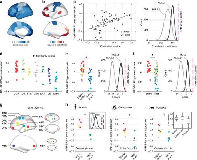
We then aimed to examine (1) whether HAR-BRAIN genes were more expressed particularly in regions of higher-order cognitive networks compared to the total set of HAR genes, and (2) to what extent HAR-BRAIN genes were more expressed in regions of higher-order cognitive networks, more than an average set of genes related to general brain processes (i.e., BRAIN genes). First, the cortical expression pattern of HAR-BRAIN genes was significantly correlated with the pattern of human cortical expansion (r(53) = 0.488, p < 0.001; Fig. 3c). Furthermore, HAR-BRAIN genes showed significantly higher expression in regions of cognitive networks as compared to the SMN/VN (t(44) = 5.136, p < 0.001, FDR corrected; Fig. 3e), with the highest expression levels again observed in the DMN (t(55) = 3.267, p = 0.002, FDR corrected, DMN versus the rest of the cortex; Fig. 3f). These effects were, respectively, ×3.2 and ×2.7 larger than the effect obtained by HAR-nonBRAIN genes (t(55) = 1.028, p = 0.309 and t(55) = 1.212, p = 0.232, separately, Supplementary Fig. 2). Notably, examining the other six functional networks separately did not show any significant elevations of HAR-BRAIN gene expression (Supplementary Table 3), suggesting the highest expression level in the DMN.
Fig. 3.
HAR-BRAIN gene expression. a Cortical gene expression of HAR-BRAIN genes. b Cortical maps of the significance level obtained by permutation tests, comparing expressions of HAR-BRAIN genes to equally sized random gene-sets taken from BRAIN (NULL1) and ECE genes (NULL2). c Association between the gene expression profile of HAR-BRAIN genes and normalized cortical expansion between human and chimpanzee (left). The correlation coefficient is significantly higher than NULL1 and NULL2 (both p < 0.001, two-sided, permutation test; right). d HAR-BRAIN gene expression within each of the seven functional networks ranked in descending order of the mean expression. Asterisks (*) indicates significantly upregulated regions as in panel b. e HAR-BRAIN gene expression in cognitive networks (DMN, FPN, and VAN) versus the SMN and VN (left), with permutation results demonstrated in the right panel (two-sided p = 0.003 and p < 0.001 for NULL1 and NULL2, respectively). f HAR-BRAIN gene expression in the DMN versus the rest of the cortex (left), with permutation results demonstrated in the right panel (two-sided p < 0.001 for both NULL1 and NULL2). g Species-homologous brain areas as presented in the PsychENCODE dataset for the human (left), chimpanzee (upper right), and macaque (lower right). h Normalized expression levels of HAR-BRAIN genes in regions of higher-order networks compared to areas of the SMN/VN in humans (p < 0.001, two-sample t test). Largest differences in gene expression are found in humans, with chimpanzees in second place, followed by macaques (p = 0.002, Jonckheere-Terpstra test). Asterisks (*) indicates two-sided p < 0.05, FDR corrected. Central marks denote the mean gene expression. Boxplot center, median; box = 1st−3rd quartiles (Q); lower whisker, Q1–1.5 × interquartile range (IQR); upper whisker, Q3 + 1.5 × IQR. Colors indicate the assignment of functional networks, as in Fig. 2b. M1C primary motor cortex, S1C primary sensory cortex, IPC inferior parietal cortex, STC superior temporal cortex, ITC inferior temporal cortex, A1C primary auditory cortex, OFC orbital frontal cortex, VFC ventral frontal cortex, DFC dorsal frontal cortex, V1C primary visual cortex, MFC medial frontal cortex. Source data provided as Source Data file
Second, we compared HAR-BRAIN gene expression with two types of null-distributions of expression differences generated by randomly selecting the same number of genes (i.e., 415) from the pool of 2979 BRAIN genes (referred to as NULL1) and 8686 ECE genes (referred to as NULL2). The elevated expression of HAR-BRAIN genes in regions of higher-order cognitive networks was significantly larger than both null distributions (p = 0.006 and p < 0.001 for NULL1 and NULL2, respectively; 10,000 permutations; Fig. 3e). The same result was observed when examining DMN regions specifically (p < 0.001 for both NULL1 and NULL2; 10,000 permutations; Fig. 3f), suggesting a specific role of HAR-BRAIN genes in differentiating DMN regions from the rest of the brain. Permutation testing based on randomly shuffling cortical areas showed similar results (Supplementary Fig. 3). An exploratory examination on gene expression in each individual further demonstrated that the differentiation of HAR-BRAIN gene expression between the DMN and the rest of the cortex significantly correlated with the ratio of brain volume of the DMN regions (r(4) = 0.839, p = 0.037; Supplementary Fig. 4).
We then examined HAR-BRAIN expression for each of the cortical regions of the 7 functional networks separately; 10 cortical areas showed significantly high expression of HAR-BRAIN genes compared to a random selection of genes out of both BRAIN (NULL1) and ECE genes (NULL2) (FDR corrected, q < 0.05; 10,000 permutations; Fig. 3b). Importantly, 7 out of these 10 regions described regions of the higher cognitive networks and 6 out of these 7 regions described DMN regions (p = 0.034 and 0.008, respectively; hypergeometric test). These findings together suggest a specific role of HAR-BRAIN genes in the architecture of cognitive functional brain networks, beyond effects of general evolutionary conserved genes and general BRAIN genes.
Chimpanzee-human comparative gene expression
Our analyses so far suggested that HAR-BRAIN genes were more expressed in highly expanded regions of higher-order cognitive networks, but did not yet provide direct information on whether HAR-BRAIN gene expression is upregulated in the human brain compared to that of other primate species. To examine this, we used gene expression data from the PsychENCODE database (http://evolution.psychencode.org/)22, which describes gene expression of 11 comparable cortical regions across the human, chimpanzee, and macaque. Due to the lower spatial sampling of cortical regions (data of 3 DMN regions available, Fig. 3g), we limited our examination to a comparison between cognitive networks and the SMN/VN. First, we replicated the observation of high expression of HAR-BRAIN genes in regions of higher-order cognitive networks compared to regions of the SMN/VN in humans (t(8) = 7.135, p < 0.001; Fig. 3h), with this effect significantly exceeding both NULL1 and NULL2 (both p < 0.001, 10,000 permutations). This confirmed our AHBA-based findings of high HAR-BRAIN gene expression in cognitive networks in the human brain. Second, the differentiating level of HAR-BRAIN gene expression between cognitive and primary areas observed in humans (Cohen’s d = 4.605) was found to be ×2.7 larger than the effects found in chimpanzees (Cohen’s d = 1.695) and ×2.8 larger compared to macaques (Cohen’s d = 1.616). Chimpanzees and macaques showed only marginally higher HAR-BRAIN gene expression in regions of higher-order cognitive networks as compared to the SMN/VN (chimpanzees: t(8) = 2.626, p = 0.030; macaques: t(8) = 2.504, p = 0.037). Furthermore, the difference in effect size between humans and chimpanzees was larger than expected based on NULL1 and NULL2 (both p < 0.001; human-macaque: NULL1, p = 0.026 and NULL 2, p = 0.090, only trend-level, not significant [n.s.]; 10,000 permutations; Supplementary Fig. 5).
Further evaluation of this cross-species effect showed a significantly decreasing step-wise relationship of differences in HAR-BRAIN gene expression between regions of higher-order networks and the SMN/VN from humans (highest) to chimpanzees and macaques (lowest differentiating expression, Jonckheere–Terpstra test, p = 0.002). To reduce the influence of a relatively large variance of expression levels within chimpanzees and macaques (Fig. 3h), we performed a leave-one-out analysis (iteratively leaving out one region at a time) and confirmed a larger mean gene expression difference between cognitive network regions and primary regions in humans in comparison to chimpanzees and macaques (Supplementary Fig. 6). These findings thus suggest that humans display upregulated expression of HAR-BRAIN genes in brain areas involved in cognitive brain function as compared to other primates.
Top strongest differentiating DMN genes
We continued by investigating the biological properties of genes showing the highest levels of expression in DMN regions out of all genes. For each gene in AHBA, we computed the upregulated level of gene expression in regions of the DMN by calculating the t-score for expressions of the selected gene in regions of the DMN against the rest of the brain. The top 200 highly expressed genes (i.e., genes showing the highest positive t-scores, referred to as DMN genes; all p < 0.004) were taken as the DMN’s most differentiating genes (Supplementary Data 3). Out of the top 200 DMN genes, we identified 37 to be HAR genes, particularly including 18 HAR-BRAIN genes, which greatly exceeded the chance level of randomly selecting 37 or 18 out of 20,734 genes (both p < 0.001, hypergeometric test). We also examined the top 53 genes with p < 0.0014 (partial Bonferroni corrected, see Supplementary Methods) and top 469 genes with p < 0.01 (uncorrected), which revealed comparable findings (Supplementary Note 2).
To investigate whether the observed effect was restricted to the DMN, an additional permutation analysis was performed by shuffling region labels and re-computing the top genes for each of these random network assignments. We found the ratio of HAR genes in the set of top DMN genes to be significantly higher than the null condition (p < 0.001, 10,000 permutations), which confirmed a dominant role of HAR genes in DMN organization. To further examine potential DMN specificity, we also selected the top 200 genes showing the largest differentiating gene expression in each of the other functional networks compared to the SMN/VN. In contrast to the DMN (revealing 18 overlapping genes with the set of HAR-BRAIN genes), the top 200 gene sets identified by the VAN, DAN, FPN, and LN, comprised, respectively, only 12,8,7 and 7 HAR-BRAIN genes.
Gene-set enrichment analysis on the set of DMN genes using hypergeometric testing in the web-based platform FUMA23 showed significant over-representation of DMN genes in Gene Ontology (GO) terms related to cellular components of dendrite (p = 2.60 × 10⁻⁵), somatodendritic compartment (p = 4.49 × 10⁻⁵), synapse (p = 2.15 × 10⁻⁴), perikaryon (p = 7.45 × 10⁻⁵) and neuron projection terminus (p = 2.26 × 10⁻⁴), as well as molecular functions of neuropeptide hormone activity and calcium activated potassium/cation channel activity (p = 1.66 × 10⁻⁵ and 1.32 × 10⁻⁴; FDR corrected; Supplementary Table 4).
GWAS on DMN functional activity
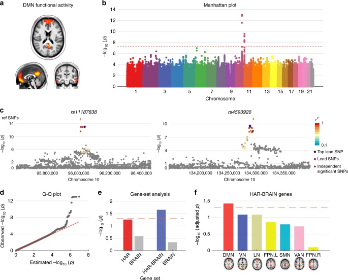
We then wanted to examine whether HAR/HAR-BRAIN genes played a role in inter-subject variation in default-mode functional activity in today’s human population. We performed a GWAS on 6,899 participants from the UK Biobank24 (see Supplementary Methods) with the amplitude of fMRI time series of the independent component analysis (ICA)-based resting-state networks (“NETMAT amplitudes 25”25 as described in https://www.fmrib.ox.ac.uk/ukbiobank/) as the phenotypes of interest. Particularly, we focused on the amplitude of the ICA component #1 that resembles the DMN (referred to as DMN amplitude, Fig. 4a). GWAS results for all single-nucleotide polymorphisms (SNP) with minor allele frequency (MAF) > 0.005 were assessed (Fig. 4b). The quantile-quantile plot showed a linkage disequilibrium score regression [LDSC] intercept of 0.999 (standard error [s.e.] = 0.006), with an inflation level of λGC = 1.005 and mean χ2 statistic = 1.012. LDSC-based SNP heritability [h2SNP] was 0.09 [s.e. = 0.06]. We observed 3 independent (r2 < 0.1) genome-wide significant SNPs (p < 5 × 10⁻⁸; using linear regression model; Fig. 4b) across 2 genomic loci (Fig. 4c). Furthermore, we annotated 19 significant SNPs (p < 5 × 10⁻⁸) with high LD (r2 ≥ 0.6) to the 3 independent SNPs using gene-mapping functions in FUMA23 (see Methods section), which resulted in a set of 12 genes (Supplementary Table 5; three genes [PLCE1, NOC3L, and SLC35G1] annotated using brain-related eQTL and Hi-C mappings). Hypergeometric testing23 showed significant enrichment of the 12 genes in the GWAS catalog26 reported gene-set “plasma clozapine-norclozapine ratio in treatment-resistant schizophrenia” (p = 1.26 × 10⁻¹²; Supplementary Table 6). None of the genes overlapped with HAR-BRAIN genes or the top 200 DMN. One gene (INPP5A) denoted as a HAR gene.
Fig. 4.
GWAS on DMN activity. a DMN component. b GWAS Manhattan plot showing –log10-transformed two-tailed p-value for all SNP (y-axis) and base-pair positions along the chromosomes (x-axis). Dotted red line indicates Bonferroni-corrected genome-wide significance (p-value < 5 × 10⁻⁸). c Regional plots of the two genomic loci (left, lead SNP: rs11187838 and right, lead SNP: rs4593926). d Q–Q plot of SNP-based p-value in panel b. Observed −log10 transformed two-tailed p-values of associations with DMN functional activity are plotted against expected null p-values for all SNPs in the GWAS. e MAGMA conditional gene-set analysis. −log10-transformed p-values of the associations between HAR/HAR-BRAIN genes and DMN functional activity conditional upon BRAIN genes. Dashed line indicates p = 0.05. f MAGMA gene-set analysis on HAR-BRAIN genes and other “NETMAT amplitude 25” phenotypes representing functional activity in the other functional networks (−log10-transformed adjusted p-values, FDR corrected). Colors indicate the assignment of functional networks, as in Fig. 2b. Dashed line indicates adjusted p = 0.05
We further investigated the potential association of HAR/HAR-BRAIN genes with variations in DMN amplitude using MAGMA linear-regression-based gene-set analysis27. We found HAR-BRAIN genes to be significantly associated with the phenotypic variation in DMN amplitude (β = 0.015, p = 0.016, FDR corrected). No significant effect was found for the set of HAR genes (β = 0.011, p = 0.051; Supplementary Table 7) or DMN genes (β = 0.005, p = 0.219). An additional conditional gene-set analysis28 including the set of BRAIN genes as a covariate, further showed a significant association of HAR-BRAIN genes with variations in DMN amplitude (β = 0.014, p = 0.022; HAR genes: β = 0.011, p = 0.055; Fig. 4e). Furthermore, no significant effect was observed when we examined the association between HAR-BRAIN genes and amplitude of other ICA components resembling the rest of the functional networks (p > 0.09; Fig. 4f and Supplementary Table 8), implicating a specific role of HAR-BRAIN genes in genetic variations of DMN functional activity. Using the normalized DMN amplitude (corrected for the mean amplitude across all networks) as the phenotype of interest showed similar results (HAR genes: β = 0.018, p = 0.003; HAR-BRAIN genes: β = 0.020, p = 0.002; Supplementary Fig. 7).
Stratified LDSC analysis29 did not result in findings of significant enrichment of genetic variance of DMN functional activity explained by SNPs overlapping with HAR regions. This is likely related to HARs occupying relatively small regions in the whole genome with a mean length of only 256 base pairs (Supplementary Fig. 8), which limits statistical power to perform such post-hoc analyses. The relatively large s.e. of the SNP heritability might be related to the limited sample size.
HAR genes, cognitive abilities, and psychiatric disorders
We then examined the potential role of HAR/HAR-BRAIN and DMN genes in human cognition by cross-referencing these genes with a recent GWAS meta-analysis on intelligence (h2SNP = 0.184 [s.e. = 0.008]) performed on 269,867 individuals30. Gene-set analysis27 revealed both sets of HAR and HAR-BRAIN genes to be significantly associated with individual variations in intelligence (HAR: β = 0.058, p = 5.43 × 10⁻¹⁰; HAR-BRAIN: β = 0.075, p = 1.22 × 10⁻¹⁵; Supplementary Table 7). Conditional gene-set analysis28 including BRAIN genes as a covariate further confirmed a significant association of both HAR and HAR-BRAIN genes with intelligence (HAR: β = 0.056, p = 1.60 × 10⁻⁹; HAR-BRAIN: β = 0.060, p = 6.34 × 10⁻¹⁰). Intelligence was not found to be specifically associated with the total set of DMN genes (β = 0.012, p = 0.061), but a significant effect was observed for the subset of 37 intersected HAR-DMN genes (β = 0.022, p = 0.009, FDR corrected).
We next linked HAR/HAR-BRAIN and DMN genes to genetic effects on social behavior, which is thought to be more advanced in humans than in other primate species16. Summary statistics of the trait “Frequency of friend/family visits” (h2SNP = 0.035 [s.e. = 0.002]) based on a GWAS analysis on 383,941 individuals in the UK Biobank were obtained from the GWAS ATLAS web tool31 (http://atlas.ctglab.nl, ID 3216). HAR/HAR-BRAIN genes were found to be significantly associated with this trait (HAR: β = 0.037, p = 1.24 × 10⁻⁷; HAR-BRAIN: β = 0.039, p = 1.01 × 10⁻⁷; Supplementary Table 7), with effects unrelated to the set of BRAIN genes (HAR: β = 0.037, p = 1.73 × 10⁻⁷; HAR-BRAIN: β = 0.036, p = 2.63 × 10⁻⁶). Furthermore, DMN genes were also significantly associated with individual variation in sociability (β = 0.012, p = 0.024; HAR-DMN genes: β = 0.014, p = 0.022, FDR corrected).
We also examined the potential association of HAR/HAR-BRAIN and DMN genes with schizophrenia, a disorder hypothesized to relate to human brain evolution32,33. We used the summary statistics of a GWAS in 33,426 schizophrenia patients and 54,065 healthy controls (h2SNP = 0.187 [0.008])34 provided by the Psychiatric Genomics Consortium (http://www.med.unc.edu/pgc/). We observed HAR/HAR-BRAIN genes to be significantly associated with genetic variants in schizophrenia (HAR: β = 0.019, p = 0.013; HAR-BRAIN: β = 0.043, p = 5.06 × 10⁻⁷; Supplementary Table 7). These results remained significant in the conditional gene-set analysis with BRAIN genes taken as a covariate (HAR: β = 0.017, p = 0.023; HAR-BRAIN: β = 0.028, p = 0.001). DMN genes were not found to be significantly associated with schizophrenia (β = 0.011, p = 0.067; HAR-DMN genes: β = 0.005, p = 0.261). In addition to common variations indicated by GWAS, we further examined the enrichment of HAR/HAR-BRAIN and DMN genes in rare variants of brain disorders using the NPdenovo database (http://www.wzgenomics.cn/NPdenovo/)35. Hypergeometric testing showed HAR and HAR-BRAIN genes to be significantly enriched in risk genes of ASD (p < 0.001 and p = 0.005, separately) and schizophrenia (p < 0.001 and p = 0.008, separately; FDR corrected). DMN genes also showed significant enrichment in risk genes of ASD (p = 0.004), but not schizophrenia (p = 0.264; Supplementary Fig. 9).
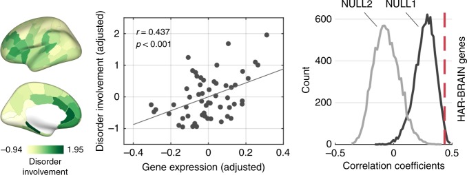
We also examined a potential association of HAR-BRAIN genes with brain changes related to psychiatric disorders. We used data from voxel-based morphometry (VBM) studies in five psychiatric brain disorders (schizophrenia, bipolar disorder, ASD, major depressive disorder [MDD] and obsessive-compulsive disorder [OCD]) and created a cortical map describing the distribution of cortical volume changes of these five psychiatric disorders (including in total of 260 VBM studies). The spatial pattern of disorder involvement across the cortex was significantly associated with the gene expression pattern of HAR-BRAIN genes (r(55) = 0.437, p < 0.001, with the cortical volume controlled; Fig. 5; r(55) = 0.221, p = 0.098 for HAR genes), an effect significantly exceeding the effect obtained by BRAIN genes (p = 0.022) and ECE genes (p < 0.001, 10,000 permutations). For an out-group analysis, the cortical pattern of HAR-BRAIN gene expression did not correlate to the disease map of five alternative, non-psychiatric disorders (amyotrophic lateral sclerosis, stroke, alcoholism, insomnia, fibromyalgia; r = 0.141, p = 0.302).
Fig. 5.
Cortical disorder involvement and HAR-BRAIN gene expression. Left panel shows brain maps of cortical involvement across five major psychiatric disorders (e.g., schizophrenia, bipolar disorder, autism spectrum disorder, major depression, and obsessive-compulsive disorder). Middle panel shows correlation between HAR-BRAIN gene expression and disorder involvement (Pearson’s r = 0.437, p < 0.001; corrected for cortical volume), and right panel shows the comparison of the correlation coefficient to null distributions generated by random BRAIN (NULL1; p = 0.022) and ECE genes (NULL2; p < 0.001, permutation test, 10,000 permutations). Source data provided as Source Data file
Discussion
Our combined comparative neuroimaging and genetic findings provide evidence of evolutionary changes in the human genome to have played a central function in the expansion and cortical organization of cognitive functional networks in the human brain, potentially in service of specialization of higher-order cognitive function in human evolution.
Our results show high levels of cortical expansion in regions of both the FPN and DMN in humans. This is compatible with prior observations of cortical expansion between macaque and human36, showing large expansion of associative prefrontal, temporal, and parietal areas in the human brain36,37. This evolutionary expansion pattern has been suggested to overlap with the pattern of cortical variation in today’s human population38, suggesting larger brains to display relatively larger multi-modal associative areas, variation further linked to inter-subject variation in cognitive abilities38. Our observations of high expression of HAR genes in these brain areas now suggest that genes linked to hominization may have had a special role in the process of cortical development of these multimodal association areas.
Our comparative analyses further show that genes associated with human brain evolution (HAR-BRAIN genes) are not equally expressed in all cortical areas but rather are more expressed in areas related to higher-order cognitive processing. HAR genes, representing conserved loci with elevated divergence in humans17,19, have been argued to function as important neuronal enhancers39 and to be key players in biological processes of nervous system development and neurogenesis, amongst others (Supplementary Table 9). HAR genes are enriched in human-evolved elements that converge on specific cell types and laminae involved in brain development and cerebral cortical expansion in the primate lineage40 and are suggested to be particularly expressed in supragranular cortical layers important for forming cortico-cortical connectivity40. Our findings of high expression of HAR genes in central cognitive networks, and most pronounced in the DMN, may thus reflect enhanced complexity of cognitive cortical areas and circuits in human brain evolution41,42.
Our results corroborate previous observations of a strong link between aspects of cellular and macroscale connectivity43,44. Association areas show transcriptional profiles enriched for genes specific to the organization of supragranular layers45, with the spatial layout of functional networks captured by coupled transcription profiles of genes enriched in supragranular layers46 and genes related to ion channel activity and synaptic function47. These observations are further in line with the notion of genes related to the resting-state brain activity of the DMN to display greater expression in neurons10. Moreover, our observation of upregulated HAR-BRAIN gene expression in cognitive networks in humans implicates an evolutionarily enhanced complexity of neuronal connectivity in cognitive networks. This might be potentially further related to humans having a longer period of neuronal progenitor expansion compared with chimpanzees and macaques contributing to a differentiated neuronal number and cortical size48.
Some of the genes found at the intersection of HAR, BRAIN, and DMN genes directly relate to the development of the human central nervous system. For example, CDH8, CDH9, and CDH10 are involved in synaptic adhesion, axon outgrowth, and guidance49, and play a role in ASD49. CBLN1 is important for synapse integrity and synaptic plasticity together with NRXN1 and GRID250. CA10 is believed to be central in the development of the central neural system by coordinating neurexins, which are presynaptic cell-adhesion molecules that bind to diverse postsynaptic ligands and who are linked to several neuropsychiatric disorders51. KCNB2 is known to be essential in regulating neurotransmitter release and neuronal excitability52.
Our findings show that genes highly expressed in the DMN contain genetic variants related to human intellectual ability and sociability. This is compatible with twin studies showing a genetic correlation between IQ and gray matter morphology of DMN regions like the medial frontal cortex and parahippocampal gyrus53. The DMN has been argued to be important for human self-projection abilities that include planning the future54, theory of mind, and navigation14, of which humans show a higher complexity compared to chimpanzees55. This central cognitive system comprises highly connected network hubs like the precuneus and inferior parietal lobule56, regions involved in multimodal information integration57, a key aspect of higher-order cognitive brain function. The observation of an association between the spatial pattern of HAR expression and cortical expansion on the one hand, and a significant involvement of HAR genes in genetic variation related to intelligence and social behavior on the other, suggests that the expansion of highly connected hub areas in support of higher-order brain function has been an important driving factor of human brain evolution.
Evolutionary pressure on cognitive networks subserving higher-order brain functions may have been accompanied by an increased risk of brain dysfunction32,33. Our comparative findings provide evidence for this hypothesis, with genes important for human brain evolution found to play a role in the development of psychiatric disorders. The pattern of cortical expression of HAR-BRAIN genes shows significant overlap with the pattern of cortical involvement across mental disorders, with particular involvement of lateral and medial prefrontal cortices. These are key ‘brain hubs’ and components of higher-order networks identified to be generally implicated in the anatomy of a wide range of brain disorders58. Our findings further suggest HAR and DMN genes to significantly relate to the genetic architecture for schizophrenia and autism, disorders that are often reported to involve disturbed DMN functional connectivity59,60. These findings are consistent with reported genetic associations between the DMN and psychiatric disorders61 and support the notion of genes related to evolutionary adaptations and brain development to potentially contribute to default-mode network involvement in brain disorders61.
Several methodological points have to be discussed. We used predefined functional networks to link data from distinct modalities. Network divisions may overlook functional heterogeneity across cortical regions and participation of brain regions in multiple networks, and several other spatial variations of networks are equally viable62 (see Supplementary Note 5 for alternative definitions of networks, Supplementary Fig. 10–11). Second, the set of HAR genes as used in this study was selected ‘as-is’ from the study of Doan et al.19. HAR-associated genes were labeled as those where HARs are within the introns, within or near (less than 1 kb) 5′ and 3′ UTRs, or are the closest flanking gene that was less than 2.1 mb away (with 70% being less than 500 kb away)19. Other mapping approaches can be used to identify and further specify HAR gene sets linked to specific functions. We examined alternative sets of 196 genes mapped from HAR using brain-related Hi-C and eQTL datasets from the PsychENCODE Consortium63, and a set of 396 genes related to ASD-linked HAR mutations identified using massively parallel reporter assays19. These alternative selections and allocations of HAR genes revealed highly consistent findings (data presented in detail in Supplementary Note 1).
Our comparative study shows that recent changes in our genome have played a central role in the expansion and function of higher-order cognitive networks in the human brain. Our findings suggest that expansion of higher-order functional networks and their cognitive properties have potentially been an important locus of change in recent human brain evolution.
Methods
Cortical expansion
In vivo MRI data from 29 adult chimpanzees and 50 adult human subjects were analyzed (see Supplementary Methods for details). Data of chimpanzees were acquired under protocols approved by the YNPRC and the Emory University Institutional Animal Care and Use Committee (IACUC, approval #: YER2001206) (see also Ethics statement). MRI data of humans were obtained from the Human Connectome Project (https://www.humanconnectome.org). T1-weighted scans of chimpanzees and humans were processed using FreeSurfer (v5.3.0; https://surfer.nmr.mgh.harvard.edu/) for tissue classification, cortical ribbon reconstruction, and brain parcellation. Pial surface reconstructions were used for vertex-to-vertex mapping across chimpanzee and humans and subsequent computation of vertex-wise and region-wise expansion (114-region subdivision of the Desikan-Killiany atlas [DK-114]64,65, 57 per hemisphere; see Supplementary Methods and Supplementary Figs. 12–13). Vertex-wise and region-wise expansion maps are available at https://www.connectomelab.nl/downloads. Validation analysis was performed using the chimpanzee-human BB38 atlas that describes homologous cortical regions between two species33 (Supplementary Fig. 14).
Gene expression
AHBA. Cortical gene expression patterns were taken from the transcriptomic data of the Allen Human Brain Atlas (AHBA, http://human.brain-map.org/static/download), including a detailed dataset of microarray gene expression data from brain samples of six human donors (all without a history of neuropsychiatric or neuropathological disorders, demographics tabulated in Supplementary Table 10). Data included expression levels of 20,734 genes represented by 58,692 probes for each cortical region of the left hemisphere4,66. Tissue samples were spatially mapped to each of the cortical regions of the FreeSurfer DK-114 atlas64,65, based on their distance to the nearest voxel within the cortical ribbon of MNI 152 template (and BB38 atlas for validation, see Supplementary Note 4). Samples were normalized to Z scores and averaged across regions (see Supplementary Methods), resulting in a subject × region × gene (6 × 57 × 20,734) data matrix. Normalized gene expression data was averaged across individual datasets to obtain a group level gene expression matrix of the size of 57 × 20,734.
PsychENCODE. Comparative cortical transcription data were obtained from the PsychENCODE database (http://evolution.psychencode.org/)22, describing batch-corrected, normalized expression levels of 16,463 genes for 11 comparable cortical areas of the human (6 subjects), chimpanzee (5 subjects), and macaque brain (5 subjects, all age and gender controlled and corrected for batch effects22, see Supplementary Methods, Supplementary Table 11, and ref. 22 for details). Gene expression data were normalized to Z scores across cortical regions within each dataset, resulting in three gene expression matrices (one for each species) of the size of n × 11 × 16,463 (n = 6/5/5 for human/chimpanzee/macaque). Data were averaged across individual datasets to obtain a group level gene expression matrix of the size of 11 × 16,463 for each species.
HAR genes. Genes located in human accelerated regions (HARs) of the genome were taken as presented by comparative genome analysis representing genomic loci with accelerated divergence in humans19. A total number of 2737 human accelerated regions were identified, representing 2143 HAR-associated genes19. One thousand seven hundred and eleven HAR-associated genes were described in the AHBA dataset and used in our analyses, referred to as HAR genes.
BRAIN genes. BRAIN genes were selected as the set of genes commonly expressed in human brain tissue using the Genotype-Tissue Expression (GTEx) database (data source: GTEx Analysis Release V6p; https://www.gtexportal.org/). The GTEx portal contains 56,238 gene expression profiles in 53 body sites collected from 7333 postmortem samples in 449 individuals. From these 56,238 genes, a total number of 2823 genes were identified as BRAIN genes showing significantly higher expressions in brain sites than non-brain sites (one-sided t-test and an FDR corrected q < 0.05 were used). Four hundred and five of these 2823 genes overlapped with the set of HAR genes, referred to as HAR-BRAIN genes.
DMN genes
For each of the 20,734 AHBA genes, a two-sided two-sample t-test was performed between expression levels of regions of the DMN and regions of the other resting-state networks. Genes showing the top 200 largest t-scores (showing p < 0.004, uncorrected) were selected and referred to as DMN genes (consistent results were obtained for the set of genes reaching p < 0.05, corrected for multiple comparisons and alternatively the set of genes reaching p < 0.01 without correction; see Supplementary Note 2 and Supplementary Tables 12–13). Enrichment of HAR genes in top DMN genes was statistically evaluated using hypergeometric test. Gene-set analysis was performed for the set of DMN genes by means of the hypergeometric test implemented in the GENE2FUNC function in FUMA (http://fuma.ctglab.nl)23 (see Supplementary Methods).
DMN GWAS
GWAS was performed on 6899 participants from the UK Biobank (July 2017 release; http://www.ukbiobank.ac.uk; including individuals of European ancestry, relatives excluded). fMRI amplitude of seven ICA-based resting-state networks (described as “NETMAT amplitudes 25” in http://big.stats.ox.ac.uk/; UK Biobank field ID: 25754; for a detailed description, see refs. 25,67 and https://www.fmrib.ox.ac.uk/ukbiobank) were taken as phenotypes of interest. We focused on the phenotype “NETMAT amplitudes 25(01)”, describing ICA component #1 resembling the DMN. In addition, ICA component #2, #3, #5, #6, #10, and #14 were examined, respectively, reflecting the VN, VAN, FPN.R, FPN.L, SMN, and LN. GWAS was conducted in PLINK v2.0068, using an additive linear regression model controlling for covariates of age, sex, twenty European-based ancestry principal components, genotyping array, and total brain volume (derived from the T1 image, linearly transformed to mean zero and variance one). Stringent quality control measures were applied to the summary statistics of the GWAS (see Supplementary Methods and ref. 30 for a detailed description of the used procedures). SNP-based results were mapped and annotated using positional mapping, eQTL mapping, and chromatin interaction mapping as implemented in the SNP2GENE function in FUMA23. MAGMA gene-set analysis was used to examine the association of HAR/HAR-BRAIN/DMN genes with phenotypic variations27,28.
Gene-set analysis
SNP-based summary statistics of three GWAS were obtained, including (1) a recent GWAS meta-analysis on intelligence in 269,867 individuals30 (https://ctg.cncr.nl/software/summary_statistics); (2) a GWAS of a social interaction related trait, “Frequency of friend/family visits”, in 383,941 individuals in the UK Biobank31 (field ID: 1031; GWAS ATLAS web tool, http://atlas.ctglab.nl/traitDB/3216); (3) a GWAS in 33,426 schizophrenia patients and 54,065 healthy controls34 as provided by the Psychiatric Genomics Consortium (http://www.med.unc.edu/pgc/). Gene annotation was performed using MAGMA27, providing gene-based p-values and effect sizes that are non-directional and reflect both positive and negative direction given phenotypic variants. Gene-set analysis was performed based on a linear regression model implemented in MAGMA27 to examine to what extent HAR/HAR-BRAIN and DMN genes are associated with phenotypic variation. Results reaching an FDR corrected q < 0.05 were taken as statistically significant (corrected for all 20 tested associations). Conditional gene-set analysis28 was used to control for the effect of BRAIN genes.
Cortical involvement in psychiatric disorders
The BrainMap database was used to assess cortical involvement across five major psychiatric disorders (schizophrenia, bipolar disorder, ASD, MDD, and OCD, including in total of 260 studies) (http://www.brainmap.org). Disease voxel-based morphometry (VBM) data of 260 case-control studies present in BrainMap were extracted using the Sleuth toolbox69 and meta-analyses were conducted for each disorder using the GingerALE toolbox70. Resulting brain maps of activation likelihood estimation (ALE) were registered to the MNI 152 template and regional ALE was computed by averaging ALEs of all voxels within each cortical region of the DK-114 atlas. Regional averaged ALE scores were transformed to Z scores and averaged into a cross-disorder cortical involvement map describing per region the level of involvement across five major psychiatric disorders (see Supplementary Methods).
Statistical analysis
Pearson’s correlation was used to examine the association of the profile of cortical gene expression with the pattern of chimpanzee-to-human cortical expansion. Two-sided two-sample t-test was used to statistically test the difference in evolutionary cortical expansion and mean gene expression of HAR and HAR-BRAIN genes between regions of higher-order cognitive networks (e.g., the DMN, FPN, and VAN) and regions of the SMN/VN. Similar analysis was conducted between each of the functional networks and the rest of the brain. Results reaching an FDR corrected q < 0.05 were taken as statistically significant (corrected for eight tests in each analysis). Cohen’s d was computed, as the difference between two means divided by a standard deviation, to indicate the effect size. Permutation testing (10,000 permutations) was used to differentiate effects of HAR-BRAIN genes from effects of general BRAIN genes (referred to as NULL1) and genes associated with evolutionarily conserved elements of the human genome (ECE genes, referred to as NULL2). ECE genes were obtained from evolutionarily conserved elements in the human genome with length larger than 200 base pairs as described in ref. 21 and were mapped to genes when they fall inside the genomic location provided by the gene27, resulting in a set of 9125 genes. For each permutation (for NULL1 and NULL2), 415 genes (the same size as the number of HAR-BRAIN genes) were randomly selected from the pool of 2979 BRAIN genes or 9125 ECE genes, separately, and the same statistics (e.g., Pearson’s correlation or t-test) were computed for this random set to generate an empirical null-distribution (i.e., noted as the NULL1 distribution for BRAIN genes and NULL2 distribution for ECE genes). The original effects were assigned a two-sided p-value by comparing to the null-distributions, according to the proportion (P) of random permutations that exceeded the original statistics of HAR-BRAIN genes (p = P × 2 if P < 0.5, otherwise p = (1−P) × 2).
Ethics statement
Data of chimpanzees were acquired under protocols approved by the YNPRC and the Emory University Institutional Animal Care and Use Committee (IACUC, approval #: YER2001206). No new chimpanzee MRI data was acquired for this study; all chimpanzee MRIs were obtained from a data archive of scans obtained prior to the 2015 implementation of U.S. Fish and Wildlife Service and National Institutes of Health regulations governing research with chimpanzees.
Reporting summary
Further information on research design is available in the Nature Research Reporting Summary linked to this article.
Supplementary information
Description of Additional Supplementary Files
Acknowledgements
The work of M.P.v.d.H. was supported by an ALW open (ALWOP.179) and VIDI (452-16-015) grant from the Netherlands Organization for Scientific Research (NWO) and a Fellowship of MQ. Y.W. was supported by the China Scholarship Council (201506040039). P.R.J. was supported by the Sophia Foundation for Scientific Research (SSWO, grant s14-27). L.L. was supported by the National Institute of Mental Health (MH100029). D.P. was supported by The Netherlands Organization for Scientific Research (NWO VICI 453-14-005). Primate work was supported by National Institutes of Health Grants P01AG026423 and National Center for Research Resources P51RR165 (superseded by the Office of Research Infrastructure Programs/OD P51OD11132) to the Yerkes National Primate Research Center, and by the National Chimpanzee Brain Resource, R24NS092988. Human neuroimaging data was kindly provided by the Human Connectome Project, WU-Minn Consortium (Principal Investigators: David Van Essen and Kamil Ugurbil; 1U54MH091657) funded by the 16 NIH Institutes and Centers that support the NIH Blueprint for Neuroscience Research; and by the McDonnell Center for Systems Neuroscience at Washington University. The Genotype-Tissue Expression (GTEx) Project was supported by the Common Fund of the Office of the Director of the National Institutes of Health, and by NCI, NHGRI, NHLBI, NIDA, NIMH, and NINDS. The genetic analyses were carried out on the Genetic Cluster Computer, which is financed by the Netherlands Scientific Organization (NWO: 480-05-003), Vrije Universiteit, Amsterdam, The Netherlands, and the Dutch Brain Foundation, and is hosted by the Dutch National Computing and Networking Services SurfSARA. We would like to thank Mats Nagel (VU Amsterdam) and Ting Qi (Max Planck Institute for Human Cognitive and Brain Sciences) for helpful discussions and suggestions.
Author contributions
Y.W. and S.C.d.L. performed the analyses. M.P.v.d.H. conceived the idea of this study and supervised analyses. L.L., T.M.P., and J.K.R. collected chimpanzee MRI data. L.H.S. and D.J.A. contributed to chimpanzee data processing. P.R.J., J.E.S., and K.W. prepared genetic data. D.P. supervised the genetic analysis pipeline. Y.W. and M.P.v.d.H. wrote the paper with contributions from all coauthors.
Data availability
Human gene expression data that support the findings of this study are available in the Allen Brain Atlas (“Complete normalized microarray datasets”, http://human.brain-map.org). Comparative gene expression data that support the findings of this study are available in the PsychENCODE (Human Brain Evolution) (“Gene expression in RPKM (batch correction by Combat)”, http://www.evolution.psychencode.org). The GTEx data that support the findings of this study are available in the GTEx Portal V6p release (“The median RPKM by tissue”, https://www.gtexportal.org). The human MRI data (in the part of the cortical expansion) that support the findings of this study are available from the Human Connectome Project (Q3 release, https://www.humanconnectome.org). The chimpanzee MRI data that support the findings of this study are available as part of the National Chimpanzee Brain Resources (MRI database, https://www.chimpanzeebrain.org). The chimpanzee-human expansion map is available at https://www.connectomelab.nl/downloads. The genotype data used for the GWAS on DMN functional amplitude that support the findings of this study are available in the UK Biobank (application 16406; https://www.ukbiobank.ac.uk). The GWAS summary statistics for “intelligence” that support the findings of this study are available from https://ctg.cncr.nl/software/summary_statistics. The GWAS summary statistics for “Frequency of friend/family visits” that support the findings of this study are available from the GWAS ATLAS webtool (http://atlas.ctg.nl/traitDB/3216). The GWAS summary statistics for “schizophrenia” that support the findings of this study are available from the Psychiatric Genomics Consortium (http://www.med.unc.edu/pgc). The cross-disorder VBM data that support the findings of this study are available in the BrainMap (Sleuth, http://www.brainmap.org). The source data underlying Figs. 2c-d, 3a-f, 3h, and 5, and Supplementary Figs 1, 2, 4, and 14 are provided as Source Data files.
Code availability
All code is available from the corresponding author upon reasonable request.
Competing interests
The authors declare no competing interests.
Footnotes
Peer review information Nature Communications thanks Hyejung Won and the other, anonymous, reviewer(s) for their contribution to the peer review of this work. Peer reviewer reports are available.
Publisher’s note Springer Nature remains neutral with regard to jurisdictional claims in published maps and institutional affiliations.
Supplementary information
Supplementary information is available for this paper at 10.1038/s41467-019-12764-8.
References
- 1.Britten RJ. Divergence between samples of chimpanzee and human DNA sequences is 5%, counting indels. Proc. Natl Acad. Sci. USA. 2002;99:13633–13635. doi: 10.1073/pnas.172510699. [DOI] [PMC free article] [PubMed] [Google Scholar]
- 2.Buckner RL, Krienen FM. The evolution of distributed association networks in the human brain. Trends Cogn. Sci. 2013;17:648–665. doi: 10.1016/j.tics.2013.09.017. [DOI] [PubMed] [Google Scholar]
- 3.Preuss, T. M. Chapter 8: The human brain: evolution and distinctive features. In: On Human Nature.(Academic Press, 2017).
- 4.Ardesch, D. J., et al. Evolutionary expansion of connectivity between multimodal association areas in the human brain compared with chimpanzees. Proc. Natl Acad. Sci. USA (2019). [DOI] [PMC free article] [PubMed]
- 5.Buckner RL, Andrews-Hanna JR, Schacter DL. The brain’s default network: anatomy, function, and relevance to disease. Ann. NY Acad. Sci. 2008;1124:1–38. doi: 10.1196/annals.1440.011. [DOI] [PubMed] [Google Scholar]
- 6.Raichle ME. The brain’s default mode network. Annu Rev. Neurosci. 2015;38:433–447. doi: 10.1146/annurev-neuro-071013-014030. [DOI] [PubMed] [Google Scholar]
- 7.Uddin LQ. Salience processing and insular cortical function and dysfunction. Nat. Rev. Neurosci. 2015;16:55–61. doi: 10.1038/nrn3857. [DOI] [PubMed] [Google Scholar]
- 8.Ge T, Holmes AJ, Buckner RL, Smoller JW, Sabuncu MR. Heritability analysis with repeat measurements and its application to resting-state functional connectivity. Proc. Natl Acad. Sci. USA. 2017;114:5521–5526. doi: 10.1073/pnas.1700765114. [DOI] [PMC free article] [PubMed] [Google Scholar]
- 9.Glahn DC, et al. Genetic control over the resting brain. Proc. Natl Acad. Sci. USA. 2010;107:1223–1228. doi: 10.1073/pnas.0909969107. [DOI] [PMC free article] [PubMed] [Google Scholar]
- 10.Wang GZ, et al. Correspondence between resting-state activity and brain gene expression. Neuron. 2015;88:659–666. doi: 10.1016/j.neuron.2015.10.022. [DOI] [PMC free article] [PubMed] [Google Scholar]
- 11.Barks SK, Parr LA, Rilling JK. The default mode network in chimpanzees (Pan troglodytes) is similar to that of humans. Cereb. Cortex. 2015;25:538–544. doi: 10.1093/cercor/bht253. [DOI] [PMC free article] [PubMed] [Google Scholar]
- 12.Mantini D, et al. Default mode of brain function in monkeys. J. Neurosci. 2011;31:12954–12962. doi: 10.1523/JNEUROSCI.2318-11.2011. [DOI] [PMC free article] [PubMed] [Google Scholar]
- 13.Miranda-Dominguez O, et al. Bridging the gap between the human and macaque connectome: a quantitative comparison of global interspecies structure-function relationships and network topology. J. Neurosci. 2014;34:5552–5563. doi: 10.1523/JNEUROSCI.4229-13.2014. [DOI] [PMC free article] [PubMed] [Google Scholar]
- 14.Buckner RL, Carroll DC. Self-projection and the brain. Trends Cogn. Sci. 2007;11:49–57. doi: 10.1016/j.tics.2006.11.004. [DOI] [PubMed] [Google Scholar]
- 15.Tulving, E. Episodic memory and autonoesis: uniquely human? In: The missing link in cognition: Origins of self-reflective consciousness. (Oxford University Press, 2005).
- 16.Tomasello M, Herrmann E. Ape and human cognition: what’s the difference? Curr. Dir. Psychol. Sci. 2010;19:3–8. doi: 10.1177/0963721409359300. [DOI] [Google Scholar]
- 17.Pollard KS, et al. An RNA gene expressed during cortical development evolved rapidly in humans. Nature. 2006;443:167–172. doi: 10.1038/nature05113. [DOI] [PubMed] [Google Scholar]
- 18.Pollard KS, et al. Forces shaping the fastest evolving regions in the human genome. PLOS Genet. 2006;2:1599–1611. doi: 10.1371/journal.pgen.0020168. [DOI] [PMC free article] [PubMed] [Google Scholar]
- 19.Doan RN, et al. Mutations in human accelerated regions disrupt cognition and social behavior. Cell. 2016;167:341–354 e312. doi: 10.1016/j.cell.2016.08.071. [DOI] [PMC free article] [PubMed] [Google Scholar]
- 20.Yeo BT, et al. The organization of the human cerebral cortex estimated by intrinsic functional connectivity. J. Neurophysiol. 2011;106:1125–1165. doi: 10.1152/jn.00338.2011. [DOI] [PMC free article] [PubMed] [Google Scholar]
- 21.Lindblad-Toh K, et al. A high-resolution map of human evolutionary constraint using 29 mammals. Nature. 2011;478:476–482. doi: 10.1038/nature10530. [DOI] [PMC free article] [PubMed] [Google Scholar]
- 22.Sousa AMM, et al. Molecular and cellular reorganization of neural circuits in the human lineage. Science. 2017;358:1027–1032. doi: 10.1126/science.aan3456. [DOI] [PMC free article] [PubMed] [Google Scholar]
- 23.Watanabe K, Taskesen E, van Bochoven A, Posthuma D. Functional mapping and annotation of genetic associations with FUMA. Nat. Commun. 2017;8:1826. doi: 10.1038/s41467-017-01261-5. [DOI] [PMC free article] [PubMed] [Google Scholar]
- 24.Sudlow Cathie, Gallacher John, Allen Naomi, Beral Valerie, Burton Paul, Danesh John, Downey Paul, Elliott Paul, Green Jane, Landray Martin, Liu Bette, Matthews Paul, Ong Giok, Pell Jill, Silman Alan, Young Alan, Sprosen Tim, Peakman Tim, Collins Rory. UK Biobank: An Open Access Resource for Identifying the Causes of a Wide Range of Complex Diseases of Middle and Old Age. PLOS Medicine. 2015;12(3):e1001779. doi: 10.1371/journal.pmed.1001779. [DOI] [PMC free article] [PubMed] [Google Scholar]
- 25.Elliott LT, et al. Genome-wide association studies of brain imaging phenotypes in UK Biobank. Nature. 2018;562:210–216. doi: 10.1038/s41586-018-0571-7. [DOI] [PMC free article] [PubMed] [Google Scholar]
- 26.Buniello A, et al. The NHGRI-EBI GWAS Catalog of published genome-wide association studies, targeted arrays and summary statistics 2019. Nucleic Acids Res. 2019;47:D1005–D1012. doi: 10.1093/nar/gky1120. [DOI] [PMC free article] [PubMed] [Google Scholar]
- 27.de Leeuw CA, Mooij JM, Heskes T, Posthuma D. MAGMA: generalized gene-set analysis of GWAS data. PLoS Comput. Biol. 2015;11:e1004219. doi: 10.1371/journal.pcbi.1004219. [DOI] [PMC free article] [PubMed] [Google Scholar]
- 28.de Leeuw CA, Stringer S, Dekkers IA, Heskes T, Posthuma D. Conditional and interaction gene-set analysis reveals novel functional pathways for blood pressure. Nat. Commun. 2018;9:3768. doi: 10.1038/s41467-018-06022-6. [DOI] [PMC free article] [PubMed] [Google Scholar]
- 29.Finucane HK, et al. Partitioning heritability by functional annotation using genome-wide association summary statistics. Nat. Genet. 2015;47:1228. doi: 10.1038/ng.3404. [DOI] [PMC free article] [PubMed] [Google Scholar]
- 30.Savage JE, et al. Genome-wide association meta-analysis in 269,867 individuals identifies new genetic and functional links to intelligence. Nat. Genet. 2018;50:912–919. doi: 10.1038/s41588-018-0152-6. [DOI] [PMC free article] [PubMed] [Google Scholar]
- 31.Watanabe K, et al. A global overview of pleiotropy and genetic architecture in complex traits. Nat. Genet. 2019;51:1339–1348. doi: 10.1038/s41588-019-0481-0. [DOI] [PubMed] [Google Scholar]
- 32.Crow TJ. Is schizophrenia the price that Homo sapiens pays for language? Schizophr. Res. 1997;28:127–141. doi: 10.1016/S0920-9964(97)00110-2. [DOI] [PubMed] [Google Scholar]
- 33.van den Heuvel, M. P. et al. Evolutionarily modifications of human brain connectivity associated with schizophrenia. Brain, In press (2019). [DOI] [PMC free article] [PubMed]
- 34.Ruderfer DM, et al. Genomic dissection of bipolar disorder and schizophrenia, including 28 subphenotypes. Cell. 2018;173:1705–1715.e1716. doi: 10.1016/j.cell.2018.05.046. [DOI] [PMC free article] [PubMed] [Google Scholar]
- 35.Li J, et al. Genes with de novo mutations are shared by four neuropsychiatric disorders discovered from NPdenovo database. Mol. Psychiatry. 2016;21:290–297. doi: 10.1038/mp.2015.40. [DOI] [PMC free article] [PubMed] [Google Scholar]
- 36.Hill J, et al. Similar patterns of cortical expansion during human development and evolution. Proc. Natl Acad. Sci. USA. 2010;107:13135–13140. doi: 10.1073/pnas.1001229107. [DOI] [PMC free article] [PubMed] [Google Scholar]
- 37.Donahue CJ, Glasser MF, Preuss TM, Rilling JK, Van Essen DC. Quantitative assessment of prefrontal cortex in humans relative to nonhuman primates. Proc. Natl Acad. Sci. USA. 2018;115:E5183–E5192. doi: 10.1073/pnas.1721653115. [DOI] [PMC free article] [PubMed] [Google Scholar]
- 38.Reardon PK, et al. Normative brain size variation and brain shape diversity in humans. Science. 2018;360:1222–1226. doi: 10.1126/science.aar2578. [DOI] [PMC free article] [PubMed] [Google Scholar]
- 39.Ryu H., et al. Massively parallel dissection of human accelerated regions in human and chimpanzee neural progenitors. Preprint at https://www.biorxiv.org/content/10.1101/256313v1 (2018).
- 40.Won, H., Huang, J., Opland, C. K., Hartl, C. L., Geschwind, D. H. Human evolved regulatory elements modulate genes involved in cortical expansion and neurodevelopmental disease susceptibility. Nat. Commun.10, 2396 (2019). [DOI] [PMC free article] [PubMed]
- 41.Jacobs B, et al. Regional dendritic and spine variation in human cerebral cortex: a quantitative golgi study. Cereb. Cortex. 2001;11:558–571. doi: 10.1093/cercor/11.6.558. [DOI] [PubMed] [Google Scholar]
- 42.Elston GN, Benavides-Piccione R, DeFelipe J. The pyramidal cell in cognition: a comparative study in human and monkey. J. Neurosci. 2001;21:RC163. doi: 10.1523/JNEUROSCI.21-17-j0002.2001. [DOI] [PMC free article] [PubMed] [Google Scholar]
- 43.van den Heuvel, M. P., Scholtens, L. H., Kahn, R. S. Multiscale neuroscience of psychiatric disorders. Biol. Psychiatry 86, 512–522 (2019). [DOI] [PubMed]
- 44.Scholtens LH, Schmidt R, de Reus MA, van den Heuvel MP. Linking macroscale graph analytical organization to microscale neuroarchitectonics in the macaque connectome. J. Neurosci. 2014;34:12192–12205. doi: 10.1523/JNEUROSCI.0752-14.2014. [DOI] [PMC free article] [PubMed] [Google Scholar]
- 45.Vertes PE, et al. Gene transcription profiles associated with inter-modular hubs and connection distance in human functional magnetic resonance imaging networks. Philos. Trans. R. Soc. Lond. B Biol. Sci. 2016;371:20150362. doi: 10.1098/rstb.2015.0362. [DOI] [PMC free article] [PubMed] [Google Scholar]
- 46.Krienen FM, Yeo BT, Ge T, Buckner RL, Sherwood CC. Transcriptional profiles of supragranular-enriched genes associate with corticocortical network architecture in the human brain. Proc. Natl Acad. Sci. USA. 2016;113:E469–E478. doi: 10.1073/pnas.1510903113. [DOI] [PMC free article] [PubMed] [Google Scholar]
- 47.Richiardi J, et al. BRAIN NETWORKS. Correlated gene expression supports synchronous activity in brain networks. Science. 2015;348:1241–1244. doi: 10.1126/science.1255905. [DOI] [PMC free article] [PubMed] [Google Scholar]
- 48.Otani T, Marchetto MC, Gage FH, Simons BD, Livesey FJ. 2D and 3D stem cell models of primate cortical development identify species-specific differences in progenitor behavior contributing to brain size. Cell Stem Cell. 2016;18:467–480. doi: 10.1016/j.stem.2016.03.003. [DOI] [PMC free article] [PubMed] [Google Scholar]
- 49.Redies C, Hertel N, Hubner CA. Cadherins and neuropsychiatric disorders. Brain Res. 2012;1470:130–144. doi: 10.1016/j.brainres.2012.06.020. [DOI] [PubMed] [Google Scholar]
- 50.Hirai H, et al. Cbln1 is essential for synaptic integrity and plasticity in the cerebellum. Nat. Neurosci. 2005;8:1534–1541. doi: 10.1038/nn1576. [DOI] [PubMed] [Google Scholar]
- 51.Sterky FH, et al. Carbonic anhydrase-related protein CA10 is an evolutionarily conserved pan-neurexin ligand. Proc. Natl Acad. Sci. USA. 2017;114:E1253–E1262. doi: 10.1073/pnas.1621321114. [DOI] [PMC free article] [PubMed] [Google Scholar]
- 52.Coetzee WA, et al. Molecular diversity of K+ channels. Ann. NY Acad. Sci. 1999;868:233–285. doi: 10.1111/j.1749-6632.1999.tb11293.x. [DOI] [PubMed] [Google Scholar]
- 53.Hulshoff Pol HE, et al. Genetic contributions to human brain morphology and intelligence. J. Neurosci. 2006;26:10235–10242. doi: 10.1523/JNEUROSCI.1312-06.2006. [DOI] [PMC free article] [PubMed] [Google Scholar]
- 54.Suddendorf T, Addis DR, Corballis MC. Mental time travel and the shaping of the human mind. Philos. Trans. R. Soc. Lond. B Biol. Sci. 2009;364:1317–1324. doi: 10.1098/rstb.2008.0301. [DOI] [PMC free article] [PubMed] [Google Scholar]
- 55.Corballis MC. Mental time travel: a case for evolutionary continuity. Trends Cogn. Sci. 2013;17:5–6. doi: 10.1016/j.tics.2012.10.009. [DOI] [PubMed] [Google Scholar]
- 56.van den Heuvel MP, Sporns O. Network hubs in the human brain. Trends Cogn. Sci. 2013;17:683–696. doi: 10.1016/j.tics.2013.09.012. [DOI] [PubMed] [Google Scholar]
- 57.Barbey AK. Network neuroscience theory of human intelligence. Trends Cogn. Sci. 2018;22:8–20. doi: 10.1016/j.tics.2017.10.001. [DOI] [PubMed] [Google Scholar]
- 58.Crossley NA, et al. The hubs of the human connectome are generally implicated in the anatomy of brain disorders. Brain. 2014;137:2382–2395. doi: 10.1093/brain/awu132. [DOI] [PMC free article] [PubMed] [Google Scholar]
- 59.Padmanabhan A, Lynch CJ, Schaer M, Menon V. The default mode network in autism. Biol. Psychiatry Cogn. Neurosci. Neuroimaging. 2017;2:476–486. doi: 10.1016/j.bpsc.2017.04.004. [DOI] [PMC free article] [PubMed] [Google Scholar]
- 60.de Lange, S. C. et al. Shared vulnerability for connectome alterations across psychiatric and neurological brain disorders. Nat. Hum. Behav.3, 1–11 (2019). [DOI] [PubMed]
- 61.Meda SA, et al. Multivariate analysis reveals genetic associations of the resting default mode network in psychotic bipolar disorder and schizophrenia. Proc. Natl Acad. Sci. USA. 2014;111:E2066–E2075. doi: 10.1073/pnas.1313093111. [DOI] [PMC free article] [PubMed] [Google Scholar]
- 62.Smith SM, et al. Correspondence of the brain’s functional architecture during activation and rest. Proc. Natl Acad. Sci. USA. 2009;106:13040–13045. doi: 10.1073/pnas.0905267106. [DOI] [PMC free article] [PubMed] [Google Scholar]
- 63.Wang DF, et al. Comprehensive functional genomic resource and integrative model for the human brain. Science. 2018;362:1266. doi: 10.1126/science.aat8464. [DOI] [PMC free article] [PubMed] [Google Scholar]
- 64.Cammoun L, et al. Mapping the human connectome at multiple scales with diffusion spectrum MRI. J. Neurosci. Methods. 2012;203:386–397. doi: 10.1016/j.jneumeth.2011.09.031. [DOI] [PubMed] [Google Scholar]
- 65.Desikan RS, et al. An automated labeling system for subdividing the human cerebral cortex on MRI scans into gyral based regions of interest. NeuroImage. 2006;31:968–980. doi: 10.1016/j.neuroimage.2006.01.021. [DOI] [PubMed] [Google Scholar]
- 66.Romme IA, de Reus MA, Ophoff RA, Kahn RS, van den Heuvel MP. Connectome disconnectivity and cortical gene expression in patients with schizophrenia. Biol. Psychiatry. 2017;81:495–502. doi: 10.1016/j.biopsych.2016.07.012. [DOI] [PubMed] [Google Scholar]
- 67.Miller KL, et al. Multimodal population brain imaging in the UK Biobank prospective epidemiological study. Nat. Neurosci. 2016;19:1523–1536. doi: 10.1038/nn.4393. [DOI] [PMC free article] [PubMed] [Google Scholar]
- 68.Purcell S, et al. PLINK: a tool set for whole-genome association and population-based linkage analyses. Am. J. Hum. Genet. 2007;81:559–575. doi: 10.1086/519795. [DOI] [PMC free article] [PubMed] [Google Scholar]
- 69.Fox PT, Lancaster JL. Mapping context and content: the BrainMap model. Nat. Rev. Neurosci. 2002;3:319–321. doi: 10.1038/nrn789. [DOI] [PubMed] [Google Scholar]
- 70.Eickhoff SB, et al. Coordinate-based activation likelihood estimation meta-analysis of neuroimaging data: a random-effects approach based on empirical estimates of spatial uncertainty. Hum. Brain Mapp. 2009;30:2907–2926. doi: 10.1002/hbm.20718. [DOI] [PMC free article] [PubMed] [Google Scholar]
Associated Data
This section collects any data citations, data availability statements, or supplementary materials included in this article.
Supplementary Materials
Description of Additional Supplementary Files
Data Availability Statement
Human gene expression data that support the findings of this study are available in the Allen Brain Atlas (“Complete normalized microarray datasets”, http://human.brain-map.org). Comparative gene expression data that support the findings of this study are available in the PsychENCODE (Human Brain Evolution) (“Gene expression in RPKM (batch correction by Combat)”, http://www.evolution.psychencode.org). The GTEx data that support the findings of this study are available in the GTEx Portal V6p release (“The median RPKM by tissue”, https://www.gtexportal.org). The human MRI data (in the part of the cortical expansion) that support the findings of this study are available from the Human Connectome Project (Q3 release, https://www.humanconnectome.org). The chimpanzee MRI data that support the findings of this study are available as part of the National Chimpanzee Brain Resources (MRI database, https://www.chimpanzeebrain.org). The chimpanzee-human expansion map is available at https://www.connectomelab.nl/downloads. The genotype data used for the GWAS on DMN functional amplitude that support the findings of this study are available in the UK Biobank (application 16406; https://www.ukbiobank.ac.uk). The GWAS summary statistics for “intelligence” that support the findings of this study are available from https://ctg.cncr.nl/software/summary_statistics. The GWAS summary statistics for “Frequency of friend/family visits” that support the findings of this study are available from the GWAS ATLAS webtool (http://atlas.ctg.nl/traitDB/3216). The GWAS summary statistics for “schizophrenia” that support the findings of this study are available from the Psychiatric Genomics Consortium (http://www.med.unc.edu/pgc). The cross-disorder VBM data that support the findings of this study are available in the BrainMap (Sleuth, http://www.brainmap.org). The source data underlying Figs. 2c-d, 3a-f, 3h, and 5, and Supplementary Figs 1, 2, 4, and 14 are provided as Source Data files.
All code is available from the corresponding author upon reasonable request.


