PLOS ONE logoLink to PLOS ONE
. 2019 Oct 25;14(10):e0224338. doi: 10.1371/journal.pone.0224338

Circulating cell-free DNA from plasma undergoes less fragmentation during bisulfite treatment than genomic DNA due to low molecular weight

Bonnita Werner 1, Nicole Laurencia Yuwono 1, Claire Henry 1, Kate Gunther 1, Robert William Rapkins 1, Caroline Elizabeth Ford 1, Kristina Warton 1,2,*
Editor: Baochuan Lin3
PMCID: PMC6814277  PMID: 31652288

Abstract

Background

Methylation patterns in circulating cell-free DNA are potential biomarkers for cancer and other pathologies. Currently, bisulfite treatment underpins most DNA methylation analysis methods, however, it is known to fragment DNA. Circulating DNA is already short, and further fragmentation during bisulfite treatment is of concern, as it would potentially reduce the sensitivity of downstream assays.

Methods

We used high molecular weight genomic DNA to compare fragmentation and recovery following bisulfite treatment with 2 commercially available kits (Qiagen). The bisulfite treated DNA was visualised on an agarose gel and quantified by qPCR. We also bisulfite treated, visualised and quantitated circulating DNA from plasma.

Results

There was no difference in DNA fragmentation between the two kits tested, however, the Epitect Fast kit gave better recovery than the standard Epitect kit, with the same conversion efficiency. We also found that bisulfite treated circulating DNA migrates as distinct bands on agarose gels, suggesting that, in contrast to genomic DNA, it remains largely intact following treatment. Bisulfite treatment of 129 and 234 base PCR products confirmed that this was due to the short length of the circulating DNA fragments. Compared to double stranded DNA, bisulfite treated single stranded DNA gives a very weak signal on gel electrophoresis.

Conclusions

DNA fragmentation during bisulfite treatment does not contribute to loss of sensitivity in methylation analysis of circulating DNA. The absence of DNA fragments below approximately 170 bases from agarose gel images of purified circulating DNA raises the possibility that these fragments are single stranded following the DNA extraction step.

Introduction

Methylation analysis of circulating cell-free DNA (cirDNA) in blood plasma offers scope for the identification of cancer biomarkers[1, 2], as well as determining the tissues types that contribute to the cirDNA pool[3, 4]. DNA methylation is particularly relevant in the field of cancer diagnostics, since it is more consistent between individual tumours than mutation, and thus enables PCR detection of tumour DNA without a priori knowledge of the tumour mutation profile[1].

Since it was first described in 1992[5], bisulfite treatment has been the mainstay of DNA methylation analysis. Bisulfite reacts with unmethylated cytosine, resulting in conversion to uracil, while methylated cytosine is reacts at a much lower rate and the majority of residues remain unchanged. Thus, the cytosine methylation status of a DNA region can be determined by comparison of the sequence before and after bisulfite treatment.

Bisulfite treatment is known to fragment DNA as a side effect of the low pH and high temperature required for complete conversion of unmethylated cytosine to uracil. When working with high molecular weight genomic DNA, the fragmentation contributes to full conversion of the sample as it creates DNA pieces within a size range that is readily denatured, that is, no larger than 2 kb[6]. However, DNA fragmentation has been of concern when working with cirDNA, because cirDNA is already highly fragmented, with most molecules occurring at a size of 167 bases, presumably reflecting its apoptotic origin[7]. The small size of cirDNA makes it difficult to both purify[8, 9], and to detect by PCR, as any DNA molecules that contain breaks within the PCR target sequence are not available for amplification[10]. Hence, exploiting DNA methylation for cancer biomarker development has been regarded as a trade-off between the high consistency of tumour methylation patterns and the decreased assay sensitivity due to fragmentation of DNA targets by bisulfite treatment.

In this study, we visualised bisulfite treated cirDNA on an agarose gel, and showed that it undergoes relatively little fragmentation compared to high molecular weight genomic DNA. We also showed that this relative stability under bisulfite treatment conditions is due to the small size of the cirDNA fragments. This is the first time that this property of cirDNA has been demonstrated and it has implications for the development of cancer detection tests and liquid biopsies that target the low molecular weight fraction of cirDNA. We also compared two commercially available bisulfite conversion kits for DNA recovery and fragmentation.

Materials and methods

Biospecimens

This study was approved by the University of New South Wales Human Research Ethics Committee, approval number HC17020. All participants gave written informed consent. Blood samples from healthy female donors with an age range of 21 to 47 years were used. Blood was drawn into 10 mL EDTA tubes (Becton Dickenson), and plasma was separated by centrifugation at 2500 xg at 4°C for 10 minutes, followed by a second spin of the plasma at 3500 xg at 4°C for 10 minutes. All blood samples were processed into plasma within 3 hours of collection and plasma was stored at -80°C until use. All plasma samples were used in experiments within 8 months of the blood collection procedure. Plasma was thawed at room temperature, and any leftover plasma unused after thawing was discarded, i.e. each plasma sample had only been frozen once. Human genomic DNA (gDNA) purchased from Roche (Cat #11691112001) and stored at 4°C as specified by the manufacturer was used as the high molecular weight DNA sample.

CirDNA extraction

cirDNA was extracted from the plasma samples using the QIAamp Circulating Nucleic Acid (CNA) kit (cat# 55114, Qiagen) according to manufacturer’s instructions, however, for gel visualisation of cirDNA, we increased the plasma input volume in order to obtain a highly concentrated sample. We have previously shown that the CNA kit protocol can be scaled up to accommodate up to 17.5 mL plasma input with no loss of purification efficiency[11]. In this study, to visualise bisulfite treated cirDNA on a gel, a total of 102 mL of plasma from 5 donors was pooled and processed in 6 extractions of 17 mL plasma, with appropriate scaling of reagents up until the elution step. At the elution step, a single 25 μL aliquot of Elution Buffer was passed over all 6 columns, followed by a second aliquot of 25 μL then a third aliquot of 25 μL. This resulted in the cirDNA from 102 mL of plasma being collected in a total elution volume of approximately 60 μL, taking into account buffer losses on the columns.

For comparison of yield from the Epitect standard and the Epitect Fast kit, cirDNA was extracted from a total of 20 mL of plasma in 4 x 5 mL extractions with 84 μL elution volumes for each extraction. The eluted DNA was pooled to obtain a total volume of ~320 μL.

Bisulfite conversion of gDNA

1.5 μg of gDNA were bisulfite treated using either the Epitect kit (Qiagen, Cat# 59104, here referred to as Epitect standard kit) or the Epitect Fast kit (Qiagen, Cat# 59824). When using the Epitect Fast kit, the 60°C incubation steps were carried out for either 10 minutes or 20 minutes, as indicated in the figure legends. No carrier RNA was added to the reactions. All bisulfite treated gDNA samples were eluted in 25 μL of Elution Buffer.

To assess the efficiency of the bisulfite conversion, samples underwent amplification of the cancer associated gene, MGMT using the MGMT Pyro Kit (Qiagen). The kit detects methylation levels for five CpG sites spanning exon 1 with a cytosine not associated with a CpG site serving as an internal control for bisulfite conversion.

Bisulfite conversion of cirDNA

For comparison of yield from Epitect standard and Epitect Fast kit, cirDNA extracted from a total of 20 mL of blood plasma and pooled in ~320 μL of Elution Buffer was treated in two separate experiments in duplicate 40 μL volumes with each of the 2 protocols (total of 4 replicates per protocol). The 60°C incubations of the Epitect fast protocol were carried out for 20 minutes. No carrier RNA was added to the reactions. Both the Epitect standard and the Epitect Fast bisulfite treatments had a 25 μL elution volume.

For gel visualisation, cirDNA extracted from 102 mL of plasma was bisulfite converted using the Epitect Fast kit, with 40 μL of purified cirDNA converted per reaction. No carrier RNA was added to the reactions. The cirDNA was eluted in 33 μL of Elution Buffer.

Bisulfite conversion of PCR products

For each PCR product, 40 μL from a total 50 μL of PCR reaction volume were bisulfite converted using the Epitect Fast kit and 20 minute 60°C incubation times. Converted DNA was eluted in 25 μL of Elution Buffer.

Quantitative PCR

All PCR reactions were carried out on a Biorad CFX96 Real Time PCR machine. Bisulfite converted DNA was measured by quantitative PCR (qPCR) of the GSTP1 (NM_000852.3) gene promoter using methylation non-specific primers (forward primer: TTTGTGAAGIGGGTGTGTAA; reverse primer: CAAATCCCCAACIAAACCTA; product size 148 bases). PCR reactions were set up to contain 1 x buffer, 0.2 mM each dNTP (New England Biolabs, Cat# N0447S), 3 mM MgCl2, 0.2 μM of each primer, 1/10 000 dilution of SYTO9 (Thermo Fisher Scientific, Cat# S34854) and 0.16 μL per reaction of Platinum Taq Polymerase (Invitrogen, Cat# 10966). Cycling was 95°C for 3 minutes, then 95°C for 5 seconds, 58°C for 20 seconds and 72°C for 30 seconds for 45 cycles, followed by a melt curve to confirm reaction specificity. A no template control with water replacing the DNA template was included in all PCR reactions, and no amplification was observed.

A standard curve made with Epitect standard kit converted DNA was used to obtain relative quantitation of genomic DNA and cirDNA. Samples containing DNA equivalent to 1.2 ng genomic DNA input converted using the two different kits were quantified against the standard curve. A total of 4 replicates from 2 separate bisulfite conversion experiments (total of 4 replicates of each protocol) was quantitated by qPCR in triplicate.

Bisulfite treated cirDNA from quadruplicate replicates of the Epitect standard and Epitect Fast kit was quantitated in triplicate by qPCR of the GSTP1 promoter against the standard curve described above.

PCR for bisulfite treatment of short DNA fragments

DNA fragments of 234 and 129 bases were generated by PCR of SNAI1 (NM_0059850) (F primer CCTCCCTGTCAGATGAGGAC; R primer CCAGGCTGAGGTATTCCTTG) and IDH1 (NM_005896) (F primer: CGGTCTTCAGAGAAGCCATT; R primer GCAAATCACATTATTGCCAAC) respectively. PCR reaction composition was as for GSTP1 above, scaled up to 50 μL reaction volume, with cycling of 95°C for 3 minutes, then 95°C for 10 seconds, 60°C for 20 seconds and 72°C for 15 seconds for 46 cycles. From each 50 μL PCR reaction, 4 μL of untreated PCR product was loaded on a gel, while 40 μL was bisulfite converted as described below.

Gel electrophoresis

Agarose gels were made up with 1% agarose (Lonza Seakem, Cat# 50002) in 40 mM Tris, 20 mM acetic acid, 1 mM EDTA, pH 8.4 buffer (TAE buffer), plus 10 μL Gel Red stain (Biotium, Cat# 41003) per 100 mL added just before the gel was poured. For gels visualising cirDNA, 0.1 μL of 100 bp DNA ladder MWM (molecular weight markers) (New England Biolabs, Cat# N3231S) was loaded, alongside the sample volumes indicated in the figure. For gels visualising PCR product 1 μL of DNA ladder MWM was loaded alongside 4 μL of control untreated PCR product and 25 μL of bis-treated PCR product, that had been obtained from 40 μl of untreated PCR product. Gels were run at 100 V for the times indicated in the figure legends.

Statistical analysis

p-values for DNA quantitation data were calculated using unpaired one-tailed t-tests with Graphpad Prism 8 software.

Results

Epitect Fast kit versus Epitect standard kit bisulfite conversion of genomic DNA and cirDNA

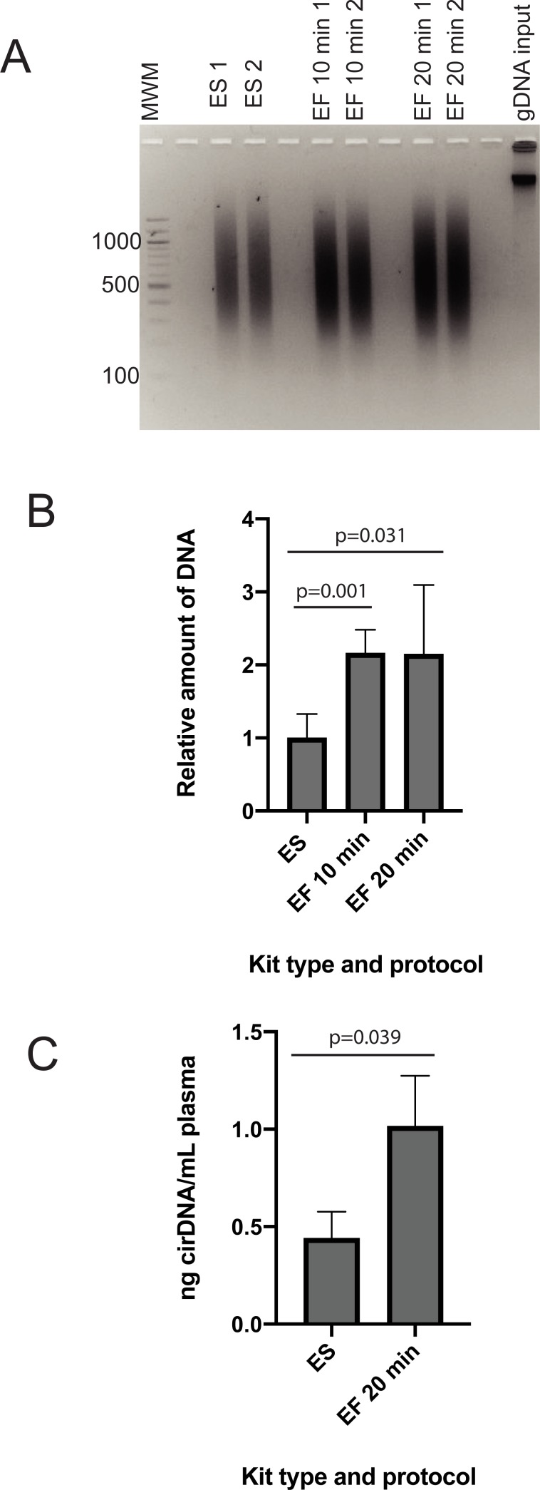
Epitect Fast kit was compared against the standard Epitect kit for recovery and size of genomic DNA after bisulfite conversion (Fig 1A and 1B). The Epitect Fast kit provides the option of carrying out the 60°C incubation steps for either 10 minutes or 20 minutes, and we compared both using genomic DNA. We used qPCR and a standard curve approach to obtain relative quantitation of Epitect standard and Epitect Fast bisulfite converted DNA. The standard curve was constructed using genomic DNA converted with the Epitect standard kit and used to quantitate bis-treated samples containing DNA equivalent to 1.2 ng genomic DNA. As expected, the sample containing Epitect standard-treated DNA returned an apparent DNA quantity of approximately 1.2 ng. The sample containing Epitect Fast-treated DNA returned an apparent DNA quantity of approximately 2.6 ng using both the 10 minute and the 20 minute conversion protocol. For the purpose of calculating the relative differences in yield shown in Fig 1B, the Epitect standard samples were set to ‘1’. Epitect Fast kit was found to give around 2-fold higher recovery. Our results on DNA recovery are similar to those of Holmes and colleagues, who reported a higher or similar recovery with the Epitect Fast kit than the Epitect standard kit, however, this was dependent on the PCR target used for quantition[12].

Fig 1.

Fig 1

(A) Agarose gel of gDNA bisulfite converted using either the ES or the EF kit. (B) Relative quantification of gDNA recovery following either ES or EF kit conversion. (C) cirDNA recovery following ES or EF kit conversion. 60°C incubation times for the EF kit are as indicated. B and C technical replicates n = 4; PCR replicates n = 3. ES–Epitect Standard kit; EF–Epitect Fast kit.

We also observed a higher recovery of cirDNA treated with the Epitect Fast kit, with the apparent cirDNA concentration in plasma of 0.44 ng/mL when using the Epitect standard kit and 1.02 ng/mL when using the Epitect Fast kit (Fig 1C).

We did not observe any decrease in the fragmentation of genomic DNA using the Epitect Fast protocol (Fig 1A). Both protocols resulted in DNA that was highly fragmented compared to the input material, with most of the sample appearing at below 1200 bases on an agarose gel. This is in contrast to the work of Holmes and colleagues, who showed a small decrease in fragmentation when using the Epitect Fast/FFPE bisulfite kit, however, the difference was very slight[12]. G-C rich sequences that contain unmethylatated cytosines are known to undergo more fragmentation than A-T rich sequences during bisulfite treatment[13], so individual loci may respond differently to conversion protocols, but we observed no difference in overall fragmentation between the two methods.

Genomic DNA samples converted using the Epitect Fast kit and the Epitect standard kit were checked for completion of bisulfite conversion using the MGMT Pyro Kit. The assay assesses 5 CpG sites within exon 1 of the tumour suppressor gene MGMT, and interrogates a cytosine nucleotide not followed by a guanine in exon 1 to determine bisulfite conversion. Both methods were found to give ≥97% bisulfite conversion. This is very similar to the results of Holmes and colleagues, who reported a 98.7% and 99.8% conversion efficiency as determined by clonal Sanger sequencing when using the Epitect standard and Epitect Fast kits respectively[12]. Based on the higher yield, Epitect Fast kit was chosen for treatment of the cirDNA samples for gel visualisation.

CirDNA undergoes less fragmentation than genomic DNA during bisulfite treatment

In pilot experiments, we consistently found that while untreated cirDNA is readily visualisable on a gel, bisulfite treated cirDNA does not produce a visible band. We tried a number of approaches to overcome this. Initially, we incubated the gel on ice prior to UV viewing, based on the premise that the bis-treated cirDNA is not visible because it is single stranded, and thus has a low affinity for Gel Red dye. Ice bath incubation, which should have increased the amount of double stranded DNA, did not result in visible bis-treated cirDNA.

We then increased the starting plasma input volume, such that the amount of bis-treated material loaded per lane was the equivalent to cirDNA extracted from 20 mL of blood plasma. This also did not result in visible bis-treated DNA.

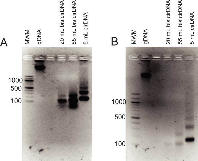
Finally, we increased the bis-treated cirDNA to the entire amount extracted from 55 mL of plasma and viewed the agarose gel under UV light after only 15 minutes of electrophoresis. With this approach, we were easily able to visualise the bis-treated cirDNA (Fig 2). With 15 minutes of electrophoresis, cirDNA from both 55 mL and 20 mL of plasma was sufficient to produce strong bands.

Fig 2.

Fig 2

Agarose gel of cirDNA and bis-treated cirDNA following (A) 15 minutes and (B) 40 minutes of electrophoresis. The amount of cirDNA in each lane corresponds to the total cirDNA extracted from the plasma volumes indicated. gDNA–untreated genomic DNA.

Several aspects of the data are notable. Firstly, distinct cirDNA bands are visible, suggesting that a large proportion of the DNA is not fragmented by bisulfite treatment, as fragmentation would likely have generated random sized DNA lengths that migrate as a smear. The cirDNA result is in contrast to genomic DNA, where all the bis-treated DNA migrated as a broad smear, between approximately 300 and 1200 bases, with no distinct bands present (Fig 1A). The bis-treated cirDNA bands migrated somewhat below the corresponding untreated cirDNA sample, with the difference most likely reflecting not change in length, but faster migration of single stranded DNA compared to double stranded DNA.

Secondly, the bis-treated DNA is largely lost from the gel image following 40 minutes of electrophoresis. Fig 2A shows that after 15 minutes, the band of bis-treated cirDNA extracted from 55 mL of plasma is much stronger than the control cirDNA extracted from 5 mL of plasma. The bis-treated cirDNA extracted from 20 mL of plasma also gives a very strong signal. In contrast, after 40 minutes (Fig 2B), the bis-treated cirDNA band from 55 mL is much weaker than the control cirDNA band, and the bis-treated cirDNA band from 20 mL is almost invisible.

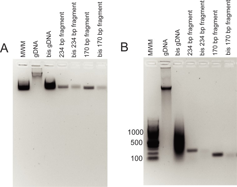
We postulated that the presence of discrete bands in bis-treated cirDNA samples was due to short DNA pieces being less susceptible to fragmentation during bisulfite treatment than long DNA pieces, and tested this by bisulfite treating PCR products of 243 and 129 bases. We found that these PCR products also underwent relatively little fragmentation, migrating as discrete bands after bisulfite treatments (Fig 3). The amount of bis-treated PCR product loaded on the gel was equivalent to 10-fold the amount of the corresponding untreated PCR product loaded, but resulted in relatively weak bands, highlighting the difficulties of visualising single-stranded DNA. The more rapid loss of the bis-treated PCR product from the gel compared to the control PCR product was confirmed by quantitation of the relative band intensities (S1 Fig).

Fig 3.

Fig 3

Agarose gel of PCR product and bis-treated PCR product following (A) 15 minutes and (B) 40 minutes of electrophoresis. Size of each PCR product is as indicated. gDNA–untreated genomic DNA; bis gDNA–bis-treated genomic DNA.

Discussion

Bisulfite treatment is well known to fragment DNA, however, we found that cirDNA persisted as discrete bands on an agarose gel after bisulfite conversion. This is due to greater stability of short DNA fragments, as demonstrated by bisulfite treatment of 234 and 129 bp PCR products. These data suggest that methylation biomarkers in cirDNA are not likely to suffer a decrease in sensitivity due to target fragmentation during bisulfite treatment. High molecular weight DNA, within the size range shown in Fig 1A and 1B, is only rarely observed in plasma samples[7], but when present will become fragmented after bisulfite treatment. This caveat applies in particular to blood samples from patients with cancer and other severe diseases, in whom higher molecular weight cirDNA and greater inter-individual variation are more likely.

We also found that the bis-treated DNA becomes less visible on the gel with increasing electrophoresis time. There are a number of mechanisms that might explain this. One is that single stranded DNA is more susceptible to DNAses than double stranded DNA and is progressively cleaved as the gel is run. The agarose gel running buffer does contain EDTA, which is generally inhibitory for DNAses, but it is possible that some DNAse activity remains. Another potential reason is that the Gel Red dye migrates towards the cathode, thus once the DNA enters the anode-most part of the gel, it is in an area where the dye concentration is insufficient to visualise single stranded DNA. However, we had previously attempted to post-stain gels to visualise bis-treated cirDNA, and this had not been effective, suggesting that dye concentration is not the main factor. A third mechanism is that the bis-treated DNA, which is single stranded, forms intramolecular and intermolecular secondary structures during migration, resulting in a sample that becomes less and less uniform with increasing electrophoresis time, and thus no longer migrates as a discrete band. If this is the case, it would suggest that the smearing visible around the bis-treated cirDNA bands is due to a range of secondary structures being present, rather than DNA fragmentation. Finally, DNAse digestion, decreased dye concentration and DNA secondary structure may all independently contribute to bis-treated cirDNA being difficult to visualise.

Our results also highlight a discrepancy between cirDNA whole genome sequencing data and cirDNA agarose gel appearance. It has been shown that single-stranded, but not double-stranded, sequencing library preparations identify a substantial fraction of cirDNA that is below 167 bases in size[14]. This was assumed to be because the protocols are better at capturing short DNA molecules, as they exclude a size restriction step[14]. However, even when a large amount of cirDNA is run on an agarose gel (Fig 2A), no DNA migrating below the main 167 base band is visible. This raises the possibility that the short DNA molecules are not only short, but also single stranded. Thus, they would be difficult to visualise on agarose gels for the same reasons, described above, that single-stranded bis-treated DNA is difficult to visualise.

If this is the case, the question arises of whether DNA fragments below 167 bases are endogenously single-stranded in blood plasma, or whether they become single-stranded as a side-effect of the DNA purification process. We believe the latter to be more likely. A commonly used method of cirDNA extraction, and the one used by us in this study, is the Circulating Nucleic Acids kit from Qiagen. This kit protocol includes a plasma proteinase K digestion step which is carried out for 30 minutes at 60°C in high concentration guanidine salt. It is likely that under these conditions, short DNA fragments become denatured. With this in mind, we ran side-by-side plasma cirDNA extractions using a 40°C digest step as well as the standard 60°C digest step. The decreased temperature resulted in a slight decrease in overall yield, but did not produce short cirDNA fragments that were visible on an agarose gel (data not shown). The reason why cirDNA fragments below 167 bases are apparent in single-stranded cirDNA sequencing libraries, but can’t be seen on a gel, remains unclear.

In conclusion, we have used agarose gel electrophoresis to show that DNA fragments below 234 bases, including cirDNA, undergo relatively little fragmentation during bisulfite treatment, and that single-stranded DNA is difficult to visualise on an agarose gel. The stability of short DNA fragments is significant for efforts to utilise methylation as a diagnostic target for cirDNA assays, since it shows that additional DNA fragmentation due to bisulfite treatment is not likely to impact assay sensitivity. High sensitivity is a critical consideration when developing cancer diagnostic and monitoring assays based on cirDNA.

Supporting information

S1 Fig. Quantitation of ssDNA on agarose gel during electrophoresis timecourse.

Agarose gel ratio of control to bisulphite treated PCR product band intensity after 15 minutes and 40 minutes of electrophoresis for (A) SNAI1 and (B) IDH1.

(AI)

S2 Fig. Raw gels.

(PDF)

Data Availability

All relevant data are within the paper.

Funding Statement

No authors were paid a salary directly via an external funding body, however, the salary of KW is from an Ovarian Cancer Research Foundation Grant, CH salary is funded by Gynaecological Oncology Fund, and RR salary is funded by Cure Brain Cancer foundation, with all salaries administered by the University of New South Wales. NY is funded by an Australian Government Research Training Program Scholarship with a Translation Cancer Research Network top up scholarship. RR is funded by. CF is funded as an employee of the University of New South Wales. The funders had no role in study design, data collection and analysis, decision to publish or preparation of the manuscript.

References

  • 1.Warton K, Samimi G. Methylation of cell-free circulating DNA in the diagnosis of cancer. Front Mol Biosci. 2015;2:13 10.3389/fmolb.2015.00013 eCollection 2015. [DOI] [PMC free article] [PubMed] [Google Scholar]
  • 2.Warton K, Mahon KL, Samimi G. Methylated circulating tumor DNA in blood: power in cancer prognosis and response. Endocr Relat Cancer. 2016;23(3):R157–71. 10.1530/ERC-15-0369 Epub 2016 Jan 13. [DOI] [PMC free article] [PubMed] [Google Scholar]
  • 3.Lehmann-Werman R, Neiman D, Zemmour H, Moss J, Magenheim J, Vaknin-Dembinsky A, et al. Identification of tissue-specific cell death using methylation patterns of circulating DNA. Proc Natl Acad Sci U S A. 2016;113(13):E1826–34. 10.1073/pnas.1519286113 Epub 2016 Mar 14. [DOI] [PMC free article] [PubMed] [Google Scholar]
  • 4.Moss J, Magenheim J, Neiman D, Zemmour H, Loyfer N, Korach A, et al. Comprehensive human cell-type methylation atlas reveals origins of circulating cell-free DNA in health and disease. Nat Commun. 2018;9(1):5068 10.1038/s41467-018-07466-6 [DOI] [PMC free article] [PubMed] [Google Scholar]
  • 5.Frommer M, McDonald LE, Millar DS, Collis CM, Watt F, Grigg GW, et al. A genomic sequencing protocol that yields a positive display of 5-methylcytosine residues in individual DNA strands. Proc Natl Acad Sci U S A. 1992;89(5):1827–31. 10.1073/pnas.89.5.1827 [DOI] [PMC free article] [PubMed] [Google Scholar]
  • 6.Clark SJ, Statham A, Stirzaker C, Molloy PL, Frommer M. DNA methylation: bisulphite modification and analysis. Nat Protoc. 2006;1(5):2353–64. 10.1038/nprot.2006.324 [DOI] [PubMed] [Google Scholar]
  • 7.Jahr S, Hentze H, Englisch S, Hardt D, Fackelmayer FO, Hesch RD, et al. DNA fragments in the blood plasma of cancer patients: quantitations and evidence for their origin from apoptotic and necrotic cells. Cancer Res. 2001;61(4):1659–65. [PubMed] [Google Scholar]
  • 8.Devonshire AS, Whale AS, Gutteridge A, Jones G, Cowen S, Foy CA, et al. Towards standardisation of cell-free DNA measurement in plasma: controls for extraction efficiency, fragment size bias and quantification. Anal Bioanal Chem. 2014;406(26):6499–512. 10.1007/s00216-014-7835-3 Epub 2014 May 24. [DOI] [PMC free article] [PubMed] [Google Scholar]
  • 9.Warton K, Graham LJ, Yuwono N, Samimi G. Comparison of 4 commercial kits for the extraction of circulating DNA from plasma. Cancer Genet. 2018;228–229:143–150. 10.1016/j.cancergen.2018.02.004 Epub Mar 6. [DOI] [PubMed] [Google Scholar]
  • 10.Andersen RF, Spindler KL, Brandslund I, Jakobsen A, Pallisgaard N. Improved sensitivity of circulating tumor DNA measurement using short PCR amplicons. Clin Chim Acta. 2015;439:97–101. 10.1016/j.cca.2014.10.011 Epub Oct 20. [DOI] [PubMed] [Google Scholar]
  • 11.Warton K, Lin V, Navin T, Armstrong NJ, Kaplan W, Ying K, et al. Methylation-capture and Next-Generation Sequencing of free circulating DNA from human plasma. BMC Genomics. 2014;15:476 10.1186/1471-2164-15-476 [DOI] [PMC free article] [PubMed] [Google Scholar]
  • 12.Holmes EE, Jung M, Meller S, Leisse A, Sailer V, Zech J, et al. Performance evaluation of kits for bisulfite-conversion of DNA from tissues, cell lines, FFPE tissues, aspirates, lavages, effusions, plasma, serum, and urine. PLoS One. 2014;9(4):e93933 10.1371/journal.pone.0093933 eCollection 2014. [DOI] [PMC free article] [PubMed] [Google Scholar]
  • 13.Olova N, Krueger F, Andrews S, Oxley D, Berrens RV, Branco MR, et al. Comparison of whole-genome bisulfite sequencing library preparation strategies identifies sources of biases affecting DNA methylation data. Genome Biol. 2018;19(1):33 10.1186/s13059-018-1408-2 [DOI] [PMC free article] [PubMed] [Google Scholar]
  • 14.Snyder MW, Kircher M, Hill AJ, Daza RM, Shendure J. Cell-free DNA Comprises an In Vivo Nucleosome Footprint that Informs Its Tissues-Of-Origin. Cell. 2016;164(1–2):57–68. 10.1016/j.cell.2015.11.050 [DOI] [PMC free article] [PubMed] [Google Scholar]

Decision Letter 0

Baochuan Lin

15 Aug 2019

PONE-D-19-20341

Circulating cell-free DNA from plasma undergoes less fragmentation during bisulphite treatment than genomic DNA due to low molecular weight

PLOS ONE

Dear Dr Warton,

Thank you for submitting your manuscript to PLOS ONE. After careful consideration, we feel that it has merit but does not fully meet PLOS ONE’s publication criteria as it currently stands. Therefore, we invite you to submit a revised version of the manuscript that addresses the points raised during the review process.

I have received the reviews of your manuscript. While your paper addresses an interesting question, the reviewers stated several concerns about your study where additional experimentation and documentation is needed.  Both reviewers voice a number of concerns regarding the used methodology for data analysis, and the limitations of the test system.  These comments need to be addressed carefully.  Please see reviewers' insightful comments below for detail.

We would appreciate receiving your revised manuscript by Sep 29 2019 11:59PM. When you are ready to submit your revision, log on to https://www.editorialmanager.com/pone/ and select the 'Submissions Needing Revision' folder to locate your manuscript file.

If you would like to make changes to your financial disclosure, please include your updated statement in your cover letter.

To enhance the reproducibility of your results, we recommend that if applicable you deposit your laboratory protocols in protocols.io, where a protocol can be assigned its own identifier (DOI) such that it can be cited independently in the future. For instructions see: http://journals.plos.org/plosone/s/submission-guidelines#loc-laboratory-protocols

Please include the following items when submitting your revised manuscript:

  • A rebuttal letter that responds to each point raised by the academic editor and reviewer(s). This letter should be uploaded as separate file and labeled 'Response to Reviewers'.

  • A marked-up copy of your manuscript that highlights changes made to the original version. This file should be uploaded as separate file and labeled 'Revised Manuscript with Track Changes'.

  • An unmarked version of your revised paper without tracked changes. This file should be uploaded as separate file and labeled 'Manuscript'.

Please note while forming your response, if your article is accepted, you may have the opportunity to make the peer review history publicly available. The record will include editor decision letters (with reviews) and your responses to reviewer comments. If eligible, we will contact you to opt in or out.

We look forward to receiving your revised manuscript.

Kind regards,

Baochuan Lin, Ph.D.

Academic Editor

PLOS ONE

Journal requirements:

When submitting your revision, we need you to address these additional requirements.

1. Please ensure that your manuscript meets PLOS ONE's style requirements, including those for file naming. The PLOS ONE style templates can be found at

http://www.journals.plos.org/plosone/s/file?id=wjVg/PLOSOne_formatting_sample_main_body.pdf and http://www.journals.plos.org/plosone/s/file?id=ba62/PLOSOne_formatting_sample_title_authors_affiliations.pdf

2. Thank you for stating the following financial disclosure:

 "The funders had no role in study design, data collection and analysis, decision to publish, or preparation of the manuscript."

a)    Please provide an amended Funding Statement that declares *all* the funding or sources of support received during this specific study (whether external or internal to your organization) as detailed online in our guide for authors at http://journals.plos.org/plosone/s/submit-now.  

b)    Please state what role the funders took in the study.  If any authors received a salary from any of your funders, please state which authors and which funder. If the funders had no role, please state: "The funders had no role in study design, data collection and analysis, decision to publish, or preparation of the manuscript."

Please include your amended statements within your cover letter; we will change the online submission form on your behalf.

3. Thank you for stating the following in the Competing Interests section:

"I have read the journal's policy and the authors of this manuscript have the following competing interests: KW - stock ownership in Guardant Health, Exact Sciences and Epigenomics AG. "

Please confirm that this does not alter your adherence to all PLOS ONE policies on sharing data and materials, by including the following statement: "This does not alter our adherence to  PLOS ONE policies on sharing data and materials.” (as detailed online in our guide for authors http://journals.plos.org/plosone/s/competing-interests).  If there are restrictions on sharing of data and/or materials, please state these. Please note that we cannot proceed with consideration of your article until this information has been declared.

Please include your updated Competing Interests statement in your cover letter; we will change the online submission form on your behalf.

Please know it is PLOS ONE policy for corresponding authors to declare, on behalf of all authors, all potential competing interests for the purposes of transparency. PLOS defines a competing interest as anything that interferes with, or could reasonably be perceived as interfering with, the full and objective presentation, peer review, editorial decision-making, or publication of research or non-research articles submitted to one of the journals. Competing interests can be financial or non-financial, professional, or personal. Competing interests can arise in relationship to an organization or another person. Please follow this link to our website for more details on competing interests: http://journals.plos.org/plosone/s/competing-interests

4. We note that you have included the phrase “data not shown” in your manuscript. Unfortunately, this does not meet our data sharing requirements. PLOS does not permit references to inaccessible data. We require that authors provide all relevant data within the paper, Supporting Information files, or in an acceptable, public repository. Please add a citation to support this phrase or upload the data that corresponds with these findings to a stable repository (such as Figshare or Dryad) and provide and URLs, DOIs, or accession numbers that may be used to access these data. Or, if the data are not a core part of the research being presented in your study, we ask that you remove the phrase that refers to these data.

5. PLOS ONE now requires that authors provide the original uncropped and unadjusted images underlying all blot or gel results reported in a submission’s figures or Supporting Information files. This policy and the journal’s other requirements for blot/gel reporting and figure preparation are described in detail at https://journals.plos.org/plosone/s/figures#loc-blot-and-gel-reporting-requirements and https://journals.plos.org/plosone/s/figures#loc-preparing-figures-from-image-files. When you submit your revised manuscript, please ensure that your figures adhere fully to these guidelines and provide the original underlying images for all blot or gel data reported in your submission. See the following link for instructions on providing the original image data: https://journals.plos.org/plosone/s/figures#loc-original-images-for-blots-and-gels.

In your cover letter, please note whether your blot/gel image data are in Supporting Information or posted at a public data repository, provide the repository URL if relevant, and provide specific details as to which raw blot/gel images, if any, are not available. Email us at plosone@plos.org if you have any questions.

[Note: HTML markup is below. Please do not edit.]

Reviewers' comments:

Reviewer's Responses to Questions

Comments to the Author

1. Is the manuscript technically sound, and do the data support the conclusions?

The manuscript must describe a technically sound piece of scientific research with data that supports the conclusions. Experiments must have been conducted rigorously, with appropriate controls, replication, and sample sizes. The conclusions must be drawn appropriately based on the data presented.

Reviewer #1: No

Reviewer #2: Partly

**********

2. Has the statistical analysis been performed appropriately and rigorously?

Reviewer #1: N/A

Reviewer #2: Yes

**********

3. Have the authors made all data underlying the findings in their manuscript fully available?

The PLOS Data policy requires authors to make all data underlying the findings described in their manuscript fully available without restriction, with rare exception (please refer to the Data Availability Statement in the manuscript PDF file). The data should be provided as part of the manuscript or its supporting information, or deposited to a public repository. For example, in addition to summary statistics, the data points behind means, medians and variance measures should be available. If there are restrictions on publicly sharing data—e.g. participant privacy or use of data from a third party—those must be specified.

Reviewer #1: Yes

Reviewer #2: Yes

**********

4. Is the manuscript presented in an intelligible fashion and written in standard English?

PLOS ONE does not copyedit accepted manuscripts, so the language in submitted articles must be clear, correct, and unambiguous. Any typographical or grammatical errors should be corrected at revision, so please note any specific errors here.

Reviewer #1: Yes

Reviewer #2: No

**********

5. Review Comments to the Author

Please use the space provided to explain your answers to the questions above. You may also include additional comments for the author, including concerns about dual publication, research ethics, or publication ethics. (Please upload your review as an attachment if it exceeds 20,000 characters)

Reviewer #1: Warton et al aim to demonstrate that cell free circulating DNA is less susceptible to fragmentation during bisulfite modification compared to genomic DNA.

The only way to assess this was visualisation on agarose gels using Gel Red Stain from Biotium.

Although these findings are generally interesting, further substantial work is required to support their conclusions:

Gel Red is an intercalating Nucleic Acid stain like Ethidium Bromide, and although less toxic, it will stain ssDNA less well despite the Biotium product description page claiming that Gel Red stains ssDNA; the aforementioned is even acknowledged on the Biotium website where it indicates that Gel Red stains ssDNA 50% less efficiently than dsDNA "Titration assays using a fluorescence microplate reader showed that the fluorescence signal of GelRed® bound to ssDNA and RNA is about half that of GelRed® bound to dsDNA." This may explain the issues of visualisation, and makes comparison with concentrations of dsDNA difficult. Alternative techniques are required to discriminate single from double-strand DNA.

In addition, it is mandatory to use alternative techniques to quantify DNA of different fragment sizes (i.e. the use of a Fragment Analyser).

The size of the cirDNA before and after bisulfite treatment should be reported (Fig 2), as the ambiguity with respect to size makes it very difficult to follow the comparison with the PCR fragment sizes used and specified in Fig 3 and the fragmentation of gDNA (300-1200 bp) (also Fig 3); the aforesaid clarification would help the discussion throughout the manuscript.

What is the nature of the “short DNA fragments” which are not found (line 67)? Do the authors mean the absence of fragmented bisulfite treated cirDNA which are smaller than the visualised sizes? It is important to accurately describe those sizes in the Results section, as “short” is relative and too vague in this context.

Reviewer #2: In their present study “Circulating cell-free DNA from plasma undergoes less fragmentation during bisulphite treatment than genomic DNA due to low molecular weight” Werner et al investigated the fragmentation of DNA caused by bisulfite reaction that is required for DNA methylation studies. The authors compared the fragmentation of high molecular weight DNA with fragmented apoptotic DNA in plasma. The manuscript is clear, concise, and well written. The results has some merit for a few researchers in the field. However, the study suffers from several limitations which should be addressed prior to its publication.

Major points

1. Test system: The authors used Human Genomic DNA from human blood (buffy coat) as a high molecular weight (>50kb according to the manufacturer) reference DNA sample. Such HMW DNA is usually not present in clinically relevant samples. Accordingly, the test system is of limited use. This should be critically discussed in the manuscript.

2. Novelty: Holmes et al. (PLoS One. 2014; 9(4): e93933.) have previously published that converting the same high molecular weight DNA using the same two Qiagen kits leads to >97% conversion; higher yields with the EpiTect Fast kit, and similar fragmentation patterns. Some of the results presented by Werner et al. are in perfect concordance with the results shown by Holmes et al.. Hence, the study by Holmes at al. should be appropriately included into the introduction and discussion of the paper.

3. Influence of extraction method on fragmentation: Both investigated kits use silica membrane spin column-based DNA purification after bisulfite conversion. This purification might have an influence on the fragmentation of HMW DNA irrespective of the bisulfite conversion. The authors should perform a control reaction in which they omit the bisulfite reagent and replace it with water in order to test the influence of the extraction method on the fragmentation.

4. Sequence-specific fragmentation: The analyzed only two loci (MGMT and GSTP1). Fragmentation of DNA due to bisulfite treatment might be sequence-specific. Do AT-rich sequences show differences regarding fragmentation as compared to GC-rich sequences which are obviously much more affected by the bisulfite reaction? This matter should be discussed.

5. In Figure 3A the authors showed that two PCR product showed the same band on the agarose gel before and after bisulfite conversion. The authors concluded that the fragmentation of the PCR product due to the bisulfite conversion is limited due to its small size and that the low concentration of the bisulfite converted PCR product is caused by the impaired visualization of ssDNA on an agarose gel. The results do not entirely support the conclusion. Since the authors used only 1% agarose gels, fragmented PCR products might not be visible. Other methods (e.g. 3.5% agarose gel, bioanalyzer, polyacrylamide gel) would be much better suited to visualize fragmented DNA. Secondly, the authors used PCR product as a surrogate DNA sample representing short DNA fragments. Even though the size is comparable to apoptotic DNA in plasma, PCR products might show a different behavior during the bisulfite conversion since the concentration of identical DNA sequences is much higher leading to a lower denaturation efficacy. It would be more convincing if the authors had compared the fragmentation of a large PCR product (~2kb) with their short ones (~200bp).

Minor points

1. Most scientist in the field use the spelling “bisulfite” instead of “bisulphite”. Using the more common spelling would increase the visibility of the article.

2. Since the nature of the bisulfite conversion is a major part of this study, its features should be described more accurately. It is incorrect that methylated cytosine are unreactive. Their conversion occurs at much lower rates compared to cytosines. It is not entirely correct that DNA degradation is caused by the high temperature alone. DNA degradation is mainly caused by depurination and depyrimidation leading to abasic sites, followed by DNA strand breaks due to N-glycoside bond cleavage. This depurination and depyrimidation is mainly due to the low pH needed for the bisulfite conversion. A high temperature additionally increases this problem.

3. Fig. 3: pb -> bp

4. Please use italicized gene names (IDH1, SNAI1, GSTP1).

5. The first paragraph extensively discusses features of gel electrophoresis regarding the visualization of small DNA fragments and in particular bisulfite converted small DNA fragments. Firstly, the scope of the study was not to describe the analytical performance of agarose gel electrophoresis, a method from the 1960s. Secondly, it is well known to people skilled in the art, that DNA disappears form an agarose gel after prolonged incubation, mainly due to diffusion. Thirdly, the authors used 1% agarose gel, a concentration that is not suited for small DNA fragments. In conclusion, the disappearance of small DNA fragments from the agarose gel after extended incubation time is most likely due to diffusion that is facilitated by the use of a too low concentrated agarose gel and prolonged incubation times. This paragraph of the discussion can be removed. In addition, Figures 2B and 3B can be removed since they contain basic knowledge that is already available to everyone in the field.

**********

6. PLOS authors have the option to publish the peer review history of their article (what does this mean?). If published, this will include your full peer review and any attached files.

If you choose “no”, your identity will remain anonymous but your review may still be made public.

Do you want your identity to be public for this peer review? For information about this choice, including consent withdrawal, please see our Privacy Policy.

Reviewer #1: No

Reviewer #2: No

[NOTE: If reviewer comments were submitted as an attachment file, they will be attached to this email and accessible via the submission site. Please log into your account, locate the manuscript record, and check for the action link "View Attachments". If this link does not appear, there are no attachment files to be viewed.]

While revising your submission, please upload your figure files to the Preflight Analysis and Conversion Engine (PACE) digital diagnostic tool, https://pacev2.apexcovantage.com/. PACE helps ensure that figures meet PLOS requirements. To use PACE, you must first register as a user. Registration is free. Then, login and navigate to the UPLOAD tab, where you will find detailed instructions on how to use the tool. If you encounter any issues or have any questions when using PACE, please email us at figures@plos.org. Please note that Supporting Information files do not need this step.

PLoS One. 2019 Oct 25;14(10):e0224338. doi: 10.1371/journal.pone.0224338.r002

Author response to Decision Letter 0


3 Sep 2019

We thank the reviewers for their careful reading of our manuscript, and thoughtful feedback. Specific responses to their comments are listed below.

Reviewer 1

Comment

Gel Red is an intercalating Nucleic Acid stain like Ethidium Bromide, and although less toxic, it will stain ssDNA less well despite the Biotium product description page claiming that Gel Red stains ssDNA; the aforementioned is even acknowledged on the Biotium website where it indicates that Gel Red stains ssDNA 50% less efficiently than dsDNA "Titration assays using a fluorescence microplate reader showed that the fluorescence signal of GelRed® bound to ssDNA and RNA is about half that of GelRed® bound to dsDNA." This may explain the issues of visualisation, and makes comparison with concentrations of dsDNA difficult. Alternative techniques are required to discriminate single from double-strand DNA.

Response

Reviewer 1 is correct in pointing out that GelRed stains ssDNA less well than it does dsDNA. Unfortunately, there is currently no DNA stain that stains ssDNA efficiently, and this was one of the main challenges with visualising the cirDNA after bisulphite conversion. We acknowledge this limitation of the methodology by stating “the bis-treated cirDNA is not visible because it is single stranded, and thus has a low affinity for Gel Red dye” (lines 319-320). We attempted to modify the technique by cooling the gels, but without success. The value and novelty of our study is that we managed to visualise cirDNA following bisulphite treatment even in the absence of dyes that efficiently stain ssDNA.

Comment

In addition, it is mandatory to use alternative techniques to quantify DNA of different fragment sizes (i.e. the use of a Fragment Analyser).

Response

We did not attempt to run the samples on a Fragment Analyser-type platform for a number of reasons. Firstly, there is not published or anecdotal data that the dyes used in this type of system have any better affinity for ssDNA than GelRed. Secondly, we found that visualising the DNA depends on a large sample input volume, and this is not feasible with a BioAnalyser/Tape Station. Finally, we had successfully visualised the bis-converted cirDNA on an agarose gel, so did not proceed to other methods. We do however appreciate the suggestion, and will do this in the future, as this type of electrophoresis system would avoid DNA diffusion out of the gel, and may provide a stronger signal.

Comment

The size of the cirDNA before and after bisulfite treatment should be reported (Fig 2), as the ambiguity with respect to size makes it very difficult to follow the comparison with the PCR fragment sizes used and specified in Fig 3 and the fragmentation of gDNA (300-1200 bp) (also Fig 3); the aforesaid clarification would help the discussion throughout the manuscript.

Response

The size of the cirDNA following bisulphite treatment cannot be determined from the agarose gel, because there are no ssDNA molecular weight markers available for comparison. However, we infer from the discrete nature of the bis-treated DNA bands that no fragmentation took place, since fragmentation is likely to have generated random sized pieces that migrate as a smear, rather than a discrete band. Lines 335-337 have now been amended to clarify this point.

Comment

What is the nature of the “short DNA fragments” which are not found (line 67)? Do the authors mean the absence of fragmented bisulfite treated cirDNA which are smaller than the visualised sizes? It is important to accurately describe those sizes in the Results section, as “short” is relative and too vague in this context.

Response

We thank the reviewer for pointing this out, and have now amended the relevant section of the abstract to specify the size range of the fragments that were previously described as ‘short’, and as well as the discussion section (lines 421, 432 and 435-436).

Reviewer 2

Major points

Comment

1. Test system: The authors used Human Genomic DNA from human blood (buffy coat) as a high molecular weight (>50kb according to the manufacturer) reference DNA sample. Such HMW DNA is usually not present in clinically relevant samples. Accordingly, the test system is of limited use. This should be critically discussed in the manuscript.

Response

We agree with reviewer 2 that high molecular weight DNA, such as is shown in Figure 1 A and B is only rarely present in the circulating DNA fraction (e.g. Jahr et al., 2001), and that circulating DNA is generally low molecular weight. Data relevant to the low molecular weight size range of circulating DNA is presented in all of the figures following Figure 1B.

In our manuscript, the results with the high molecular weight DNA serve several functions. Most importantly, they provide a positive control for DNA fragmentation in the bisulphite conversion protocols we carried out, showing that the low molecular weight DNA didn’t remain intact simply because the protocol was not applied correctly.

Secondly, they allowed a comparison of the conversion efficiency of the two bisulphite kits tested. This could not be done on cirDNA, because the quantity that can readily be obtained from plasma is too low for analysis with the MGMT Pyro kit used to measure efficiency.

Finally, a lane with control high molecular weight DNA is included in all gels that show low molecular weight DNA results, firstly, for reference, and secondly, to aid us in interpretation of results in case any unexpected HMW bands appeared in other lanes.

In response to the reviewer comment, we have modified the discussion to include a sentence of the limitations of using high molecular weight DNA in this context (lines 383-386).

Comment

2. Novelty: Holmes et al. (PLoS One. 2014; 9(4): e93933.) have previously published that converting the same high molecular weight DNA using the same two Qiagen kits leads to >97% conversion; higher yields with the EpiTect Fast kit, and similar fragmentation patterns. Some of the results presented by Werner et al. are in perfect concordance with the results shown by Holmes et al.. Hence, the study by Holmes at al. should be appropriately included into the introduction and discussion of the paper.

Response

We have now included a reference to Holmes et al, 2014 when presenting the results (lines 272-275, 294-296 and 305-308). We note that Holmes et al only examined high molecular weight genomic DNA input, and all our data regarding cirDNA is novel.

Comment

3. Influence of extraction method on fragmentation: Both investigated kits use silica membrane spin column-based DNA purification after bisulfite conversion. This purification might have an influence on the fragmentation of HMW DNA irrespective of the bisulfite conversion. The authors should perform a control reaction in which they omit the bisulfite reagent and replace it with water in order to test the influence of the extraction method on the fragmentation.

Response

The suggestion that interaction with the spin column might contribute to fragmentation of the DNA is interesting, although we note that spin column purification of genomic DNA from cell line and tissue samples does not generally result in a fragmented sample. We agree that isolating steps of the bisulphite conversion protocol to identify the one that is responsible for DNA degradation is worthwhile, but unfortunately beyond the scope of our current study.

Comment

4. Sequence-specific fragmentation: The analyzed only two loci (MGMT and GSTP1). Fragmentation of DNA due to bisulfite treatment might be sequence-specific. Do AT-rich sequences show differences regarding fragmentation as compared to GC-rich sequences which are obviously much more affected by the bisulfite reaction? This matter should be discussed.

Response

We have now included a discussion of the influence of DNA sequence on bisulphite induced fragmentation (line 296- 299).

5. In Figure 3A the authors showed that two PCR product showed the same band on the agarose gel before and after bisulfite conversion. The authors concluded that the fragmentation of the PCR product due to the bisulfite conversion is limited due to its small size and that the low concentration of the bisulfite converted PCR product is caused by the impaired visualization of ssDNA on an agarose gel. The results do not entirely support the conclusion. Since the authors used only 1% agarose gels, fragmented PCR products might not be visible. Other methods (e.g. 3.5% agarose gel, bioanalyzer, polyacrylamide gel) would be much better suited to visualize fragmented DNA.

Response

We agree with Reviewer 2 that a high concentration (e.g. 3.5%) agarose gel would have been more appropriate to visualise the bis-treated DNA in Figure 2. We did initially run the bis treated cirDNA samples on 3% agarose gels, but weren’t able to see the DNA (data not shown), in retrospect, most likely due to a combination of insufficient input and running the gel for too long. We eventually turned to low (1%) concentration gels in an attempt to decrease the resolution of low molecular weight fragments, thereby compressing the anticipated smear of fragmented cirDNA and making it more readily visible. The finding that the bis-treated cirDNA was a discrete band was unanticipated. Repeating the experiment with a higher concentration gel would not substantially change our main finding that short DNA fragments, including cirDNA, undergo less degradation during bisulfite treatment. As the experiment consumes well over 100 mL of donor blood, we feel that running the same type of sample on a higher concentration gel is not justified.

Comment

Secondly, the authors used PCR product as a surrogate DNA sample representing short DNA fragments. Even though the size is comparable to apoptotic DNA in plasma, PCR products might show a different behavior during the bisulfite conversion since the concentration of identical DNA sequences is much higher leading to a lower denaturation efficacy. It would be more convincing if the authors had compared the fragmentation of a large PCR product (~2kb) with their short ones (~200bp).

Response

The reviewer is suggesting that short PCR fragments and short cirDNA fragments resist bis fragmentation via two different mechanisms – high self complementarity and short length respectively. We agree that PCR products might show a lower denaturation efficiency due to a high concentration of complementary sequence, however, the denaturation conditions are very stringent (95°C), and sufficient to denature even high molecular weight DNA as is evidenced by the near-complete efficiency of unmethylated cytosine conversion in gDNA. Given that the PCR products are also completely denatured during the bis conversion, a more parsimonious explanation is that the stability of both PCR products and cirDNA is due to short length. We do acknowledge that self-complementarity might contribute to PCR product stability, but testing this experimentally is beyond the scope of the present study.

Minor Points

Comment

1. Most scientist in the field use the spelling “bisulfite” instead of “bisulphite”. Using the more common spelling would increase the visibility of the article.

Response

We have changed the spelling of “bisulphite” to “bisulfite” throughout the manuscript.

Comment

2. Since the nature of the bisulfite conversion is a major part of this study, its features should be described more accurately. It is incorrect that methylated cytosine are unreactive. Their conversion occurs at much lower rates compared to cytosines. It is not entirely correct that DNA degradation is caused by the high temperature alone. DNA degradation is mainly caused by depurination and depyrimidation leading to abasic sites, followed by DNA strand breaks due to N-glycoside bond cleavage. This depurination and depyrimidation is mainly due to the low pH needed for the bisulfite conversion. A high temperature additionally increases this problem.

Response

We have amended the manuscript to include additional information on bisulphite conversion (line 91- 97).

Comment

3. Fig. 3: pb -> bp

Response

Figure 3 has now been amended to fix the error.

Comment

4. Please use italicized gene names (IDH1, SNAI1, GSTP1).

Response

Gene names have now been italicised throughout the manuscript.

Comment

5. The first paragraph extensively discusses features of gel electrophoresis regarding the visualization of small DNA fragments and in particular bisulfite converted small DNA fragments. Firstly, the scope of the study was not to describe the analytical performance of agarose gel electrophoresis, a method from the 1960s.

Response

While agarose gel electrophoresis has been in use for a long time, its application to bisulphite treated circulating DNA, which is single stranded, is entirely novel. Our discussion of the performance of the technique relates almost entirely to single stranded DNA and is relevant to the new results presented in the paper.

Comment

Secondly, it is well known to people skilled in the art, that DNA disappears form an agarose gel after prolonged incubation, mainly due to diffusion.

Response

We entirely agree that DNA bands on a gel decrease in intensity after prolonged electrophoresis, due to a mixture of diffusion and migrating beyond the gel stain. Our observation is that this effect much more pronounced with single stranded than with double stranded DNA.

We draw the reviewers attention to the following feature of Figure 2 – the dinucleosome fragment from the 20 mL bis cirDNA has a stronger band than the 200 and 300 base pair molecular weight markers after 15 minutes of electrophoresis, but is almost invisible after 40 minutes of gel electrophoresis, while the 200 and 300 base pair molecular weight marker bands remain clear.

To support this observation we have quantitated the relative intensities of the control and bis-treated PCR products shown in Figure 3 after 15 minutes and 40 minutes of electrophoresis. Our data, now included in the manuscript as supplementary Figure 1, clearly show that there is a much greater difference between the bis treated and the control PCR products after 40 minutes than after 15 minutes. Specifically, the control product is around 2-5 times more intense after 15 minutes, but around 15 times more intense after 40 minutes of electrophoresis. Hence, we conclude that while both single stranded and double stranded DNA are lost from agarose gels in the course of electrophoresis, the single stranded DNA is lost much more rapidly.

Comment

Thirdly, the authors used 1% agarose gel, a concentration that is not suited for small DNA fragments. In conclusion, the disappearance of small DNA fragments from the agarose gel after extended incubation time is most likely due to diffusion that is facilitated by the use of a too low concentrated agarose gel and prolonged incubation times. This paragraph of the discussion can be removed. In addition, Figures 2B and 3B can be removed since they contain basic knowledge that is already available to everyone in the field.

Response

As described above, the 1% gel was used in an attempt to compress the smear of fragmented low molecular weight DNA that we anticipated after bis-treatment of cirDNA. We have addressed the disappearance of the small DNA bands in our response to the previous comment, and with Supplementary Figure 1. Figures 2 B and 3B illustrate the more rapid loss of single stranded DNA, for both cirDNA and PCR products, which accounts for bisulphite treated cirDNA never having been visualised on a gel until now. As such, these figures are important to the study.

Attachment

Submitted filename: Werner 2019 Bis DNA PLOS One respsone to reviewers-Warton.docx

Decision Letter 1

Baochuan Lin

19 Sep 2019

PONE-D-19-20341R1

Circulating cell-free DNA from plasma undergoes less fragmentation during bisulphite treatment than genomic DNA due to low molecular weight

PLOS ONE

Dear Dr Warton,

Thank you for submitting your manuscript to PLOS ONE. After careful consideration, we feel that it has merit but does not fully meet PLOS ONE’s publication criteria as it currently stands. Therefore, we invite you to submit a revised version of the manuscript that addresses the points raised during the review process.

One of the reviewers still has a few concerns regarding the manuscript that need further clarification, please see reviewer #3 comments.

We would appreciate receiving your revised manuscript by Nov 03 2019 11:59PM. When you are ready to submit your revision, log on to https://www.editorialmanager.com/pone/ and select the 'Submissions Needing Revision' folder to locate your manuscript file.

If you would like to make changes to your financial disclosure, please include your updated statement in your cover letter.

To enhance the reproducibility of your results, we recommend that if applicable you deposit your laboratory protocols in protocols.io, where a protocol can be assigned its own identifier (DOI) such that it can be cited independently in the future. For instructions see: http://journals.plos.org/plosone/s/submission-guidelines#loc-laboratory-protocols

Please include the following items when submitting your revised manuscript:

  • A rebuttal letter that responds to each point raised by the academic editor and reviewer(s). This letter should be uploaded as separate file and labeled 'Response to Reviewers'.

  • A marked-up copy of your manuscript that highlights changes made to the original version. This file should be uploaded as separate file and labeled 'Revised Manuscript with Track Changes'.

  • An unmarked version of your revised paper without tracked changes. This file should be uploaded as separate file and labeled 'Manuscript'.

Please note while forming your response, if your article is accepted, you may have the opportunity to make the peer review history publicly available. The record will include editor decision letters (with reviews) and your responses to reviewer comments. If eligible, we will contact you to opt in or out.

We look forward to receiving your revised manuscript.

Kind regards,

Baochuan Lin, Ph.D.

Academic Editor

PLOS ONE

[Note: HTML markup is below. Please do not edit.]

Reviewers' comments:

Reviewer's Responses to Questions

Comments to the Author

1. If the authors have adequately addressed your comments raised in a previous round of review and you feel that this manuscript is now acceptable for publication, you may indicate that here to bypass the “Comments to the Author” section, enter your conflict of interest statement in the “Confidential to Editor” section, and submit your "Accept" recommendation.

Reviewer #2: All comments have been addressed

Reviewer #3: (No Response)

**********

2. Is the manuscript technically sound, and do the data support the conclusions?

The manuscript must describe a technically sound piece of scientific research with data that supports the conclusions. Experiments must have been conducted rigorously, with appropriate controls, replication, and sample sizes. The conclusions must be drawn appropriately based on the data presented.

Reviewer #2: (No Response)

Reviewer #3: No

**********

3. Has the statistical analysis been performed appropriately and rigorously?

Reviewer #2: (No Response)

Reviewer #3: N/A

**********

4. Have the authors made all data underlying the findings in their manuscript fully available?

The PLOS Data policy requires authors to make all data underlying the findings described in their manuscript fully available without restriction, with rare exception (please refer to the Data Availability Statement in the manuscript PDF file). The data should be provided as part of the manuscript or its supporting information, or deposited to a public repository. For example, in addition to summary statistics, the data points behind means, medians and variance measures should be available. If there are restrictions on publicly sharing data—e.g. participant privacy or use of data from a third party—those must be specified.

Reviewer #2: (No Response)

Reviewer #3: Yes

**********

5. Is the manuscript presented in an intelligible fashion and written in standard English?

PLOS ONE does not copyedit accepted manuscripts, so the language in submitted articles must be clear, correct, and unambiguous. Any typographical or grammatical errors should be corrected at revision, so please note any specific errors here.

Reviewer #2: (No Response)

Reviewer #3: Yes

**********

6. Review Comments to the Author

Please use the space provided to explain your answers to the questions above. You may also include additional comments for the author, including concerns about dual publication, research ethics, or publication ethics. (Please upload your review as an attachment if it exceeds 20,000 characters)

Reviewer #2: (No Response)

Reviewer #3: There are several major concerns about this study. 1. The clinical meaning is not clear. How the community can use the findings? 2. Circulating DNA from patients with cancer or other severe diseases could be very different from healthy individuals. 3. The small sample size used can not describe sufficiently inter-individual variation.

**********

7. PLOS authors have the option to publish the peer review history of their article (what does this mean?). If published, this will include your full peer review and any attached files.

If you choose “no”, your identity will remain anonymous but your review may still be made public.

Do you want your identity to be public for this peer review? For information about this choice, including consent withdrawal, please see our Privacy Policy.

Reviewer #2: No

Reviewer #3: No

[NOTE: If reviewer comments were submitted as an attachment file, they will be attached to this email and accessible via the submission site. Please log into your account, locate the manuscript record, and check for the action link "View Attachments". If this link does not appear, there are no attachment files to be viewed.]

While revising your submission, please upload your figure files to the Preflight Analysis and Conversion Engine (PACE) digital diagnostic tool, https://pacev2.apexcovantage.com/. PACE helps ensure that figures meet PLOS requirements. To use PACE, you must first register as a user. Registration is free. Then, login and navigate to the UPLOAD tab, where you will find detailed instructions on how to use the tool. If you encounter any issues or have any questions when using PACE, please email us at figures@plos.org. Please note that Supporting Information files do not need this step.

PLoS One. 2019 Oct 25;14(10):e0224338. doi: 10.1371/journal.pone.0224338.r004

Author response to Decision Letter 1


29 Sep 2019

We thank Reviewer 3 for the feedback on our manuscript. Specific responses to the comments are listed below.

1. The clinical meaning is not clear. How the community can use the findings?

As described in the introduction, DNA methylation is a target of clinical assay development, particularly in the field of cancer diagnosis and monitoring. We are reporting a technical observation regarding the difference in fragmentation susceptibility between long and short DNA during methylation analysis protocols. In response to the reviewers comment, we have modified the introduction to clarify that our results apply to cancer detection tests and liquid biopsies targeting methylation of the low molecular weight fraction of cirDNA (line 104). This is the DNA size range that our results pertain to. We also note that our results were contrary to expectations (i.e. we expected bisulfite treatments to fragment cirDNA), and as such expand the understanding of the method for the research community developing PCR assays that target methylated cirDNA.

2. Circulating DNA from patients with cancer or other severe diseases could be very different from healthy individuals.

Reviewer 3 states correctly the circulating DNA size distribution might vary in cancer and other disease states. We have modified our discussion to emphasize that our results apply to DNA in the size range of ~170 bases, and cannot be extrapolated to disease states that change the size distribution of cirDNA to include high molecular weight fragments (lines 331-333). However, short DNA fragments are expected to behave in the same way, regardless of the additional presence of long fragments (please see comment below).

3. The small sample size used can not describe sufficiently inter-individual variation.

We are not aiming to describe individual variation in DNA susceptibility to fragmentation, rather, we aim to report at a mechanistic level, how a commonly used molecular biology technique (bisulfite conversion) affects DNA fragments in the size range of ~170 bases, including cirDNA. We have shown that this applies to DNA from two very different sources, that is cirDNA from a biological source (plasma) and a synthetic piece of DNA (i.e. PCR product). There are major differences between these two types of DNA e.g.: PCR product has uniform complementary sequence while cirDNA has diverse sequences; PCR product and cirDNA are likely to have different purification carry-over contamination, yet both types of DNA are affected by bisulfite treatment in the same way, as both are short. This suggests that inter-individual variation cirDNA other than size is not likely to change the effect.

Having said that, it would be interesting to examine inter-individual variation, however this is beyond the scope of this study. Since the effect we described is based on DNA length, it would be expected that cirDNA samples from individuals with longer DNA will undergo more fragmentation. However, this does not affect our conclusion that short DNA fragments are not fragmented during bisulphite treatment.

Attachment

Submitted filename: Werner 2019 Bis DNA PLOS One respsone to reviewers-Warton.docx

Decision Letter 2

Baochuan Lin

11 Oct 2019

Circulating cell-free DNA from plasma undergoes less fragmentation during bisulphite treatment than genomic DNA due to low molecular weight

PONE-D-19-20341R2

Dear Dr. Warton,

We are pleased to inform you that your manuscript has been judged scientifically suitable for publication and will be formally accepted for publication once it complies with all outstanding technical requirements.

Within one week, you will receive an e-mail containing information on the amendments required prior to publication. When all required modifications have been addressed, you will receive a formal acceptance letter and your manuscript will proceed to our production department and be scheduled for publication.

Shortly after the formal acceptance letter is sent, an invoice for payment will follow. To ensure an efficient production and billing process, please log into Editorial Manager at https://www.editorialmanager.com/pone/, click the "Update My Information" link at the top of the page, and update your user information. If you have any billing related questions, please contact our Author Billing department directly at authorbilling@plos.org.

If your institution or institutions have a press office, please notify them about your upcoming paper to enable them to help maximize its impact. If they will be preparing press materials for this manuscript, you must inform our press team as soon as possible and no later than 48 hours after receiving the formal acceptance. Your manuscript will remain under strict press embargo until 2 pm Eastern Time on the date of publication. For more information, please contact onepress@plos.org.

With kind regards,

Baochuan Lin, Ph.D.

Academic Editor

PLOS ONE

Additional Editor Comments (optional):

Reviewers' comments:

Acceptance letter

Baochuan Lin

17 Oct 2019

PONE-D-19-20341R2

Circulating cell-free DNA from plasma undergoes less fragmentation during bisulphite treatment than genomic DNA due to low molecular weight

Dear Dr. Warton:

I am pleased to inform you that your manuscript has been deemed suitable for publication in PLOS ONE. Congratulations! Your manuscript is now with our production department.

If your institution or institutions have a press office, please notify them about your upcoming paper at this point, to enable them to help maximize its impact. If they will be preparing press materials for this manuscript, please inform our press team within the next 48 hours. Your manuscript will remain under strict press embargo until 2 pm Eastern Time on the date of publication. For more information please contact onepress@plos.org.

For any other questions or concerns, please email plosone@plos.org.

Thank you for submitting your work to PLOS ONE.

With kind regards,

PLOS ONE Editorial Office Staff

on behalf of

Dr. Baochuan Lin

Academic Editor

PLOS ONE

Associated Data

    This section collects any data citations, data availability statements, or supplementary materials included in this article.

    Supplementary Materials

    S1 Fig. Quantitation of ssDNA on agarose gel during electrophoresis timecourse.

    Agarose gel ratio of control to bisulphite treated PCR product band intensity after 15 minutes and 40 minutes of electrophoresis for (A) SNAI1 and (B) IDH1.

    (AI)

    S2 Fig. Raw gels.

    (PDF)

    Attachment

    Submitted filename: Werner 2019 Bis DNA PLOS One respsone to reviewers-Warton.docx

    Attachment

    Submitted filename: Werner 2019 Bis DNA PLOS One respsone to reviewers-Warton.docx

    Data Availability Statement

    All relevant data are within the paper.


    Articles from PLoS ONE are provided here courtesy of PLOS

    RESOURCES