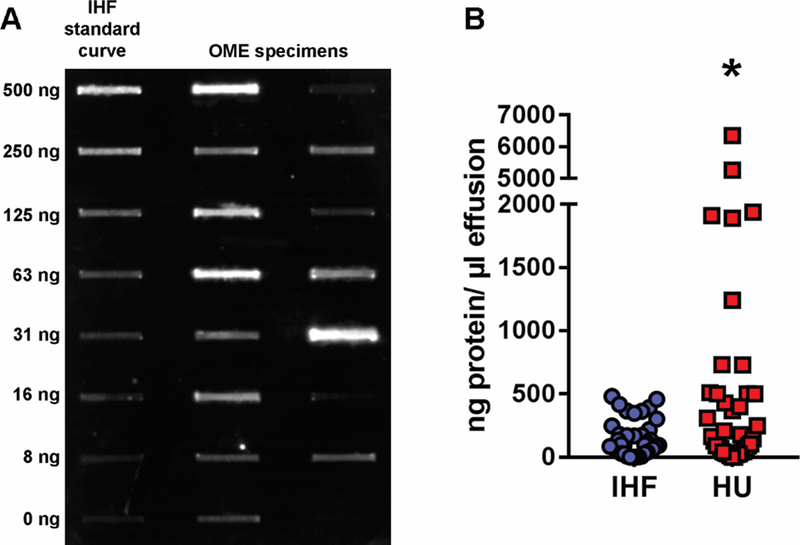
Figure 1. Quantitation of IHF and HU in effusions recovered from children with chronic OME.
Caption: Panel A, representative immunoblot to demonstrate varied amount of IHF detected in a panel of 16 OME specimens. Panel B, quantitation of IHF (blue circles) and HU (red circles). Among the 38 OME specimens collected, 95% were positive for both IHF and HU proteins by quantitative immunoblot analysis, and overall, a significantly greater amount of HU was detected, compared to IHF (P≤ 0.05). These data indicated that the immunoblot was highly sensitive in ability to both reveal the presence of DNABII proteins within the chronic OME samples, as well as quantitate them. *, P≤ 0.05

