Abstract
Combination of insect pathogenic fungi and microbial metabolites is a prospective method for mosquito control. The effect of the entomopathogenic fungus Metarhizium robertsii J.F. Bischoff, S.A. Rehner & Humber and avermectins on the survival and physiological parameters of Aedes aegypti (Linnaeus, 1762) larvae (dopamine concentration, glutathione S-transferase (GST), nonspecific esterases (EST), acid proteases, lysozyme-like, phenoloxidase (PO) activities) was studied. It is shown that the combination of these agents leads to a synergistic effect on mosquito mortality. Colonization of Ae. aegypti larvae by hyphal bodies following water inoculation with conidia is shown for the first time. The larvae affected by fungi are characterized by a decrease in PO and dopamine levels. In the initial stages of toxicosis and/or fungal infection (12 h posttreatment), increases in the activity of insect detoxifying enzymes (GST and EST) and acid proteases are observed after monotreatments, and these increases are suppressed after combined treatment with the fungus and avermectins. Lysozyme-like activity is also most strongly suppressed under combined treatment with the fungus and avermectins in the early stages posttreatment (12 h). Forty-eight hours posttreatment, we observe increases in GST, EST, acid proteases, and lysozyme-like activities under the influence of the fungus and/or avermectins. The larvae affected by avermectins accumulate lower levels of conidia than avermectin-free larvae. On the other hand, a burst of bacterial CFUs is observed under treatment with both the fungus and avermectins. We suggest that disturbance of the responses of the immune and detoxifying systems under the combined treatment and the development of opportunistic bacteria may be among the causes of the synergistic effect.
Keywords: Synergism, Lysozyme-like activity, Mosquito, Acid proteases, Phenoloxidase, Dopamine, Fungal colonization, Detoxifying enzymes, Biocontrol
Introduction
Mosquitoes are obligate intermediate hosts for a variety of pathogens that cause human mortality and morbidity worldwide. Aedes aegypti is considered to be an important vector of human diseases such as dengue and yellow fever, chikungunya, and Zika infections (Tolle, 2009; Bhatt et al., 2013), and its control is therefore an objective to prevent the transmission of these diseases. Chemical insecticides are still the most important element in mosquito control programs, despite direct and indirect toxic effects on nontarget organisms, including humans. In addition, chemicals induce resistance in a number of vector species (Vontas, Ranson & Alphey, 2010; Ranson & Lissenden, 2016; Smith, Kasai & Scott, 2016). Therefore, there is a need for alternative nonchemical vector control approaches. Classical biological control based on using various microorganisms, such as entomopathogenic fungi and bacteria, is a frequent tool for addressing this issue.
Among the biological agents employed for mosquito larvae control, bacteria from the genus Bacillus are the most widely used. In addition, products of the entomophatogenic fungi Metarhizium anisopliae s.l., and Beauveria bassiana s.l. are actively being developed for use against mosquito adults and larvae (Butt et al., 2013; Greenfield et al., 2015; Ortiz-Urquiza, Luo & Keyhani, 2015). It should be noted that mosquitoes and other insects can develop resistance to the Bacillus thuringiensis Berliner biological larvicide (Tilquin et al., 2008; Paris et al., 2011; Boyer et al., 2012). However, the resistance of insects to entomopathogenic fungi develops very slowly (Dubovskiy et al., 2013). Various species of mosquito larvae present different susceptibilities to Metarhizium, among which Ae. aegypti is the least susceptible (Greenfield et al., 2015; Garrido-Jurado et al., 2016). Thus, a concentration of conidia that is effective for Ae. aegypti control would affect a range of nontarget aquatic invertebrates. Recent studies have found that some nontarget aquatic species are more sensitive to fungal metabolites (Garrido-Jurado et al., 2016) and conidia (Belevich et al., 2017) than target mosquito species. To reduce toxic effects on the aquatic environment and increase efficacy against mosquitoes, entomopathogenic fungi may be combined with other biocontrol agents or low doses of natural insecticides. For example, combined treatment with Metarhizium and mosquito predator species (Toxorhynchites) has shown additive or synergistic effects on the mortality of Ae. aegypti (Alkhaibari et al., 2018). However, few studies have been carried out to determine the effect of combined treatment with entomopathogenic fungi and other insecticides or plant or microbial metabolites as a potential tool for improving mosquito larvae control. Synergistic effects between entomopathogenic fungi and some chemical insecticides (temephos, spinosad) (Shoukat et al., 2018; Vivekanandhan et al., 2018) or biological agents (Azadirachta indica) A. Juss (Badiane et al., 2017) on the mortality of mosquito larvae have been found. However, the physiological and biochemical aspects of this synergism were not considered.
One type of promising insecticide that can be effectively used for mosquito vector control is the avermectins. Avermectins are a class of macrocyclic lactones isolated from the soil actinomycete Streptomices avermitilis (ex Burg et al.) Kim and Goodfellow (Drinyaev et al., 1999) and include several commercial derivatives (ivermectin, abamectin, doramectin and eprinomectin) with the same mode of action—activation of glutamate-gated chloride channels, followed by uncontrolled influx of chloride ions into the cells, which leads to paralysis and death of the organism (Campbell et al., 1983). At the same time, avermectins are relatively safe for humans (Crump & Omura, 2011). Previous studies have shown that avermectins are efficient for the control of Culex quinquefasciatus Say (Freitas et al., 1996; Alves et al., 2004), Anopheles albimanus, Wiedemann An. stephensi Liston (Dreyer, Morin & Vaughan, 2018), and An. gambiae Giles (Alout et al., 2014; Chaccour et al., 2017). However, most of these studies have been carried out with adult mosquitoes feeding on blood containing ivermectin. Avermectins exhibit a relatively short half-life period, which limits their ability to kill mosquitoes but may be compensated by the application of multiple treatments or use of higher concentrations. However, these approaches may contribute to the development of mosquito larvae resistance to avermectins (Su et al., 2017). We hypothesize that the interaction of entomopathogenic fungi with avermectins can have a stable insecticidal effect at relatively low concentrations and is a promising combination for safe and effective mosquito control.
It is important that entomopathogenic fungi such as Metarhizium are adapted to terrestrial hosts and that in mosquito larvae, the fungi do not adhere to the cuticle surface and do not germinate through integuments into the hemocoel. Conidia ingested by mosquito larvae do not penetrate the gut wall (Butt et al., 2013). Thus, a “classic” host-pathogen interaction does not occur, and larval mortality is associated with stress induced by spore-bound proteases on the surface of ingested conidia (Butt et al., 2013). These authors suggest that fungal proteases cause an increase in the activity of caspases in mosquitoes, which leads to apoptosis, autolysis of tissues and death of the larvae. The activation of detoxifying enzymes and antimicrobial peptides (AMPs) occurs in larvae infected with the fungus but is not sufficient to protect the larvae from death. As a rule, mosquito larvae die showing symptoms of bacterial decomposition after treatment with Metarhizium and Beauveria (Scholte et al., 2004). Therefore, this pathogenesis can be considered mixed (both bacterial and fungal).
During this process, particular superfamilies of enzymes such as glutathione-S-transferases (GST) and nonspecific esterases (EST) are usually involved in the biochemical transformation of xenobiotics (Li, Schuler & Berenbaum, 2007). Various hormones such as biogenic amines are involved in insect stress reactions. Among them, the role of the neurotransmitter dopamine (which serves as a neurohormone as well) in this process remain poorly understood. It is known that dopamine mediates phagocytosis and is involved in the activation of the pro-phenoloxidase (proPO) cascade, thus playing an important role in fungal and bacterial pathogenesis as well as in the development of toxicoses caused by insecticides (Delpuech, Frey & Carton, 1996; Gorman, An & Kanost, 2007; Wu et al., 2015). In addition, both pathogens and toxicants can lead to changes in the antimicrobial activity of insects and the bacterial load that can affect the susceptibility of insects to pathogenic fungi (Wei et al., 2017; Ramirez et al., 2018; Polenogova et al., 2019). It should be noted that the above mentioned physiological reactions in mosquito larvae under the combined action of entomopathogenic fungi and insecticides have not yet been studied.
The aims of this study were (1) to determine the susceptibility of Aedes aegypti larvae to combined treatment with avermectins and Metarhizium robertsii and (2) estimate their immune and detoxificative responses to M. robertsii and avermectins either alone or their combination.
Materials & Methods
Insecticides and fungi
The entomopathogenic fungus Metarhizium robertsii (strain MB-1) from the collection of microorganisms of the Institute of Systematics and Ecology of Animals SB RAS was used in this work. The conidia of the fungus were grown on autoclaved millet for 10 days at 26 °C in the dark, followed by drying and sifting (Belevich et al., 2017). The industrial product “Phytoverm” 0.2% (SPC “Pharmbiomed”, Russia) was used in these experiments and includes a complex of natural avermectins (A1a (9%), A2a (18%), B1a (46%), B2a (27%)) produced by Streptomyces avermitilis.
Insect maintenance and toxicity tests
Aedes aegypti larvae from the collection of the Institute of Systematics and Ecology of Animals SB RAS were maintained in tap water in the laboratory at 24 °C (±1 °C) under a natural photoperiod (approximately 16:8 light:dark). The larvae were fed Tetramin Junior fish food (Tetra, Germany). The susceptibility of Ae. aegypti to both avermectins and the conidia of M. robertsii was tested in 200 ml plastic containers containing 100 ml of water with 15 larvae. Third-4th-instar larvae were used in the experiment. The experiment involved four treatments: control, fungus, avermectins, and fungus + avermectins. The fungal conidia and avermectins were suspended in distilled water, vortexed and applied separately or together to the containers with mosquito larvae at a volume of 2 ml per container. The final conidial concentration for infection was 1 ×10⁶ conidia/ml. The final concentration of avermectins was 0.00001%/ml. The control was treated with the same amount of distilled water. Mortality was assessed daily for 6 days. Ten replicates with 15 larvae were performed for each treatment.
Light microscopy and colonization assessment
Forty-eight hours posttreatment (pt), mosquito larvae (n = 3 for each treatment) were collected in 2% glutaraldehyde containing 0.1 M Na-cacodylate buffer (pH 7.2) and were maintained at 4 °C for 1–24 h. Semi-thin sections were stained with crystal violet and basic fuchsin and were observed with a phase-contrast microscope (Axioskop 40, Carl Zeiss, Germany).
To assess fungal colonization, 48 h pt and newly dead larvae (4–6 days pt) were cut open, and their internal contents were squeezed onto a glass side. The contents were examined for the presence/absence of hyphal bodies using light microscopy (n = 30 for each treatment). Newly dead larvae were placed on moistened filter paper in Petri dishes (n = 30) to determine the germination and surface sporulation of Metarhizium.
Total larval body supernatant
Mosquito larvae bodies of individual 4th-instar larvae of Ae. aegypti were collected in 50 µl of cool (+4 °C) 0.01 M PBS (50 mM, pH 7.4, 150 mM NaCl) with 0.1 mM N-phenylthiourea (PTU) to measure GST, EST and acid protease activities or without PTU to measure phenoloxidase (PO) activity. Then, the samples were sonicated in an ice bath with three 10 s bursts using a Bandelin Sonopuls sonicator. The sample solution was centrifuged at 20.000 g for 5 min at +4 °C. The obtained supernatant was directly used to determine enzyme activities.
Detection of phenoloxidase, glutathione-S-transferase and esterase activity
The activities of PO, GST and EST were measured at 12 and 48 h after exposure (n = 20 per treatment for each enzyme).
PO activity was assayed by using a method modified from that described by Ashida & Söderhäll (1984). The PO activity of the larval homogenates was determined spectrophotometrically on the basis of the formation of dopachrome at a wavelength of 490 nm. Aliquots of the samples (10 µl) were added to microplate wells containing 200 µl of 10 mM 3.4-dihydroxyphenylalanine and incubated at 28 °C in the dark for 45 min. The PO activity was measured kinetically every 5 min and the time point was chosen according to Michaelis constant.
The activity of EST was measured using the method of Prabhakaran & Kamble (1995) with some modifications. Aliquots of the samples (3 µl) were added to microplate wells containing 200 µl of 0.01% p-nitrophenylacetate and incubated for 10 min at 28 °C. The activity of EST was determined spectrophotometrically at a wavelength of 410 nm on the basis of the formation of nitrophenyl. The EST activity was measured kinetically every 2 min and the time point was chosen according to Michaelis constant.
The measurement of GST activity was carried out according to the method of Habig, Pabst & Jakoby (1974) with some modifications. Aliquots of the samples (7 µl) were added to microplate wells containing 200 µl of 1 mM glutathione and 5 µl of 1 mM 2.4-Dinitrochlorobenzene and incubated at 28°C for 12 min. The activity of GST was determined spectrophotometrically on the basis of the formation of 5-(2.4-dinitrophenyl)-glutathione at a 340 nm wavelength. The GST activity was measured kinetically every 3 min and the time point was chosen according to Michaelis constant.
Enzymes activity was measured in units of the transmission density (ΔA) of the incubation mixture during the reaction per 1 min and 1mg of protein. The protein concentration in the samples was determined by the method of Bradford (1976). To generate the calibration curve, bovine serum albumin was used.
Dopamine concentration measurements
Dopamine concentrations were measured at the 12 and 48 h pt in individual larval bodies (n = 10 per treatment). Mosquito larvae were homogenized in 30 µl of phosphate buffer and incubated in a Biosan TS 100 Thermoshaker for 10 min at 28 °C and 600 rpm, then incubated at room temperature for 20 min and centrifuged at 4 °C and 10.000 g for 10 min. The supernatants were transferred to clean tubes and centrifuged with the same settings for 5 min. Before transfer to the chromatograph, the samples were filtered.
Dopamine concentrations were measured by an external standard method using an Agilent 1,260 Infinity high-performance liquid chromatograph with an EsaCoulochem III electrochemical detector (cell model 5010A, potential 300 mV) according to the method of Gruntenko et al. (2005) with some modifications. Dopamine hydrochloride (Sigma-Aldrich) was used as a standard. Separation was performed in a ZorbaxSB-C18 column (4.6–250 mm, particles 5 µm) in isocratic mode. Mobile phase: 90% buffer (200 mg/l 1-OctaneSulfonicAcid (Sigma-Aldrich), 3.5 g/l KH2PO4) and 10% acetonitrile. The flow rate was 1 ml/min. Chromatogram processing was performed using ChemStation software, and the amount of dopamine was determined by comparing the peak areas of the standard and the sample.
Acid proteases
Acid protease activity was measured using method described by Anson (1938) with modifications. Fifty µl of the homogenate supernatant was added to 250 µl of 0.1 M acetate buffer (pH 4.6) containing 0.3% hemoglobin (Sigma, CAS number 9008-02-0). The samples were incubated for 60 min at 27 °C, and the reaction was stopped by adding 500 µl of 5% TCA and cooling on ice for 10 min at 4 °C. The samples were centrifuged at 14,000 g for 5 min at 4 °C, and the enzyme activity was determined spectrophotometrically at a wavelength of 280 nm in a 96-well plate reader.
Lysozyme-like activity
Lysozyme-like activity in the mosquito homogenate was determined through analysis of the lytic zone by diffusion into agar. Ten milliliters of Nutrient Agar (NA) (HiMedia, India) and Micrococcus lysodeikticus bacteria (1 × 10⁷ cells/ml) were added to Petri dishes. The agar was perforated to create 2 mm-diameter wells, which were then filled with 3 µl of full-body homogenate, followed by incubation at 37 °C for 24 h. Series of dilutions of chicken egg white lysozyme (EWL) (Sigma) (0.5 mg/ml, 0.2 mg/ml, 0.1 mg/ml, 0.005 mg/ml, 0.001 mg/ml) were added to each dish, allowing us to obtain a calibration curve based on these standards. Lytic activity was determined by measuring the diameter of the clear zone around each well and expressed as the equivalent of EWL (mg/ml) (Mohrig & Messner, 1968).
CFU counts of Metarhizium and cultivated bacteria in infected larvae
Homogenates of the mosquitoes (3 larvae per sample) were suspended in 1 ml of sterile aqueous Tween-20 (0.03%), and the suspensions were then diluted 50-fold. Next, 100 µl aliquots were inoculated onto the surface of modified Sabouraud agar (10 g peptone, 40 g D-glucose anhydrous, 20 g agar, 1 g yeast extract) supplemented with an antibiotic cocktail (acetyltrimethyl ammonium bromide 0.35 g/L; cycloheximide 0.05 g/L; tetracycline 0.05 g/L; streptomycin 0.6 g/L; PanReacAppliChem, Germany) for the inhibition of bacteria and saprotrophic fungi. The Petri dishes were maintained at 28 °C in the dark. The colonies were then counted after 7 days.
For the estimation of cultivated bacterial CFU counts, homogenates of larvae (3 larvae per sample) were suspended in 1 ml of 0.1 M phosphate buffer. Then, the suspension was diluted to 10−2, 10−3 and 10−4. Aliquots of 100 µl of the larval dilutions were inoculated onto the surface of blood agar media (HiMedia, Mumbai, India). The Petri dishes were maintained at 28 °C. The colonies were counted after 48 h. Three samples of each treatment were used in the analysis.
Statistical analysis
Data were analyzed using GraphPad Prism v.4.0 (GraphPad Software Inc., USA), Statistica 8 (StatSoft Inc., USA), PAST 3 (Hammer, Harper & Ryan, 2001) and AtteStat 12.5 (Gaidyshev, 2004). Differences between synergistic and additive effects were determined by comparing the expected and observed insect mortality using the χ2 criterion (Robertson & Preisler, 1992). The expected mortality from dual treatment was calculated by the formula PE = P0 + (1 − P0) × (P1) + (1 − P0) × (1 − P1) × (P2), where PE is the expected mortality after combined treatment with fungus and avermectins, P0 is mortality in the control groups, P1 is the mortality posttreatment with M. robertsii, P2 is the mortality posttreatment with avermectins. The χ2 values were calculated by the formula χ2 = (L0 − LE)2/LE + (D0 − DE)2/DE, where L0 is the observed number of survived larvae, LE is the expected number of surviving larvae, D0 is the observed number of dead larvae, and DE is the expected number of dead larvae. This formula was used to test the hypothesis of independence (1 df: P = 0.05). Additive effect was indicated if χ2 < 3.84. A synergistic effect was indicated if χ2 > 3.84 and observed mortality greater than the expected one. A value of 3.84 corresponds to P < 0.05 with a degree of freedom = 1. The Kaplan–Meier test was used to calculate the median lethal time (presented as LT50 ± SE). A log-rank test was used to quantify differences in mortality dynamics. As the distribution of the physiological parameters except for the dopamine concentration deviated from a normal distribution (Shapiro–Wilk test, P < 0.05), we used the nonparametric equivalent of a two-way ANOVA: the Scheirer-Ray-Hare test (Scheirer, Ray & Hare, 1976), followed by Dunn’s post hoc test. The data on dopamine concentrations passed the normality test (Shapiro–Wilk test, P > 0.05) and were analyzed by two-way ANOVA followed by Tukey’s post hoc test. Differences between Metarhizium CFU counts were compared by t-tests.
Results
Synergy between avermectins and the fungus
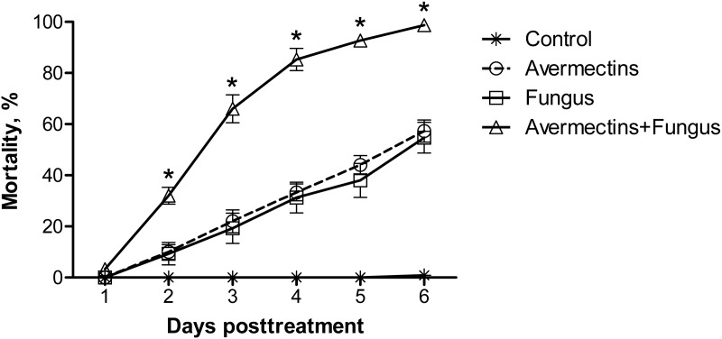
Significant differences in the dynamics of larval mortality between the treatments were observed (log-rank test: χ2 = 397.3, df = 3, P < 0.0001; Fig. 1). Treatment with avermectins or conidia of M. robertsii led to 57 and 55% mortality, respectively, whereas combined treatment led to 99% mortality at the 6th day pt. Mortality in the control treatment did not exceed 1%. The median lethal time post-combined treatment (3 ± 0.1 d) occurred twice as fast as under treatment with avermectins (6 ± 0.3 d) or the fungus (6 d ± inf.) alone (χ2 > 113.7, df = 1, P < 0.0001).
Figure 1. Mortality dynamics of Ae. aegypti larvae after treatment with M. robertsii (1 × 10⁶ conidia/ml), avermectins (0.00001%) and their combination.
The control was treated with distilled water. The asterisks (*) indicate a synergistic effect (χ2 > 18.5, df = 1, P < 0.001, see Table S1).
From the 2nd to the 6th day pt, the avermectins and fungus interacted synergistically (χ2 > 18.5, df = 1, P < 0.001, ESM Table S1). These effects were consistently observed in four independent experiments.
Colonization assay
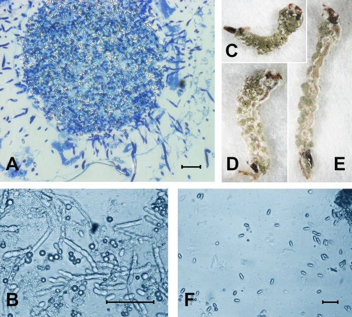
At 48 h pt, we observed mass accumulation of Metarhizium conidia in the gut lumen (Fig. 2A). Germinated conidia were not detected in larvae at 48 h pt (n = 12). However, in one sample (combined treatment), hyphal bodies were detected in the hemocoel (Fig. 2A). In the newly dead larvae after the fungal and combined treatments (4–6 days), we detected colonization of the hemocoel with hyphal bodies (Fig. 2B). Under the combined treatment, 83% hypha-positive larvae were found, while in the fungal treatment, 90% hypha-positive larvae were recorded. No significant differences between these treatments were observed (χ2 = 0.58, df = 1, P = 0.45, n = 30 larvae per treatment). No hyphal bodies were detected in the fungus-free treatments. A total of 70% and 60% of larvae were overgrown with Metarhizium under incubation in moist chambers (Fig. 2C) after treatment with the fungus or the mixture (avermectins + fungus), respectively. Only nongerminated conidia, but no hyphal bodies, were detected in the water in which treated larvae were maintained (Fig. 2D).
Figure 2. The colonization of Ae. aegypti by M. robertsii.
(A) Accumulation of conidia in the gut and colonization of the hemocoel by hyphal bodies. (B) Colonization of the fat body. (C–E) Mosquito larvae with surface conidiation of Metarhizium in a moist chamber. (F) Nongerminated conidia in a sample of water in which infected larvae were maintained. Scale bar: 20 µm.
Phenoloxidase activity
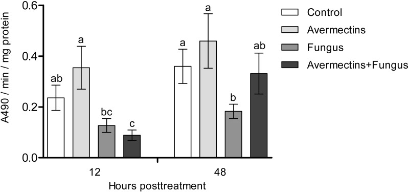
At 12 h pt, we registered a significant decrease in PO activity under the influence of fungal infection (Scheirer-Ray-Hare test, effect of fungus: H1.52 = 12.6, P = 0.00038; Fig. 3). Avermectins did not significantly change PO activity (H1.52 = 0.3, P = 0.54). A stronger decrease in enzyme activity was observed after combined treatment, but a significant factor interaction was not revealed (H1.52 = 1.9, P = 0.16). At 48 h pt, we detected a significant increase in PO activity under the influence of avermectins (H1.32 = 5.33, P = 0.02). The effect of the fungus was not significant (H1.32 = 0.7, P = 0.39), but a tendency toward an interaction between the factors was revealed (H1.32 = 3.2, P = 0.07). This is explained by the inhibition of PO activity by the fungus alone (Dunn’s test, P = 0.01, P = 0.04, compared to the control and avermectin treatments, respectively) and by the tendency of increased enzyme activity after combined treatment.
Figure 3. Activity of PO in the whole-body homogenates of Ae. aegypti larvae after treatment with M. robertsii, avermectins and their combination.
In the control treatment, equal amounts of water were added. Error bars represent the standard error of the mean. Significant differences are indicated with different letters within one time point (Dunn’s test, P < 0.05).
Dopamine concentration
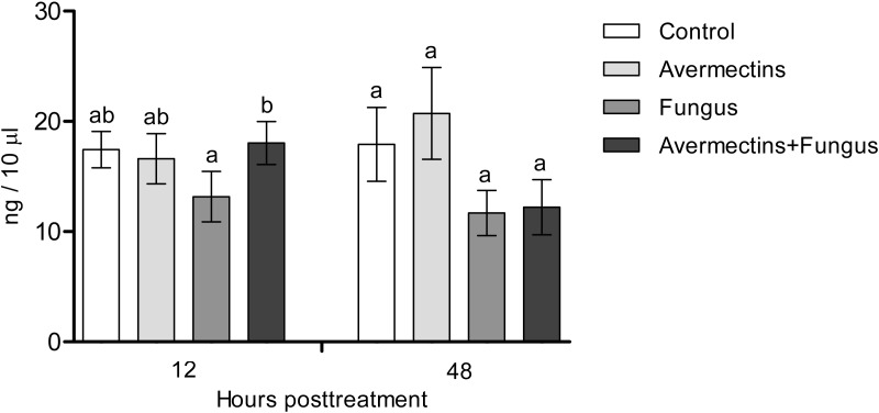
The effects of the fungus or avermectins on the dopamine concentration at 12 h pt were not significant (F1.31 = 1.2, P = 0.27; Fig. 4), although a trend toward a factor interaction was revealed (F1.31 = 3.3, P = 0.07). This was due to a clear tendency to decrease the dopamine concentration after treatment with the fungus alone (HSD Tukey test, P = 0.07 compared to fungus-free treatments) but not with the combination of the fungus and avermectins (P = 0.47 compared to fungus-free treatments). At 48 h pt, we observed a significant decrease in the dopamine concentration under the influence of the fungus (F1.31 = 4.62, P = 0.03); however, there were no significant differences between the treatments (HSD Tukey test, p = 0.1). No significant interaction effects between the factors on the dopamine concentration at 48 h pt were detected (F1.31 = 0.0, P = 1.0).
Figure 4. Dopamine concentration in whole-body homogenates of Ae. aegypti larvae after treatment with M. robertsii, avermectins and their combination.
In the control treatment, equal amounts of water were added. Error bars represent the standard error of the mean. Significant differences are indicated by different letters within one time point (HSD Tukey test, P < 0.05).
Detoxifying enzymes
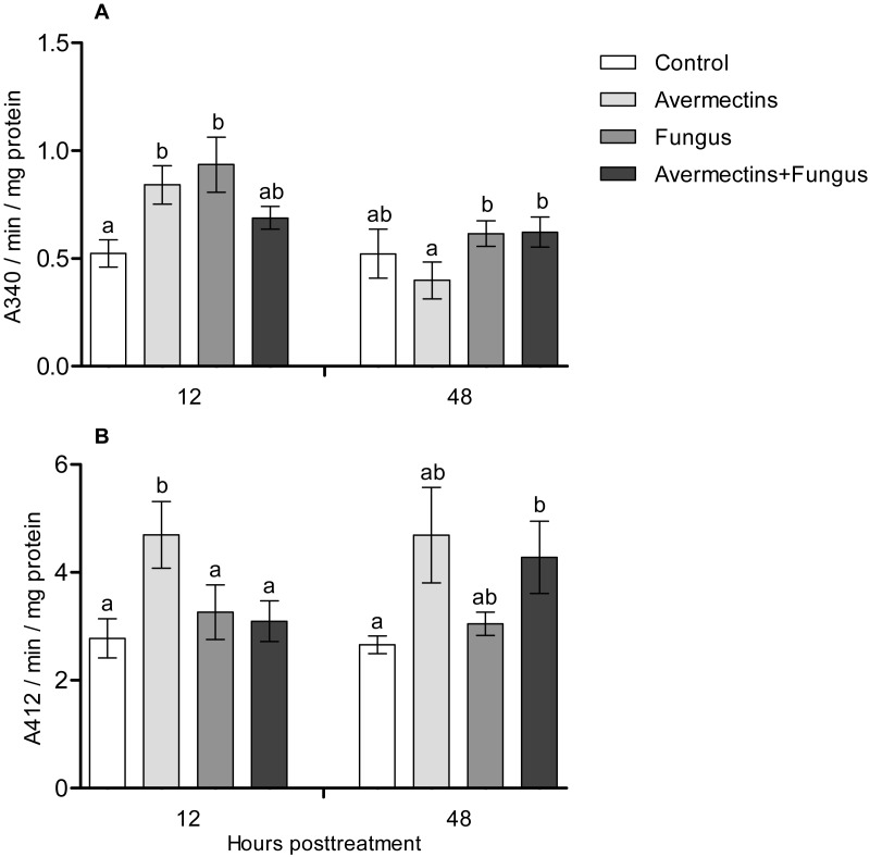
At 12 h pt, an interaction effect between the two factors (avermectins and the fungus) on GST activity was observed (H1.44 = 8.1, P = 0.0043, Fig. 5A). Avermectins and the fungus alone significantly (1.5–2-fold) increased GST activity compared to untreated larvae (Dunn’s test, P = 0.007, P = 0.001, respectively), but after combined treatment, the enzyme activity did not significantly differ from that in the control. Similar patterns were registered for EST activity at 12 h pt (Fig. 5B). In this case, EST was activated under the influence of avermectins alone (Dunn’s test, P = 0.002, compared to control), but fungal infection inhibited this activation. In particular, EST activity in the fungal and combined treatments did not differ from that in the control (Dunn’s test, P = 0.28, P = 0.34, respectively).
Figure 5. GST (A) and EST (B) activity in whole-body homogenates of Ae. aegypti larvae after treatment with M. robertsii, avermectins and their combination.
In the control treatment, equal amounts of water were added. Error bars represent the standard error of the mean. Significant differences are indicated by different letters within one time point (Dunn’s test, P < 0.05).
At 48 h pt, we observed a significant increase in GST activity under the influence of avermectins (H1.32 = 4.2, P = 0.03). The effect of the fungus as well as the interaction between the factors on GST activity at this time point was not significant (H1.32 = 2.5, P = 0.1 and H1.32 = 0.61, P = 0.43, respectively). EST activity nonsignificantly increased under the influence of avermectins (H1.32 = 1.85, P = 0.17). The effect of the fungus on enzyme activity was not significant (H1.32 = 0.001, P = 0.96), and no significant interactions between the factors were detected (H1.32 = 0.49, P = 0.48).
Acid protease activity
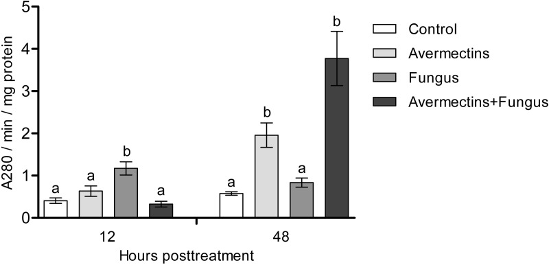
At 12 h pt, a significant interaction between avermectins and the fungus on acid protease activity was observed (H1.48 = 14.8; P = 0.00011; Fig. 6). In particular, protease activity was increased after fungal treatment alone (3-fold compared to control, P = 0.0003) but not after combined treatment. At 48 h pt, protease activity was strongly increased under the influence of avermectins (H1.43 = 27.4, P = 0.000016), and a trend toward an increase in enzyme activity was registered under the influence of the fungus (H1.43 = 3.18, P = 0.07). No significant interaction between the factors was revealed, although a trend toward the highest increase in protease activity was registered after the combined treatment.
Figure 6. Acid protease activity in whole-body homogenates of Ae. aegypti larvae after treatment with M. robertsii, avermectins and their combination.
In the control treatment, equal amounts of water were added. Error bars represent the standard error of the mean. Significant differences are indicated by different letters within one time point (Dunn’s test, P < 0.05).
Lysozyme-like activity
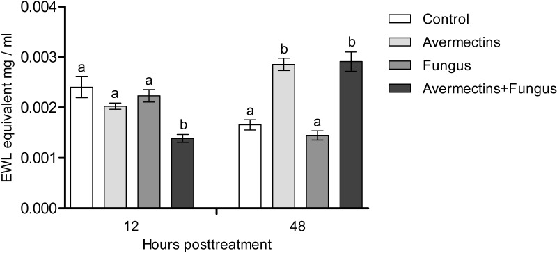
At 12 h pt, we recorded a decrease in lysozyme-like activity under the influence of both fungal infection and avermectins (effect of fungus: H1.56 = 6.46, P = 0.011; effect of avermectins: H1.56 = 14.04, P = 0.00017; Fig. 7). The greatest decrease was observed after the combined treatment (Dunn’s test, P < 0.001, compared with the other treatments). At 48 h pt, a sharp (1.7–2.0-fold) increase in lysozyme-like activity was recorded under the influence of avermectins (H1.116 = 68.6, P < 0.0000001). The effect of the fungus on the level of the enzyme at 48 h pt was not significant. No significant interaction effect between the factors on the level of lysozyme was observed at 12 and 48 h pt (H = 0.23, P = 0.63).
Figure 7. Lysozyme-like activity in whole-body homogenates of Ae. aegypti larvae after treatment with M. robertsii, avermectins and their combination.
In the control treatment, equal amounts of water were added. Error bars represent the standard error of the mean. Significant differences are indicated by different letters within one time point (Dunn’s test, P < 0.05).
Fungal and bacterial CFUs
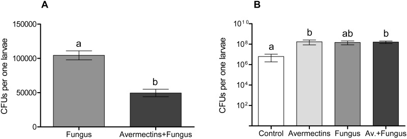
The plating of mosquito larval homogenates on modified Sabouraud agar showed significant differences in the Metarhizium CFUs between treatment with the fungus either alone or combined with avermectins (Fig. 8A). The Metarhizium CFU count in the fungal treatment was twice as high as that in the combined treatment (t = 6.4, df = 18, P = 0.001). Homogenates of the larvae from the fungus-free treatments (avermectin alone and control) did not form any fungal colonies.
Figure 8. Colony forming units of M. robertsii (A) and cultivable bacteria (B) in whole-body homogenates of Ae. aegypti larvae after treatment with M. robertsii, avermectins and their combination.
Error bars show min and max values. Significant differences are indicated by different letters (t-test, P < 0.001, for fungal CFU, and Dunn’s test, P < 0.05, for bacterial CFU).
The plating of larval homogenates on blood agar showed a significant (17–75-fold) increase in bacterial CFUs after treatment with the fungus and avermectins. A significant effect was registered for avermectins (H1.19 = 4.8, P = 0.03; Fig. 8B) but not for the fungus (H1.19 = 1.9, P = 0.17). However, a clear tendency toward an increase in CFUs was registered after treatment with the fungus alone (Dunn’s test, P = 0.054, compared to control). No significant interaction effect between the fungus and avermectins on bacterial CFUs was revealed (H1.19 = 1.9, P = 0.17).
Discussion
We showed a synergistic effect between avermectins and Metarhizium fungi on aquatic invertebrates for the first time. A similar effect was shown previously only in terrestrial insects (Colorado potato beetle, cotton moth) (Anderson et al., 1989; Asi et al., 2010; Tomilova et al., 2016), which are characterized by a completely different mode of fungal penetration (through the exo-skeleton). The accumulation of conidia of the fungus mainly in the gut lumen of mosquitoes coincides with studies of other researchers (Butt et al., 2013). However, we report the first observation of colonization of Ae. aegypti larvae after inoculation with Metarhizium conidia. It was previously suggested that only blastospores (and not conidia) are able to germinate from the gut lumen into the hemocoel of mosquito larvae (Alkhaibari et al., 2016; Alkhaibari et al., 2018). Interestingly, the larvae treated with avermectins accumulated a lower amount of conidia, but this dose was sufficient for a synergistic effect on mortality. It is likely that reduced accumulation of conidia was due to disturbance of feeding. For example, decrease in quantity of consumed food under the influence of avermectins was shown for terrestrial insects (Akhanaev et al., 2017).
We observed a decrease in PO activity and dopamine levels under the influence of the fungus, whereas in terrestrial arthropods, these enzymes are activated during mycoses (Ling & Yu, 2005; Yassine, Kamareddine & Osta, 2012; Yaroslavtseva et al., 2017; Chertkova, Grizanova & Dubovskiy, 2018). It has been suggested that dopamine release is associated with the general stress reactions related to the insect’s responses to pathogens (Hirashima, Sukhanova & Rauschenbach, 2000; Chertkova, Grizanova & Dubovskiy, 2018). In addition, dopamine is involved in the modulation of energetic metabolism and general defense mechanisms such as phagocytosis (Wu et al., 2015). PO is involved in the inactivation of fungal propagules in the cuticle and hemocoel (Butt et al., 2016). Dopamine is involved in the PO cascade (Andersen, 2010); however, synchronous and unidirectional changes in the levels of PO and dopamine are not always observed during infections (E Chertkova, 2016, personal observations). Since we observed differentiation of fungal infection structures, we suggest that some fungal metabolites inhibit the PO cascade of Ae. aegypti larvae. It was shown on terrestrial insects that Metarhizium secondary metabolites (e.g., destruxins) may reduce the number of PO-positive hemocytes (Huxham, Lackie & McCorkindale, 1989) and these metabolites may upregulate serine protease inhibitors, which inhibit proPO cascade (Pal, Leger & Wu, 2007). Alkhaibari et al. (2018) noted a short-term increase in PO activity in the whole-body homogenates of Culex quinquefasciatus larvae after infection with conidia or blastospores of M. brunneum Petch (4–6 h pt). It is possible that this reaction depends on species of mosquitoes as well as strain of the pathogen. Especially, inhibition of hemolymph melanization under M. robertsii infections was dependent from the production of secondary metabolites by different strains (Wang et al., 2012).
We observed activation of detoxifying enzymes (GST, EST) in Ae. aegypti larvae at the early stages of toxicosis and infection (12 h pt) under mono-treatments with the fungus and avermectins. However, the combined treatment led to inhibition of the activation of GST and EST. A similar effect was observed at 12 h pt for antibacterial (lysozyme-like) and acid protease activities. Combined treatment leads to either inhibition or containment of the activation of these enzymes. GST and EST are used by insects to inactivate toxic products formed by insecticide-induced toxicoses (DeSilva et al., 1997; Boyer et al., 2006; Aponte et al., 2013) as well as under mycosis (Dubovskiy et al., 2012). Especially Tang et al. (2019) showed that up-regulation of GSTz2 decreased the susceptibility of tephritid fruit fly Bactrocera dorsalis (Hendel) to abamectin. Moreover, GST may participate in inactivation of fungal secondary metabolites (Loutelier, Cherton & Lange, 1994) and reactive oxygen species (Sherratt & Hayes, 2002). Lysozyme inhibits the reproduction of Gram-positive bacteria (Abdou et al., 2007; Gandhe, Janardhan & Nagaraju, 2007; Chapelle et al., 2009), which (e.g., Microbacteriaceae) are among the dominant bacteria in Ae. aegypti larvae (Coon et al., 2014). It should also be noted that at 12 h pt of Ae. aegypti with blastospores of M. brunneum, a decrease in the expression levels of genes encoding defensins and cecropins (Alkhaibari et al., 2016), which inhibit the growth of both Gram-positive and Gram-negative bacteria and fungi, was observed (Jozefiak & Engberg, 2017). The inhibition of acid protease activity under combined treatment may indicate disorders in food consumption and absorption. Disruption of food absorption and starvation can increase mortality from both fungi and insecticides (Furlong & Groden, 2003). Thus, we assume that the physiological causes of the observed synergism lie in the initial stages of the development of infection and toxicosis.
In the later stages (48 h pt), we mainly observed activation of the enzymes (PO, GST, EST, acid proteases and lysozyme-like activity), which apparently indicates destructive processes in tissues and organs under the action of both avermectins and fungi. The increase in PO activity on the second day after treatment with avermectins was probably due to the destruction of hemocytes and the release of intracellular proPO components. We have previously shown the cytotoxic effect of avermectins on hemocytes, leading to their death (Tomilova et al., 2016). Additionally, the cytostatic and cytotoxic effects of the avermectins complex on various cells of warm-blooded animals are well known (Sivkov, Yakovlev & Chashov, 1998; Kokoz et al., 1999; Korystov et al., 1999; Maioli et al., 2013). Increase in PO activity under the influence of avermectins could also be symptom linked with proliferation of bacteria (Fig. 8). The enhancement of PO is observed under development of various bacterioses and caused by damages of insect’s tissues as well as by recognition of bacterial cell wall compounds, formation of hemocyte nodules and their melanization (Bidla et al., 2009; Tokura et al., 2014; Dubovskiy et al., 2016). An increase in GST under mycoses usually correlates with the severity of the infectious process (Dubovskiy et al., 2012; Tomilova et al., 2019) and confirms the results obtained by Butt et al. (2013) when studying the pathogenesis of M. brunneum in Ae. aegypti larvae. An increase in lysozyme-like activity under the action of avermectins could have occurred due to tissue destruction accompanied by the release of lysosome contents containing lysozyme (Zachary & Hoffmann, 1984). The activation of proteases at 48 h pt under the influence of avermectins is correlated with the increase in PO and lysozyme-like activity, which also indicates destructive changes in the tissues.
We observed an increase in the number of cultivated bacteria in the larvae when treated with both avermectins and fungi. This effect may be associated with impaired intestinal peristalsis as well as changes in the level of PO and antibacterial activity in the initial stages of toxicosis and fungal infection. Similar effects have been observed in terrestrial insects following topical infection by fungi (Wei et al., 2017; Ramirez et al., 2018; Polenogova et al., 2019) and are associated with the redistribution of immune responses between the cuticle and the gut. In mosquito larvae, the fungus comes into direct contact with gut microbiota, which may exhibit fungistatic properties (Sivakumar et al., 2017; Zhang et al., 2018, etc.) or, alternatively, may act as synergists of fungi, as shown by Wei et al. (2017) in the adults of the mosquito Anopheles stephensi. It is possible that conflicting data on the colonization of mosquito larvae by Metarhizium fungi are associated with differences in bacterial communities, which requires further research. In any case, fungi cannot successfully complete colonization in an aquatic environment, and bacterial decomposition is observed in mosquito larvae, whereas surface conidiation occurs in the air environment.
Conclusions
In conclusion, this is the first study of the survival and physiological reactions of mosquito larvae under the combined action of avermectins and entomopathogenic fungi. The synergism observed under the combined action of these agents appears to be associated with physiological changes in the early stages of toxicosis and infection. In particular, inhibition of the activity of a number of enzymes is observed under the combined treatment associated with the detoxifying and immune systems.
Colonization of Aedes aegypti larvae by the fungus Metarhizium robertsii is shown for the first time in this study. Further investigations may be focused on studying the role of endosymbiotic mosquito bacteria in the development of toxicoses and mycoses as well as the development of preparative forms based on fungi and avermectins for mosquito control in natural conditions.
Supplemental Information
Sheet 1 (Mortality) contains data using for Kaplan-Meier Survival Analysis: Log-Rank. Sheet 2 (PO) contains data using for analysis of phenoloxidase activity. Sheet 3 (DA) contains data using for analysis of dopamine concentration. Sheet 4 (GST) contains data using for analysis of Glutathione S-transferase activity. Sheet 5 (EST) contains data using for analysis of esterase activity. Sheet 6 (Protease) contains data using for analysis of protease activity. Sheet 7 (Lysozyme) contains data using for analysis of lysozyme activity. Sheet 8 (CFUs) contains data using for analysis of fungal and bacterial CFUs
*, Significant synergistic effects (χ2 > 18.5, df = 1, P < 0.001) calculated as described by Robertson and Preisler (1992)
Acknowledgments
The authors are grateful to Dr. V.A. Shilo (Karasuk Station of the ISEA) for help in organizing the experiments, to Dr. AA Alekseev for determining the exact ratio of avermectins and their isomers in an industrial product “Phytoverm” using HPLC, to Dr. AA Miller for preparation of semi-thin sections of the mosquito larvae.
Funding Statement
This work was supported by the Russian Science Foundation (project No. 18-74-00090). The funders had no role in study design, data collection and analysis, decision to publish, or preparation of the manuscript.
Additional Information and Declarations
Competing Interests
The authors declare there are no competing interests.
Author Contributions
Yuriy A. Noskov conceived and designed the experiments, performed the experiments, analyzed the data, prepared figures and/or tables, approved the final draft.
Olga V. Polenogova performed the experiments, authored or reviewed drafts of the paper, approved the final draft.
Olga N. Yaroslavtseva, Olga E. Belevich, Yuriy A. Yurchenko and Ekaterina A. Chertkova performed the experiments, authored or reviewed drafts of the paper, approved the final draft.
Natalya A. Kryukova analyzed the data, authored or reviewed drafts of the paper, approved the final draft.
Vadim Yu Kryukov conceived and designed the experiments, analyzed the data, prepared figures and/or tables, authored or reviewed drafts of the paper, approved the final draft.
Viktor V. Glupov conceived and designed the experiments, contributed reagents/materials/analysis tools, authored or reviewed drafts of the paper, approved the final draft.
Data Availability
The following information was supplied regarding data availability:
The raw data of the survival, dopamine concentration, glutathione S-transferase, nonspecific esterases, acid proteases, lysozyme-like and phenoloxidase activities are available in the Supplemental Files.
References
- Abdou et al. (2007).Abdou AM, Higashiguchi S, Aboueleinin AM, Kim M, Ibrahim HR. Antimicrobial peptides derived from hen egg lysozyme with inhibitory effect against Bacillus species. Food Control. 2007;18(2):173–178. doi: 10.1016/j.foodcont.2005.09.010. [DOI] [Google Scholar]
- Akhanaev et al. (2017).Akhanaev YB, Tomilova OG, Yaroslavtseva ON, Duisembekov BA, Kryukov VY, Glupov VV. Combined action of the entomopathogenic fungus Metarhizium robertsii and avermectins on the larvae of the colorado potato beetle Leptinotarsa decemlineata (Say) (Coleoptera, Chrysomelidae) Entomological Review. 2017;97(2):158–165. doi: 10.1134/S0013873817020026. [DOI] [Google Scholar]
- Alkhaibari et al. (2016).Alkhaibari AM, Carolino AT, Yavasoglu SI, Maffeis T, Mattoso TC, Bull JC, Samuels RI, Butt TM. Metarhizium brunneum blastospore pathogenesis in Aedes aegypti larvae: attack on several fronts accelerates mortality. PLOS Pathogens. 2016;12(7):e1005715. doi: 10.1371/journal.ppat.1005715. [DOI] [PMC free article] [PubMed] [Google Scholar]
- Alkhaibari et al. (2018).Alkhaibari AM, Maffeis T, Bull JC, Butt TM. Combined use of the entomopathogenic fungus, Metarhizium brunneum, and the mosquito predator, Toxorhynchites brevipalpis, for control of mosquito larvae: is this a risky biocontrol strategy? Journal of Invertebrate Pathology. 2018;153:38–50. doi: 10.1016/j.jip.2018.02.003. [DOI] [PMC free article] [PubMed] [Google Scholar]
- Alout et al. (2014).Alout H, Krajacich BJ, Meyers JI, Grubaugh ND, Brackney DE, Kobylinski KC, Diclaro 2nd JW, Bolay FK, Fakoli LS, Diabaté A, Dabiré RK, Bougma RW, Foy BD. Evaluation of ivermectin mass drug administration for malaria transmission control across different West African environments. Malaria Journal. 2014;13(1) doi: 10.1186/1475-2875-13-417. Article 417. [DOI] [PMC free article] [PubMed] [Google Scholar]
- Alves et al. (2004).Alves SN, Serrão JE, Mocelin G, Melo ALD. Effect of ivermectin on the life cycle and larval fat body of Culex quinquefasciatus. Brazilian Archives of Biology and Technology. 2004;47(3):433–439. doi: 10.1590/S1516-89132004000300014. [DOI] [Google Scholar]
- Andersen (2010).Andersen SO. Insect cuticular sclerotization: a review. Insect Biochemistry and Molecular Biology. 2010;40(3):166–178. doi: 10.1016/j.ibmb.2009.10.007. [DOI] [PubMed] [Google Scholar]
- Anderson et al. (1989).Anderson TE, Hajek AE, Roberts DW, Preisler HK, Robertson JL. Colorado potato beetle (Coleoptera: Chrysomelidae): effects of combinations of Beauveria bassiana with insecticides. Journal of Economic Entomology. 1989;82(1):83–89. doi: 10.1093/jee/82.1.83. [DOI] [Google Scholar]
- Anson (1938).Anson ML. The estimation of pepsin, trypsin, papain, and cathepsin with hemoglobin. The Journal of General Physiology. 1938;22(1):Article 79. doi: 10.1085/jgp.22.1.79. [DOI] [PMC free article] [PubMed] [Google Scholar]
- Aponte et al. (2013).Aponte HA, Penilla RP, Dzul-Manzanilla F, Che-Mendoza A, López AD, Solis F, Manrique-Saide P, Ranson H, Lenhart A, McCall PJ, Rodríguez AD. The pyrethroid resistance status and mechanisms in Aedes aegypti from the Guerrero state, Mexico. Pesticide Biochemistry and Physiology. 2013;107(2):226–234. doi: 10.1016/j.pestbp.2013.07.005. [DOI] [Google Scholar]
- Ashida & Söderhäll (1984).Ashida M, Söderhäll K. The prophenoloxidase activating system in crayfish. Comparative Biochemistry and Physiology Part B: Comparative Biochemistry. 1984;77(1):21–26. doi: 10.1016/0305-0491(84)90217-7. [DOI] [Google Scholar]
- Asi et al. (2010).Asi MR, Bashir MH, Afzal M, Ashfaq M, Sahi ST. Compatibility of entomopathogenic fungi, Metarhizium anisopliae and Paecilomyces fumosoroseus with selective insecticides. Pakistan Journal of Botany. 2010;42(6):4207–4214. [Google Scholar]
- Badiane et al. (2017).Badiane TS, Ndione RD, Toure M, Seye F, Ndiaye M. Larvicidal and Synergic Effects of two Biopesticides (Azadirachta indica and Metarhizium anisopliae) against Larvae of Culex quinquefasciatus (Diptera, Culicidae) (Say, 1823) International Journal of Sciences. 2017;6:109–115. doi: 10.18483/ijSci.1258. [DOI] [Google Scholar]
- Belevich et al. (2017).Belevich OE, Yurchenko YA, Glupov VV, Kryukov VY. Effect of entomopathogenic fungus Metarhizium robertsii on non-target organisms, water bugs (Heteroptera: Corixidae, Naucoridae, Notonectidae) Applied Entomology and Zoology. 2017;52:439–445. doi: 10.1007/s13355-017-0494-z. [DOI] [Google Scholar]
- Bhatt et al. (2013).Bhatt S, Gething PW, Brady OJ, Messina JP, Farlow AW, Moyes CL, Drake JM, Brownstein JS, Hoen AG, Sankoh O, Myers MF, George DB, Jaenisch T, Wint GRW, Simmons CP, Scott TW, Farrar JJ, Hay SI. The global distribution and burden of dengue. Nature. 2013;496(7446):504–507. doi: 10.1038/nature12060. [DOI] [PMC free article] [PubMed] [Google Scholar]
- Bidla et al. (2009).Bidla G, Hauling T, Dushay MS, Theopold U. Activation of insect phenoloxidase after injury: endogenous versus foreign elicitors. Journal of Innate Immunity. 2009;1(4):301–308. doi: 10.1159/000168009. [DOI] [PubMed] [Google Scholar]
- Boyer et al. (2006).Boyer S, David JP, Rey D, Lemperiere G, Ravanel P. Response of Aedes aegypti (Diptera: Culicidae) larvae to three xenobiotic exposures: larval tolerance and detoxifying enzyme activities. Environmental Toxicology and Chemistry: An International Journal. 2006;25(2):470–476. doi: 10.1897/05-267R2.1. [DOI] [PubMed] [Google Scholar]
- Boyer et al. (2012).Boyer S, Paris M, Jego S, Lempérière G, Ravanel P. Influence of insecticide Bacillus thuringiensis subsp. israelensis treatments on resistance and enzyme activities in Aedes rusticus larvae (Diptera: Culicidae) Biological Control. 2012;62:75–81. doi: 10.1016/j.biocontrol.2012.02.001. [DOI] [Google Scholar]
- Bradford (1976).Bradford MM. A rapid and sensitive method for the quantitation of microgram quantities of protein utilizing the principle of protein-dye binding. Analytical Biochemistry. 1976;72(1–2):248–254. doi: 10.1016/0003-2697(76)90527-3. [DOI] [PubMed] [Google Scholar]
- Butt et al. (2016).Butt TM, Coates CJ, Dubovskiy IM, Ratcliffe NA. Advances in genetics. Vol. 94. Academic Press; 2016. Entomopathogenic fungi: new insights into host–pathogen interactions; pp. 307–364. [DOI] [PubMed] [Google Scholar]
- Butt et al. (2013).Butt TM, Greenfield BP, Greig C, Maffeis TG, Taylor JW, Piasecka J, Dudley E, Abdulla A, Dubovskiy IM, Garrido-Jurado I, Quesada-Moraga E. Metarhizium anisopliae pathogenesis of mosquito larvae: a verdict of accidental death. PLOS ONE. 2013;8(12):e81686. doi: 10.1371/journal.pone.0081686. [DOI] [PMC free article] [PubMed] [Google Scholar]
- Campbell et al. (1983).Campbell WC, Fisher MH, Stapley EO, Albers-Schonberg G, Jacob TA. Ivermectin: a potent new antiparasitic agent. Science. 1983;221(4613):823–828. doi: 10.1126/science.6308762. [DOI] [PubMed] [Google Scholar]
- Chaccour et al. (2017).Chaccour CJ, Hammann F, Alustiza M, Castejon S, Tarimo BB, Abizanda G, IrigoyenBarrio Á, Martí Soler H, Moncada R, Bilbao JI, Aldaz A. Cytochrome P450/ABC transporter inhibition simultaneously enhances ivermectin pharmacokinetics in the mammal host and pharmacodynamics in Anopheles gambiae. Scientific Reports. 2017;7(1):8535. doi: 10.1038/s41598-017-08906-x. [DOI] [PMC free article] [PubMed] [Google Scholar]
- Chapelle et al. (2009).Chapelle M, Girard PA, Cousserans F, Volkoff NA, Duvic B. Lysozymes and lysozyme-like proteins from the fall armyworm, Spodoptera frugiperda. Molecular Immunology. 2009;47(2-3):261–269. doi: 10.1016/j.molimm.2009.09.028. [DOI] [PubMed] [Google Scholar]
- Chertkova, Grizanova & Dubovskiy (2018).Chertkova EA, Grizanova EV, Dubovskiy IM. Bacterial and fungal infections induce bursts of dopamine in the haemolymph of the Colorado potato beetle Leptinotarsa decemlineata and greater wax moth Galleria mellonella. Journal of Invertebrate Pathology. 2018;153:203–206. doi: 10.1016/j.jip.2018.02.020. [DOI] [PubMed] [Google Scholar]
- Coon et al. (2014).Coon KL, Vogel KJ, Brown MR, Strand MR. Mosquitoes rely on their gut microbiota for development. Molecular Ecology. 2014;23(11):2727–2739. doi: 10.1111/mec.12771. [DOI] [PMC free article] [PubMed] [Google Scholar]
- Crump & Omura (2011).Crump A, Omura S. Ivermectin, wonder drug’ from Japan: the human use perspective. Proceedings of the Japan Academy, Series B. 2011;87(2):13–28. doi: 10.2183/pjab.87.13. [DOI] [PMC free article] [PubMed] [Google Scholar]
- Delpuech, Frey & Carton (1996).Delpuech JM, Frey F, Carton Y. Action of insecticides on the cellular immune reaction of Drosophila melanogaster against the parasitoid Leptopilina boulardi. Environmental Toxicology and Chemistry: An International Journal. 1996;15(12):2267–2271. doi: 10.1002/etc.5620151221. [DOI] [Google Scholar]
- DeSilva et al. (1997).DeSilva D, Hemingway J, Ranson H, Vaughan A. Resistance to insecticides in insect vectors of disease: estα3, a novel amplified esterase associated with amplified estβ1 from insecticide resistant strains of the mosquito Culex quinquesfasciatus. Experimental Parasitology. 1997;87(3):253–259. doi: 10.1006/expr.1997.4245. [DOI] [PubMed] [Google Scholar]
- Dreyer, Morin & Vaughan (2018).Dreyer SM, Morin KJ, Vaughan JA. Differential susceptibilities of Anopheles albimanus and Anopheles stephensi mosquitoes to ivermectin. Malaria Journal. 2018;17(1) doi: 10.1186/s12936-018-2296-3. Article 148. [DOI] [PMC free article] [PubMed] [Google Scholar]
- Drinyaev et al. (1999).Drinyaev VA, Kruglyak EB, Mosin VA, Ter-Simonyan VG, Novik TS, Rusakov SV, Berezina NS, Berezkina NE, Sterlina TS. Physicochemical and biological properties of aversectin C. Applied Biochemistry and Microbiology. 1999;35(2):177–182. [Google Scholar]
- Dubovskiy et al. (2016).Dubovskiy IM, Kryukova NA, Glupov VV, Ratcliffe NA. Encapsulation and nodulation in insects. Invertebrate Survival Journal. 2016;13(1):229–246. [Google Scholar]
- Dubovskiy et al. (2012).Dubovskiy IM, Slyamova ND, Kryukov VY, Yaroslavtseva ON, Levchenko MV, Belgibaeva AB, Adilkhankyzy A, Glupov VV. The activity of nonspecific esterases and glutathione-S-transferase in Locusta migratoria larvae infected with the fungus Metarhizium anisopliae (Ascomycota, Hypocreales) Entomological Review. 2012;92(1):27–31. doi: 10.1134/S0013873812010022. [DOI] [Google Scholar]
- Dubovskiy et al. (2013).Dubovskiy IM, Whitten MMA, Yaroslavtseva ON, Greig C, Kryukov VY, Grizanova EV, Mukherjee K, Vilcinskas A, Glupov VV, Butt TM. Can insects develop resistance to insect pathogenic fungi? PLOS ONE. 2013;8(4):e60248. doi: 10.1371/journal.pone.0060248. [DOI] [PMC free article] [PubMed] [Google Scholar]
- Freitas et al. (1996).Freitas RMCD, Faria MDA, Alves SN, Melo ALD. Effects of ivermectin on Culex quinquefasciatus larvae. Revista do Instituto de Medicina Tropical de São Paulo. 1996;38(4):293–298. doi: 10.1590/S0036-46651996000400010. [DOI] [PubMed] [Google Scholar]
- Furlong & Groden (2003).Furlong MJ, Groden E. Starvation induced stress and the susceptibility of the Colorado potato beetle, Leptinotarsa decemlineata, to infection by Beauveria bassiana. Journal of Invertebrate Pathology. 2003;83(2):127–138. doi: 10.1016/S0022-2011(03)00066-1. [DOI] [PubMed] [Google Scholar]
- Gaidyshev (2004).Gaidyshev IP. Solution of scientific and engineering tasks by means of Excel, VBA and C/C++ St. BKhV–Petersburg; Petersburg: 2004. [Google Scholar]
- Gandhe, Janardhan & Nagaraju (2007).Gandhe AS, Janardhan G, Nagaraju J. Immune upregulation of novel antibacterial proteins from silkmoths (Lepidoptera) that resemble lysozymes but lack muramidase activity. Insect Biochemistry and Molecular Biology. 2007;37(7):655–666. doi: 10.1016/j.ibmb.2007.03.013. [DOI] [PubMed] [Google Scholar]
- Garrido-Jurado et al. (2016).Garrido-Jurado I, Alkhaibari A, Williams SR, Oatley-Radcliffe DL, Quesada-Moraga E, Butt TM. Toxicity testing of Metarhizium conidia and toxins against aquatic invertebrates. Journal of Pest Science. 2016;89(2):557–564. doi: 10.1007/s10340-015-0700-0. [DOI] [Google Scholar]
- Gorman, An & Kanost (2007).Gorman MJ, An C, Kanost MR. Characterization of tyrosine hydroxylase from Manduca sexta. Insect Biochemistry and Molecular Biology. 2007;37(12):1327–1337. doi: 10.1016/j.ibmb.2007.08.006. [DOI] [PMC free article] [PubMed] [Google Scholar]
- Greenfield et al. (2015).Greenfield BP, Peace A, Evans H, Dudley E, Ansari MA, Butt TM. Identification of Metarhizium strains highly efficacious against Aedes, Anopheles and Culex larvae. Biocontrol Science and Technology. 2015;25(5):487–502. doi: 10.1080/09583157.2014.989813. [DOI] [Google Scholar]
- Gruntenko et al. (2005).Gruntenko NE, Karpova EK, Adonyeva NV, Chentsova NA, Faddeeva NV, Alekseev AA, Auschenbach IY. Juvenile hormone, 20-hydroxyecdysone and dopamine interaction in Drosophila virilis reproduction under normal and nutritional stress conditions. Journal of Insect Physiology. 2005;51(4):417–425. doi: 10.1016/j.jinsphys.2005.01.007. [DOI] [PubMed] [Google Scholar]
- Habig, Pabst & Jakoby (1974).Habig WH, Pabst MJ, Jakoby WB. Glutathione S-transferases. The first enzymatic step in mercapturic acid formation. Journal of Biological Chemistry. 1974;249(22):7130–7139. [PubMed] [Google Scholar]
- Hammer, Harper & Ryan (2001).Hammer O, Harper DA, Ryan PD. Palaeontological statistics software package for education and data analysis. Palaeontologia Electronica. 2001;4(1):1–9. [Google Scholar]
- Hirashima, Sukhanova & Rauschenbach (2000).Hirashima A, Sukhanova MJ, Rauschenbach IY. Biogenic amines in Drosophila virilis under stress conditions. Bioscience, Biotechnology, and Biochemistry. 2000;64(12):2625–2630. doi: 10.1271/bbb.64.2625. [DOI] [PubMed] [Google Scholar]
- Huxham, Lackie & McCorkindale (1989).Huxham IM, Lackie AM, McCorkindale NJ. Inhibitory effects of cyclodepsipeptides, destruxins, from the fungus Metarhizium anisopliae, on cellular immunity in insects. Journal of Insect Physiology. 1989;35(2):97–105. doi: 10.1016/0022-1910(89)90042-5. [DOI] [Google Scholar]
- Jozefiak & Engberg (2017).Jozefiak A, Engberg RM. Insect proteins as a potential source of antimicrobial peptides in livestock production. A review. Journal of Animal and Feed Sciences. 2017;26(2):87–99. doi: 10.22358/jafs/69998/2017. [DOI] [Google Scholar]
- Kokoz et al. (1999).Kokoz YM, Tsyganova VG, Korystova AF, Grichenko AS, Zenchenko KI, Drinyaev VA, Mosin VA, Kruglyak EB, Sterlina TS, Victorov AV. Selective cytostatic and neurotoxic effects of avermectins and activation of the GABA α receptors. Bioscience Reports. 1999;19(6):535–546. doi: 10.1023/A:1020262811459. [DOI] [PubMed] [Google Scholar]
- Korystov et al. (1999).Korystov YN, Mosin VA, Shaposhnikova VV, Levitman MK, Kudryavtsev AA, Kruglyak EB, Sterlina TS, Viktorov AV, Drinyaev VA. A comparative study of the effects of aversectin C, abamectin and ivermectin on apoptosis of rat thymocytes induced by radiation and dexamethasone. Acta Veterinaria Brno. 1999;68(1):23–29. doi: 10.2754/avb199968010023. [DOI] [Google Scholar]
- Li, Schuler & Berenbaum (2007).Li X, Schuler MA, Berenbaum MR. Molecular mechanisms of metabolic resistance to synthetic and natural xenobiotics. Annual Review of Entomology. 2007;52:231–253. doi: 10.1146/annurev.ento.51.110104.151104. [DOI] [PubMed] [Google Scholar]
- Ling & Yu (2005).Ling E, Yu XQ. Prophenoloxidase binds to the surface of hemocytes and is involved in hemocyte melanization in Manduca sexta. Insect Biochemistry and Molecular Biology. 2005;35(12):1356–1366. doi: 10.1016/j.ibmb.2005.08.007. [DOI] [PubMed] [Google Scholar]
- Loutelier, Cherton & Lange (1994).Loutelier C, Cherton JC, Lange C. Conjugation reactions of cyclodepsipeptide to glutathionyl adducts by direct ‘in-beam’ synthesis under negative-ion fast-atom bombardment conditions. Rapid Communications in Mass Spectrometry. 1994;8(10):844–848. doi: 10.1002/rcm.1290081010. [DOI] [PubMed] [Google Scholar]
- Maioli et al. (2013).Maioli MA, De Medeiros HC, Guelfi M, Trinca V, Pereira FT, Mingatto FE. The role of mitochondria and biotransformation in abamectin-induced cytotoxicity in isolated rat hepatocytes. Toxicology in Vitro. 2013;27(2):570–579. doi: 10.1016/j.tiv.2012.10.017. [DOI] [PubMed] [Google Scholar]
- Mohrig & Messner (1968).Mohrig W, Messner B. Lysozyme as antibacterial agent in honey and bees venom. Acta biologica et medica Germanica. 1968;21(1) Article 85. [PubMed] [Google Scholar]
- Ortiz-Urquiza, Luo & Keyhani (2015).Ortiz-Urquiza A, Luo Z, Keyhani NO. Improving mycoinsecticides for insect biological control. Applied Microbiology and Biotechnology. 2015;99(3):1057–1068. doi: 10.1007/s00253-014-6270-x. [DOI] [PubMed] [Google Scholar]
- Pal, Leger & Wu (2007).Pal S, Leger RJS, Wu L. Fungal peptide Destruxin A plays a specific role in suppressing the innate immune response in Drosophila melanogaster. Journal of Biological Chemistry. 2007;282(12):8969–8977. doi: 10.1074/jbc.M605927200. [DOI] [PubMed] [Google Scholar]
- Paris et al. (2011).Paris M, Tetreau G, Laurent F, Lelu M, Despres L, David JP. Persistence of Bacillus thuringiensis israelensis (Bti) in the environment induces resistance to multiple Bti toxins in mosquitoes. Pest Management Science. 2011;67(1):122–128. doi: 10.1002/ps.2046. [DOI] [PubMed] [Google Scholar]
- Polenogova et al. (2019).Polenogova OV, Kabilov MR, Tyurin MV, Rotskaya UN, Krivopalov AV, Morozova VV, Mozhaitseva K, Kryukova NA, Alikina T, Kryukov VY, Glupov VV. Parasitoid envenomation alters the Galleria mellonella midgut microbiota and immunity, thereby promoting fungal infection. Scientific Reports. 2019;9(1):4012. doi: 10.1038/s41598-019-40301-6. [DOI] [PMC free article] [PubMed] [Google Scholar]
- Prabhakaran & Kamble (1995).Prabhakaran SK, Kamble ST. Purification and characterization of an esterase isozyme from insecticide resistant and susceptible strains of German cockroach, Blattella germanica (L.) Insect Biochemistry and Molecular Biology. 1995;25(4):519–524. doi: 10.1016/0965-1748(94)00093-E. [DOI] [PubMed] [Google Scholar]
- Ramirez et al. (2018).Ramirez JL, Muturi EJ, Dunlap C, Rooney AP. Strain-specific pathogenicity and subversion of phenoloxidase activity in the mosquito Aedes aegypti by members of the fungal entomopathogenic genus Isaria. Scientific Reports. 2018;8(1):9896. doi: 10.1038/s41598-018-28210-6. [DOI] [PMC free article] [PubMed] [Google Scholar]
- Ranson & Lissenden (2016).Ranson H, Lissenden N. Insecticide resistance in African Anopheles mosquitoes: a worsening situation that needs urgent action to maintain malaria control. Trends in Parasitology. 2016;32(3):187–196. doi: 10.1016/j.pt.2015.11.010. [DOI] [PubMed] [Google Scholar]
- Robertson & Preisler (1992).Robertson JL, Preisler HK. Pesticide bioassays with arthropods. CRC; Boca Raton: 1992. [DOI] [Google Scholar]
- Scheirer, Ray & Hare (1976).Scheirer CJ, Ray WS, Hare N. The analysis of ranked data derived from completely randomized factorial designs. Biometrics. 1976;32(2):429–434. doi: 10.2307/2529511. [DOI] [PubMed] [Google Scholar]
- Scholte et al. (2004).Scholte EJ, Knols BG, Samson RA, Takken W. Entomopathogenic fungi for mosquito control: a review. Journal of Insect Science. 2004;4(1):19. doi: 10.1093/jis/4.1.19. [DOI] [PMC free article] [PubMed] [Google Scholar]
- Sherratt & Hayes (2002).Sherratt PJ, Hayes JD. Enzyme systems that metabolise drugs and other xenobiotics. John Wiley & Sons Ltd; Hoboken: 2002. Glutathione S-transferases; pp. 319–352. [Google Scholar]
- Shoukat et al. (2018).Shoukat RF, Freed S, Ahmad KW, Rehman AU. Assessment of binary mixtures of entomopathogenic fungi and chemical insecticides on biological parameters of culex pipiens (diptera: culicidae) under laboratory and field conditions. Pakistan Journal of Zoology. 2018;50(1):299–299. doi: 10.17582/journal.pjz/2018.50.1.299.309. [DOI] [Google Scholar]
- Sivakumar et al. (2017).Sivakumar G, Rangeshwaran R, Yandigeri MS, Mohan M, Venkatesan T, Ballal CR, Verghese A. Characterization and role of gut bacterium Bacillus pumilus on nutrition and defense of leafhopper (Amrasca biguttula biguttula) of cotton. Indian Journal of Agricultural Sciences. 2017;87:534–539. [Google Scholar]
- Sivkov, Yakovlev & Chashov (1998).Sivkov GS, Yakovlev VV, Chashov IA. Influence of the animal’s immune response indicatirs. Veterinarnaya Medicina. 1998;5:29–31. (in Russian) [Google Scholar]
- Smith, Kasai & Scott (2016).Smith LB, Kasai S, Scott JG. Pyrethroid resistance in Aedes aegypti and Aedes albopictus: important mosquito vectors of human diseases. Pesticide Biochemistry and Physiology. 2016;133:1–12. doi: 10.1016/j.pestbp.2016.03.005. [DOI] [PubMed] [Google Scholar]
- Su et al. (2017).Su T, Thieme J, Ocegueda C, Ball M, Cheng ML. Resistance to Lysinibacillus sphaericus and other commonly used pesticides in Culex pipiens (Diptera: Culicidae) from Chico, California. Journal of Medical Entomology. 2017;55(2):423–428. doi: 10.1093/jme/tjx235. [DOI] [PubMed] [Google Scholar]
- Tang et al. (2019).Tang G, Xiong Y, Liu Y, Song Z, Yang Y, Shen G, Wang J-J, Jiang H. The transcription factor MafB regulates the susceptibility of Bactrocera dorsalis to avermectin via GSTz2. Frontiers in Physiology. 2019;10 doi: 10.3389/fphys.2019.01068. Article 1068. [DOI] [PMC free article] [PubMed] [Google Scholar]
- Tilquin et al. (2008).Tilquin M, Paris M, Reynaud S, Despres L, Ravanel P, Geremia RA, Gury J. Long lasting persistence of Bacillus thuringiensis subsp. israelensis (Bti) in mosquito natural habitats. PLOS ONE. 2008;3(10):e3432. doi: 10.1371/journal.pone.0003432. [DOI] [PMC free article] [PubMed] [Google Scholar]
- Tokura et al. (2014).Tokura A, Fu GS, Sakamoto M, Endo H, Tanaka S, Kikuta S, Tabunoki H, Sato R. Factors functioning in nodule melanization of insects and their mechanisms of accumulation in nodules. Journal of Insect Physiology. 2014;60:40–49. doi: 10.1016/j.jinsphys.2013.11.003. [DOI] [PubMed] [Google Scholar]
- Tolle (2009).Tolle MA. Mosquito-borne diseases. Current Problems in Pediatric and Adolescent Health Care. 2009;39(4):97–140. doi: 10.1016/j.cppeds.2009.01.001. [DOI] [PubMed] [Google Scholar]
- Tomilova et al. (2016).Tomilova OG, Kryukov VY, Duisembekov BA, Yaroslavtseva ON, Tyurin MV, Kryukova NA, Skorokhod V, Dubovskiy IM, Glupov VV. Immune-physiological aspects of synergy between avermectins and the entomopathogenic fungus Metarhizium robertsii in Colorado potato beetle larvae. Journal of Invertebrate Pathology. 2016;140:8–15. doi: 10.1016/j.jip.2016.08.008. [DOI] [PubMed] [Google Scholar]
- Tomilova et al. (2019).Tomilova OG, Yaroslavtseva ON, Ganina MD, Tyurin MV, Chernyak EI, Senderskiy IV, Senderskiy IV, Noskov YA, Polenogova OV, Akhanaev YB, Kryukov VY, Glupov VV. Changes in antifungal defence systems during the intermoult period in the Colorado potato beetle. Journal of Insect Physiology. 2019;116:106–117. doi: 10.1016/j.jinsphys.2019.05.003. [DOI] [PubMed] [Google Scholar]
- Vivekanandhan et al. (2018).Vivekanandhan P, Karthi S, Shivakumar MS, Benelli G. Synergistic effect of entomopathogenic fungus Fusarium oxysporum extract in combination with temephos against three major mosquito vectors. Pathogens and Global Health. 2018;112(1):37–46. doi: 10.1080/20477724.2018.1438228. [DOI] [PMC free article] [PubMed] [Google Scholar]
- Vontas, Ranson & Alphey (2010).Vontas J, Ranson H, Alphey L. Transcriptomics and disease vector control. BMC Biology. 2010;8(1):52. doi: 10.1186/1741-7007-8-52. [DOI] [PMC free article] [PubMed] [Google Scholar]
- Wang et al. (2012).Wang B, Kang Q, Lu Y, Bai L, Wang C. Unveiling the biosynthetic puzzle of destruxins in Metarhizium species. Proceedings of the National Academy of Sciences of the United States of America. 2012;109(4):1287–1292. doi: 10.1073/pnas.1115983109. [DOI] [PMC free article] [PubMed] [Google Scholar]
- Wei et al. (2017).Wei G, Lai Y, Wang G, Chen H, Li F, Wang S. Insect pathogenic fungus interacts with the gut microbiota to accelerate mosquito mortality. Proceedings of the National Academy of Sciences of the United States of America. 2017;114(23):5994–5999. doi: 10.1073/pnas.1703546114. [DOI] [PMC free article] [PubMed] [Google Scholar]
- Wu et al. (2015).Wu SF, Xu G, Stanley D, Huang J, Ye GY. Dopamine modulates hemocyte phagocytosis via a D1-like receptor in the rice stem borer, Chilo suppressalis. Scientific Reports. 2015;5:12247. doi: 10.1038/srep12247. [DOI] [PMC free article] [PubMed] [Google Scholar]
- Yaroslavtseva et al. (2017).Yaroslavtseva ON, Dubovskiy IM, Khodyrev VP, Duisembekov BA, Kryukov VY, Glupov VV. Immunological mechanisms of synergy between fungus Metarhizium robertsii and bacteria Bacillus thuringiensis ssp. morrisoni on Colorado potato beetle larvae. Journal of Insect Physiology. 2017;96:14–20. doi: 10.1016/j.jinsphys.2016.10.004. [DOI] [PubMed] [Google Scholar]
- Yassine, Kamareddine & Osta (2012).Yassine H, Kamareddine L, Osta MA. The mosquito melanization response is implicated in defense against the entomopathogenic fungus Beauveria bassiana. PLOS pathogens. 2012;8(11):e1003029. doi: 10.1371/journal.ppat.1003029. [DOI] [PMC free article] [PubMed] [Google Scholar]
- Zachary & Hoffmann (1984).Zachary D, Hoffmann D. Lysozyme is stored in the granules of certain haemocyte types in Locusta. Journal of Insect Physiology. 1984;30(5):405–411. doi: 10.1016/0022-1910(84)90098-2. [DOI] [Google Scholar]
- Zhang et al. (2018).Zhang F, Sun XX, Zhang XC, Zhang S, Lu J, Xia YM, Huang YH, Wang XJ. The interactions between gut microbiota and entomopathogenic fungi: a potential approach for biological control of Blattella germanica (L.) Pest Management Science. 2018;74(2):438–447. doi: 10.1002/ps.4726. [DOI] [PubMed] [Google Scholar]
Associated Data
This section collects any data citations, data availability statements, or supplementary materials included in this article.
Supplementary Materials
Sheet 1 (Mortality) contains data using for Kaplan-Meier Survival Analysis: Log-Rank. Sheet 2 (PO) contains data using for analysis of phenoloxidase activity. Sheet 3 (DA) contains data using for analysis of dopamine concentration. Sheet 4 (GST) contains data using for analysis of Glutathione S-transferase activity. Sheet 5 (EST) contains data using for analysis of esterase activity. Sheet 6 (Protease) contains data using for analysis of protease activity. Sheet 7 (Lysozyme) contains data using for analysis of lysozyme activity. Sheet 8 (CFUs) contains data using for analysis of fungal and bacterial CFUs
*, Significant synergistic effects (χ2 > 18.5, df = 1, P < 0.001) calculated as described by Robertson and Preisler (1992)
Data Availability Statement
The following information was supplied regarding data availability:
The raw data of the survival, dopamine concentration, glutathione S-transferase, nonspecific esterases, acid proteases, lysozyme-like and phenoloxidase activities are available in the Supplemental Files.


