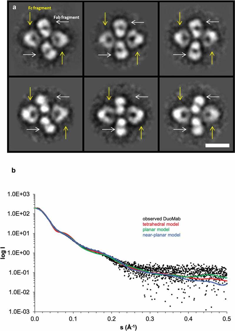
Figure 5.

Tetrameric scaffolds of DuoMabs are confirmed by class averages derived from micrographs recorded with negative stain transmission electron microscopy on antigen cMET binding DuoMabs (a, top row), as well as such binding antigen ErbB3 (a, bottom row). The Fc parts can be discriminated from Fabs because of their triangular shape and hole (yellow arrows in a). The Fabs are found in side as well as top views, with or without their central hole (white arrows in a). Scale bar represents 10 nm; all boxes are 31 by 31 nm. Small-angle X-ray scattering (SAXS) based on the ErbB3-specific DuoMab indicates that DuoMabs have a near-planar structure (b). Fit of calculated scattering profiles of different DuoMab models to the observed scattering profile of DuoMab: tetrahedral homology model (red, χ2 = 4.8), planar homology model (green, χ2 = 2.0), near-planar model obtained by a combination of rigid body and flexible modeling (blue, χ2 = 1.5).
