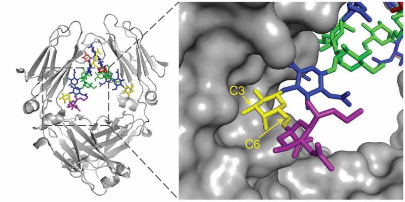
Figure 7.

Crystal structure of the α2,6-disialylated glycan in the CH2 domain (PDB id: 4BYH). The 3-arm is pointing inward, and the 6-arm is pointing outward. Sialic acid on the 3-arm is not observable because of its flexibility. Galactose (yellow) carbon positions are labeled. While sialic acid (purple) linked to the 6-position are exposed to the solvent, linking to the 3-position will likely cause steric effect as it points toward the protein backbone.
