

This article has been corrected: Due to errors in image processing, the representative image of TRAP staining in RANKL+HP d1 is incorrect. The proper Figure 1 is shown below. The authors declare that these corrections do not change the results or conclusions of this paper.
Original article: Oncotarget. 2018; 9:1868–1884. 1868-1884. https://doi.org/10.18632/oncotarget.22930
Figure 1. Hypericin suppresses RANKL-induced osteoclastogenesis.
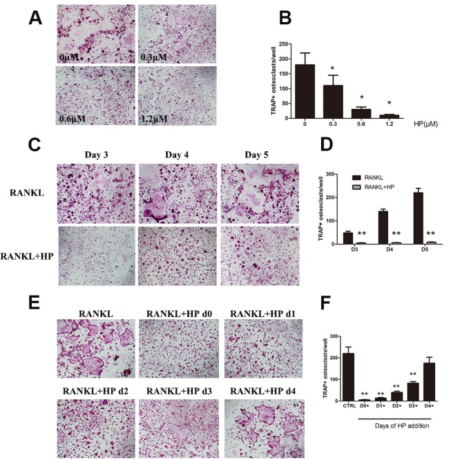
(A) Effects of HP on RANKL-induced osteoclast differentiation. RAW264.7 cells (3 × 10³ cells/well) were stimulated with RANKL (50 ng/mL) or were untreated (controls), followed by treatment with the indicated doses of HP. After 5-7 days, cells were fixed and stained for measurement of TRAP expression. The cells were photographed (original magnification, 100×). (B) The TRAP-positive multinucleated (> 3 nuclei) osteoclasts were counted. Columns represent the mean results of experiments carried out in triplicate, whereas bars represent the standard deviation (SD). (C) RAW264.7 cells (3 × 10³ cells/well) were incubated in a medium supplemented with either RANKL (50 ng/mL) or RANKL and HP (1.2 μmol/L) for 3, 4, or 5 days and then stained for measurement of TRAP expression to examine osteoclast formation. TRAP-positive cells were photographed (original magnification, 100×). (D) The TRAP-positive multinucleated (> 3 nuclei) osteoclasts were counted. Columns represent the mean results of experiments carried out in triplicate, whereas bars represent the SD. (E) RAW264.7 cells (5 × 10³ cells/well) were incubated with RANKL (50 ng/mL), and then HP (1.2 μmol/L) was added on day 0, 1, 2, 3, or 4. After five days, cells were stained for measurement of TRAP expression. The cells were photographed (original magnification, 100×). (F) The TRAP-positive multinucleated osteoclasts were counted. Columns represent the mean results of experiments carried out in triplicate, whereas bars represent the SD.