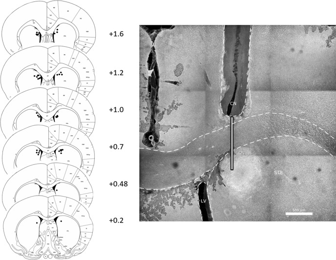
Figure 4.
Left panel, Infusion sites in aDMS. Numbers are anterior-posterior distance in mm from bregma. Right panel, Example cannula placement. Shown is the right hemisphere, and guide cannula (CN) track. Guide cannula tips were above the DMS. The thin white bar shows where the infusion cannula (which protruded 1 mm below the tip of the guide cannula) was. STR, striatum; LV, lateral ventricle. Scale bar = 500 μm.

